Choroidal Vasculopathy: Clinical Presentation, Diagnostic Evaluation, Management, and Outcomes – A Case Report
OthersPage Navigation
Abstract
Choroidal vasculopathy, commonly referred to as polypoidal choroidal vasculopathy (PCV), is a retinal disorder characterized by abnormal branching vascular networks and aneurysmal dilatations arising from the choroidal circulation beneath the retinal pigment epithelium. The condition is considered a subtype of neovascular age-related macular degeneration and is an important cause of vision loss, particularly in middle-aged and elderly individuals. Patients may present with blurred vision, metamorphopsia, recurrent subretinal hemorrhage, or sudden visual deterioration.
We present the case of a 64-year-old male who presented with progressive diminution of vision and distortion of images in the left eye. Multimodal retinal imaging including optical coherence tomography and indocyanine green angiography revealed features consistent with polypoidal choroidal vasculopathy. The patient underwent intravitreal anti-vascular endothelial growth factor therapy with significant anatomical and visual improvement during follow-up.
This case highlights the importance of early diagnosis, multimodal imaging, and timely retinal intervention in preventing irreversible visual loss associated with choroidal vasculopathy.
Introduction
Polypoidal choroidal vasculopathy is a chronic retinal vascular disorder involving abnormal choroidal blood vessels located beneath the retinal pigment epithelium. The disease is characterized by branching vascular networks with terminal aneurysmal or polyp-like dilatations that may leak fluid or bleed, leading to retinal damage and progressive visual impairment.
The condition most commonly affects individuals above 50 years of age and is more prevalent in Asian and African populations. Although the exact pathogenesis remains incompletely understood, several contributing factors have been identified, including:
- Age-related degeneration
- Choroidal vascular abnormalities
- Hypertension
- Smoking
- Genetic predisposition
- Chronic vascular endothelial dysfunction
Polypoidal choroidal vasculopathy may clinically resemble neovascular age-related macular degeneration, but it differs in disease behavior, imaging characteristics, and treatment response.
Patients may present with:
- Blurred central vision
- Metamorphopsia
- Scotomas
- Recurrent retinal hemorrhage
- Sudden visual loss
Without treatment, repeated leakage and hemorrhage can result in fibrosis, retinal scarring, and permanent vision loss. Early identification using advanced retinal imaging plays an important role in preserving visual function.
Case Report
Patient History
A 64-year-old male presented to the ophthalmology outpatient department with:
- Progressive blurring of vision in the left eye for 3 months
- Distortion of straight lines
- Difficulty reading
- Mild central visual shadow
The patient denied ocular trauma, redness, or pain.
Past medical history revealed:
- Hypertension for 12 years
- Type 2 diabetes mellitus
- Chronic smoking history
- Dyslipidemia
There was no previous history of retinal surgery or glaucoma.
Clinical Examination
General Examination
- Blood pressure: 150/90 mmHg
- Pulse rate: 84/min
- Afebrile
- Systemically stable
Ophthalmic Examination
Visual Acuity
- Right eye: 6/9
- Left eye: 6/36
Anterior Segment Examination
- Mild age-related cataract changes
- No active anterior segment inflammation
Fundus Examination
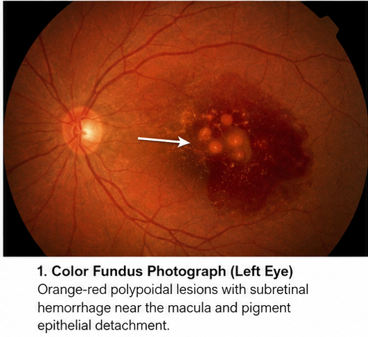
Left eye demonstrated:
- Orange-red subretinal nodular lesions
- Subretinal hemorrhage near the macula
- Pigment epithelial detachment
- Retinal pigment epithelium alterations
Right eye fundus was largely unremarkable.
Clinical Evaluation
Differential Diagnosis
Based on the clinical presentation, the following conditions were considered:
- Neovascular age-related macular degeneration
- Central serous chorioretinopathy
- Choroidal neovascular membrane
- Retinal angiomatous proliferation
- Polypoidal choroidal vasculopathy
The presence of subretinal hemorrhage and nodular vascular lesions strongly suggested polypoidal choroidal vasculopathy.
Investigations
Routine Laboratory Tests
- Complete blood count: Normal
- Blood glucose: Mildly elevated
- Renal function tests: Normal
- Lipid profile: Elevated cholesterol levels
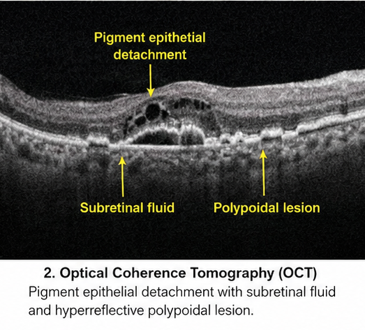
Optical Coherence Tomography (OCT)
OCT revealed:
- Pigment epithelial detachment
- Subretinal fluid accumulation
- Irregular retinal pigment epithelium elevation
- Hyperreflective polypoidal lesions
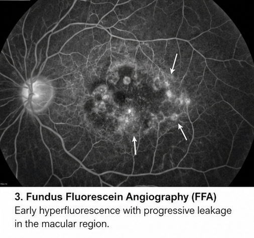
Fundus Fluorescein Angiography
Findings included:
- Areas of occult leakage
- Irregular choroidal vascular pattern
- Late staining near the macular region
Indocyanine Green Angiography (ICGA)
ICGA demonstrated:
- Branching vascular network
- Multiple polypoidal dilatations
- Active choroidal leakage
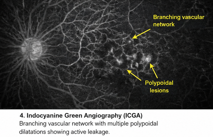
These findings confirmed the diagnosis of polypoidal choroidal vasculopathy.
Diagnosis
Based on clinical examination and retinal imaging findings, the diagnosis of Polypoidal Choroidal Vasculopathy with Active Choroidal Neovascular Activity was established.
Management and Outcome
Initial Management
The patient was advised:
- Strict blood pressure control
- Smoking cessation
- Glycemic optimization
- Regular retinal follow-up
Definitive Treatment
The patient underwent:
- Intravitreal anti-VEGF injections
Therapy was administered under aseptic precautions without procedural complications.
Follow-Up and Clinical Course
At 1 Month
- Reduction in subretinal fluid
- Improvement in visual distortion
- Visual acuity improved to 6/24
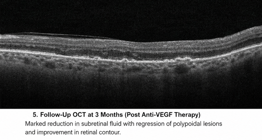
At 3 Months
- Significant regression of polypoidal lesions
- Minimal residual hemorrhage
- Stable retinal anatomy
At 6 Months
- Visual acuity improved to 6/18
- No fresh hemorrhage
- Improved reading ability and daily functioning
Discussion
Pathophysiology
Polypoidal choroidal vasculopathy results from abnormalities in the inner choroidal vasculature leading to aneurysmal vascular dilatations beneath the retinal pigment epithelium.
Important mechanisms include:
- Choroidal vascular remodeling
- Increased vascular permeability
- Endothelial dysfunction
- Chronic inflammation
- Degenerative retinal pigment epithelial changes
Repeated episodes of leakage and hemorrhage contribute to progressive retinal damage and visual decline.
Epidemiology
Key epidemiological features include:
- More common in individuals above 50 years
- Higher prevalence among Asian populations
- Male predominance in some studies
- Strong association with hypertension and smoking
- Important cause of severe central vision loss
Clinical Manifestations
Patients may present with:
- Blurred central vision
- Metamorphopsia
- Scotomas
- Reduced contrast sensitivity
- Sudden visual loss due to hemorrhage
Massive submacular hemorrhage may occur in advanced disease.
Diagnostic Considerations
Diagnosis requires multimodal retinal imaging.
Important diagnostic tools include:
- Fundus examination
- Optical coherence tomography
- Fundus fluorescein angiography
- Indocyanine green angiography
- OCT angiography
ICGA remains the gold standard for identifying polypoidal lesions and branching vascular networks.
Treatment Modalities
Medical Management
Conservative measures include:
- Control of systemic hypertension
- Smoking cessation
- Glycemic control
- Monitoring for recurrence
Interventional Therapy
Definitive treatment options include:
- Intravitreal anti-VEGF therapy
- Photodynamic therapy
- Combination therapy
- Laser photocoagulation in selected cases
Anti-VEGF therapy has become the primary treatment modality because of favorable anatomical and visual outcomes.
Complications
Potential complications include:
- Recurrent subretinal hemorrhage
- Retinal fibrosis
- Pigment epithelial tears
- Persistent macular edema
- Severe visual loss
- Retinal scarring
Delayed diagnosis may lead to irreversible retinal damage and permanent impairment of central vision.
Prognosis
Prognosis depends on:
- Early diagnosis
- Extent of hemorrhage
- Macular involvement
- Treatment response
- Frequency of recurrence
- Baseline visual acuity
Prompt retinal intervention significantly improves long-term visual outcomes and reduces retinal damage.
In this case, early multimodal imaging and anti-VEGF therapy resulted in substantial anatomical recovery and improvement in visual acuity.
Conclusion
Polypoidal choroidal vasculopathy is an important retinal vascular disorder that can lead to progressive and irreversible visual loss if untreated. Early recognition of symptoms such as blurred vision, metamorphopsia, and subretinal hemorrhage is essential for timely diagnosis.
This case highlights the critical role of multimodal retinal imaging, particularly indocyanine green angiography and optical coherence tomography, in establishing an accurate diagnosis and guiding treatment decisions. Early intervention with intravitreal anti-VEGF therapy can significantly improve retinal anatomy, preserve vision, and reduce disease recurrence.
Comprehensive long-term follow-up and management of systemic vascular risk factors remain important components of successful patient care.
References
- Yannuzzi LA, Wong DWK, Sforzolini BS, et al. Polypoidal choroidal vasculopathy and neovascularized age-related macular degeneration. https://pubmed.ncbi.nlm.nih.gov/11425670/
- Cheung CMG, Lai TYY, Ruamviboonsuk P, et al. Polypoidal choroidal vasculopathy: definition, pathogenesis, diagnosis, and management. https://pubmed.ncbi.nlm.nih.gov/28650920/
- Koh A, Lee WK, Chen LJ, et al. EVEREST study: efficacy and safety of verteporfin photodynamic therapy in polypoidal choroidal vasculopathy. https://pubmed.ncbi.nlm.nih.gov/22133795/
- Ciardella AP, Donsoff IM, Huang SJ, et al. Polypoidal choroidal vasculopathy. https://pubmed.ncbi.nlm.nih.gov/15177963/
- Wong CW, Yanagi Y, Lee WK, et al. Age-related macular degeneration and polypoidal choroidal vasculopathy in Asians. https://pubmed.ncbi.nlm.nih.gov/28237963/
- American Academy of Ophthalmology. Polypoidal Choroidal Vasculopathy Overview https://www.aao.org/education/topic-detail/polypoidal-choroidal-vasculopathy-pcv--asia-pacifi
Read more such content on @ Hidoc Dr | Medical Learning App for Doctors
Recommended News For You
Recommended Articles For You
Featured News
Featured Articles
Featured Events
Featured KOL Videos
1.
Novel ADC Improves Survival in Metastatic TNBC
2.
An Examine More Into the Acceptance of CRISPR/Cas9 Gene Therapy for Sickle Cell Illness.
3.
Celebrity Cancers Stoking Fear? Cisplatin Shortage Ends; Setback for Anti-TIGIT
4.
Pancreatic cancer RNA vaccine shows durable T cell immunity
5.
Healthcare in the Mix in President Biden's Farewell Address
1.
Interpreting Iron Studies: What Your Blood Results Really Mean
2.
Unveiling New Hope: Potential Therapeutic Targets in Hematological Malignancies
3.
Feline Anemia: Diagnosis and Treatment with Focus on Rasburicase Complications
4.
Andexanet for Factor Xa Inhibitor-Associated Acute Intracerebral Hemorrhage
5.
Biologic Therapies for Cutaneous Immune-Related Adverse Events in the Era of Immune Checkpoint Inhibitors
1.
Asian Symposium on Advancement in Hematology and Oncology
2.
Asian Symposium on Advancement in Hematology and Oncology
3.
Asian Symposium on Advancement in Hematology and Oncology
4.
International Cancer Conference
5.
Asian Symposium on Advancement in Hematology and Oncology
1.
Redefining Treatment Pathways in Relapsed/Refractory Adult B-Cell ALL
2.
Breaking Down PALOMA-2: How CDK4/6 Inhibitors Redefined Treatment for HR+/HER2- Metastatic Breast Cancer
3.
Untangling The Best Treatment Approaches For ALK Positive Lung Cancer - Part I
4.
Cost Burden/ Burden of Hospitalization For R/R ALL Patients
5.
Untangling The Best Treatment Approaches For ALK Positive Lung Cancer - Part VI

Address :
Hidoc Dr. Inc. | Delaware C Corp | 1309 Coffeen Ave. Suite 1200, Sheridan WY, 82801
Phone :
+1-415-463-3094
Email :
anishagadia@hidoc.co
© Copyright 2026 Hidoc Dr. Inc.
Terms & Conditions - LLP | Inc. | Privacy Policy - LLP | Inc. | Account Deactivation

To get started please enter your email ID
Welcome to Hidoc Dr.
Join to enhance your clinical skills and gain specialized in-depth medical knowledge