Acute Glomerulonephritis Presenting With Hematuria, Edema, and Hypertension: A Case Report
OthersPage Navigation
Introduction
Glomerulonephritis refers to a group of disorders characterized by inflammation and injury of the renal glomeruli. The clinical presentation varies according to the underlying cause and severity of glomerular involvement. Patients may present with microscopic or gross hematuria, proteinuria, edema, hypertension, and varying degrees of impairment in renal function.
Acute glomerulonephritis commonly manifests as an acute nephritic syndrome characterized by hematuria, reduced glomerular filtration, fluid retention, and hypertension. Infection-related glomerulonephritis is an important cause and may develop during or following bacterial infections. Post-streptococcal glomerulonephritis represents the classical form and may occur following streptococcal pharyngitis or skin infection.
Evaluation requires careful assessment of the clinical history, urine findings, renal function, complement levels, and evidence of a preceding or ongoing infection. Early recognition is important because severe disease may lead to acute kidney injury, significant fluid overload, hypertensive complications, or rapidly progressive loss of renal function.
Case Report
A 28-year-old man presented to the outpatient department with a five-day history of progressively increasing swelling around the eyes and both lower limbs, associated with reduced urine output and dark-colored urine.
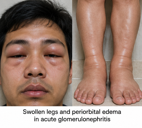
The patient had initially noticed puffiness around the eyes on waking in the morning. During the following three days, swelling gradually developed around both ankles and lower legs. He also observed that his urine had become dark brown and that the overall volume of urine had decreased.
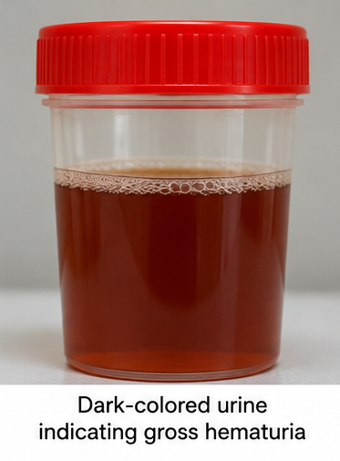
He complained of mild headache, generalized weakness, loss of appetite, and a sensation of heaviness in the lower limbs. There was no history of severe abdominal pain, vomiting, diarrhea, dysuria, urinary frequency, flank pain, or visible blood clots in the urine.
On further questioning, the patient reported an episode of sore throat with fever approximately two weeks before the onset of the current symptoms. The illness had improved with symptomatic treatment, and he had not undergone further medical evaluation.
There was no previous history of kidney disease, recurrent urinary tract infection, nephrolithiasis, diabetes mellitus, or hypertension. He had no known autoimmune disease and denied recent use of potentially nephrotoxic medications.
On examination, the patient was conscious and oriented. He had mild bilateral periorbital edema and bilateral pitting pedal edema extending to the lower legs.
His blood pressure was elevated at 156/96 mmHg. Pulse rate was 86 beats/minute and regular. There was no significant respiratory distress.

Cardiovascular and abdominal examinations did not reveal any major abnormality.
The combination of acute edema, hypertension, oliguria, and dark-colored urine following a recent upper respiratory infection raised suspicion of an acute nephritic syndrome, most likely acute post-infectious glomerulonephritis.
Investigations
Urinalysis demonstrated significant hematuria with mild-to-moderate proteinuria. Microscopic examination of the urinary sediment revealed numerous red blood cells, including dysmorphic erythrocytes, along with red blood cell casts, supporting a glomerular source of bleeding.
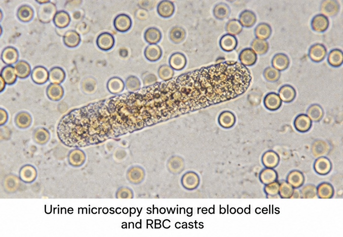
Complete blood count showed no major hematological abnormality apart from mild leukocytosis. Serum creatinine and blood urea nitrogen were moderately elevated, indicating acute impairment of renal function. Serum electrolytes were monitored closely because of the risk of abnormalities associated with reduced renal filtration.
Serum complement C3 was reduced, while evaluation for evidence of a preceding streptococcal infection was supportive of a recent infectious trigger.
Renal ultrasonography demonstrated normal-sized kidneys without hydronephrosis or evidence of urinary tract obstruction.
Based on the temporal relationship with the preceding respiratory infection, nephritic urinary sediment, reduced complement level, edema, hypertension, and impaired renal function, a clinical diagnosis of acute post-infectious glomerulonephritis was made.
Renal biopsy was not considered immediately necessary because the clinical course and laboratory findings were consistent with an acute infection-related process and renal function was closely monitored.
Differential Diagnosis
IgA Nephropathy
IgA nephropathy commonly presents with microscopic or gross hematuria and may be associated with an upper respiratory infection. However, hematuria typically develops during or shortly after the infection. In the present case, the interval between respiratory illness and renal manifestations, together with reduced complement levels, favored post-infectious glomerulonephritis.
Lupus Nephritis
Lupus nephritis may present with hematuria, proteinuria, hypertension, and renal impairment. However, the patient had no clinical history suggestive of systemic lupus erythematosus or other systemic autoimmune manifestations.
Rapidly Progressive Glomerulonephritis
Rapidly progressive glomerulonephritis is characterized by rapid deterioration in renal function and may be associated with crescent formation on renal biopsy. The relatively stable clinical condition and subsequent improvement in renal parameters made this less likely.
Acute Interstitial Nephritis
Acute interstitial nephritis can cause acute kidney injury, particularly following exposure to certain medications or infections. However, prominent glomerular hematuria with red blood cell casts and the nephritic clinical picture favored glomerulonephritis.
Urinary Tract Disease
Urinary stones, urinary tract infection, and structural urinary abnormalities can cause hematuria. However, these conditions do not typically produce dysmorphic erythrocytes and red blood cell casts accompanied by edema and hypertension.
Management and Outcome
The patient was managed with close monitoring of renal function, blood pressure, urine output, fluid balance, and serum electrolytes.
Dietary sodium restriction was advised to help control edema and hypertension. Fluid intake was adjusted according to urine output and clinical volume status.
A diuretic was administered for clinically significant fluid retention and edema. Antihypertensive treatment was initiated because blood pressure remained elevated during the initial observation period.
Because of the history of recent infection, the patient was evaluated for evidence of persistent infection. Management focused primarily on supportive treatment of the renal manifestations.
During the following several days, urine output gradually increased. Periorbital and lower-limb edema progressively decreased, and blood pressure showed improvement with treatment.
Serial renal function testing demonstrated stabilization followed by a gradual decline in serum creatinine. The urine became visibly clearer, although microscopic hematuria persisted during the early recovery period.
The patient did not develop severe hyperkalemia, pulmonary edema, hypertensive emergency, or progressive renal failure requiring renal replacement therapy.
At follow-up, edema had resolved substantially, blood pressure was controlled, and renal function continued to improve. The patient was advised to continue regular monitoring of blood pressure, serum creatinine, estimated glomerular filtration rate, and urinalysis until complete clinical and biochemical recovery.
Discussion
Glomerulonephritis represents glomerular injury produced through a variety of immune-mediated and inflammatory mechanisms. Clinical manifestations depend on the extent and pattern of glomerular damage.
Acute nephritic syndrome is one of the characteristic presentations and includes hematuria, proteinuria, reduced renal function, oliguria, edema, and hypertension. Hematuria of glomerular origin may be accompanied by dysmorphic red blood cells and red blood cell casts on urinary sediment examination.
Infection-related glomerulonephritis encompasses several forms of glomerular injury associated with bacterial and other infections. Post-streptococcal glomerulonephritis remains the classical example, although the spectrum of infection-related glomerulonephritis has expanded considerably.
Glomerular injury is largely mediated by immune and complement mechanisms. Complement deposition, with or without immunoglobulin deposition, produces an inflammatory response within the glomeruli. Reduced serum C3 can therefore provide an important diagnostic clue in the appropriate clinical setting.

The temporal relationship between infection and renal manifestations can also assist diagnosis. Post-streptococcal glomerulonephritis typically develops after an interval following the infectious episode, whereas IgA nephropathy commonly produces visible hematuria during or shortly after a respiratory or gastrointestinal infection.
Management of uncomplicated post-infectious glomerulonephritis is predominantly supportive. Careful control of fluid status and blood pressure is important. Sodium restriction and diuretic therapy may be required when clinically significant edema or volume overload is present.
Renal function, serum potassium, blood pressure, body weight, and urine output should be monitored because severe glomerular dysfunction may result in fluid overload, electrolyte disturbances, and acute kidney injury.
Kidney biopsy may be required when the presentation is atypical, complement abnormalities persist, renal function deteriorates rapidly, proteinuria is unusually severe, or the diagnosis remains uncertain. Histopathological assessment can help differentiate infection-related glomerulonephritis from IgA nephropathy, C3 glomerulopathy, lupus nephritis, and crescentic glomerulonephritis.
In the present case, the preceding respiratory infection followed by edema, gross hematuria, oliguria, hypertension, nephritic urinary sediment, reduced complement, and impaired renal function was consistent with acute post-infectious glomerulonephritis. Supportive management resulted in progressive improvement without the need for dialysis.
Conclusion
Acute glomerulonephritis should be considered in patients presenting with the combination of hematuria, edema, hypertension, oliguria, and impaired renal function.
A detailed history may reveal a preceding respiratory or cutaneous infection that provides an important clue to infection-related glomerular disease. Urinalysis demonstrating dysmorphic red blood cells and red blood cell casts supports glomerular hematuria, while complement testing and assessment of renal function help establish the clinical pattern and severity.
Management of uncomplicated post-infectious glomerulonephritis is primarily supportive and includes careful fluid management, sodium restriction, control of edema and hypertension, and serial monitoring of renal function.
In this patient, early recognition of the nephritic presentation, appropriate evaluation, and supportive management were associated with progressive improvement in edema, blood pressure, urine output, and renal function.
References
- Khalighi MA, Chang A. Infection-Related Glomerulonephritis. Glomerular Dis. 2021;1(2):82–91. https://pmc.ncbi.nlm.nih.gov/articles/PMC9677723/
- Kidney Disease: Improving Global Outcomes (KDIGO). Chapter 9: Infection-related glomerulonephritis. Kidney Int Suppl. 2012;2(2):200–208. https://pmc.ncbi.nlm.nih.gov/articles/PMC4089740/
- Satoskar AA, Parikh SV, Nadasdy T. Epidemiology, pathogenesis, treatment and outcomes of infection-associated glomerulonephritis. Nat Rev Nephrol. 2020;16:32–50. https://pubmed.ncbi.nlm.nih.gov/31705188/
- Kanjanabuch T, Kittikowit W, Eiam-Ong S. An update on acute postinfectious glomerulonephritis worldwide. Nat Rev Nephrol. 2009;5(5):259–269. https://pubmed.ncbi.nlm.nih.gov/19384331/
- Takayasu M, Hirayama K, Shimohata H, et al. Clinico-Pathogenic Similarities and Differences between Infection-Related Glomerulonephritis and C3 Glomerulopathy. Int J Mol Sci. 2023;24(9):8097. https://pmc.ncbi.nlm.nih.gov/articles/PMC10179079/
Read more such content on @ Hidoc Dr | Medical Learning App for Doctors
Recommended News For You
Recommended Articles For You
Featured News
Featured Articles
Featured Events
Featured KOL Videos
1.
Tumor characteristics found to differ for melanomas in children, teens and young adults
2.
Unexplained CAR-T Death; Americans Spurn Sunscreen; Improving Diversity in Trials
3.
"A lot" of aspirin reduces the risk of ovarian cancer, regardless of genetic factors, according to the JAMA study.
4.
Approval in Endometrial Cancer Expanded; Masks at Cancer Centers; NPR Reporter Dies
5.
Is Lenalidomide Resistance in Myeloma Reversible with JAK Inhibition?
1.
Modern Oncology Unveiled with Treatment Innovations and Personalized Cancer Care
2.
Screening for Early Therapy-Related Cancer Susceptibility
3.
Unlocking the Potential of Bicalutamide 50 mg: A Revolutionary New Treatment for Prostate Cancer
4.
Skin-Sparing Mastectomy: Balancing Cancer Control with Cosmetic Results
5.
Reducing Modifiable Childhood Cancer Exposure Risks
1.
International Conference on Innovations in Critical Care for Oncology and Cardiology
2.
International Conference on Oncology, Cardiology and Critical Care Policy
3.
International Conference on Oncology, Cancer Prevention and Public Health
4.
International Conference on Cancer Nursing and Rehabilitation Strategies
5.
International Conference on Best Practices in Oncology, Cardiology and Critical Care
1.
Dacomitinib Case Presentation: Baseline Treatment and Current Status
2.
Targeting Oncologic Drivers with Dacomitinib: Further Discussion on Lung Cancer Treatment
3.
Should We Use DARA Up Front As First-Line Therapy in MM?
4.
Iron Deficiency Anemia: Ferric Maltol As a New Treatment Option- A New Perspective
5.
Importance of Cancer Screening and Early Detection

Address :
Hidoc Dr. Inc. | Delaware C Corp | 1309 Coffeen Ave. Suite 1200, Sheridan WY, 82801
Phone :
+1-415-463-3094
Email :
anishagadia@hidoc.co
© Copyright 2026 Hidoc Dr. Inc.
Terms & Conditions - LLP | Inc. | Privacy Policy - LLP | Inc. | Account Deactivation

To get started please enter your email ID
Welcome to Hidoc Dr.
Join to enhance your clinical skills and gain specialized in-depth medical knowledge