External Hordeolum of the Upper Eyelid: A Case Report of Successful Conservative Management
OthersPage Navigation
Introduction
A stye, medically termed a hordeolum, is an acute, localized inflammatory lesion of the eyelid, usually associated with bacterial infection of an eyelid gland. It commonly presents as a painful, erythematous, localized swelling near the eyelid margin. Hordeola are frequently encountered in primary care, ophthalmology, and emergency clinical practice.
A hordeolum may be classified as external or internal according to the involved eyelid structure. An external hordeolum usually develops from infection of the glands of Zeis or Moll associated with an eyelash follicle, whereas an internal hordeolum involves the meibomian glands located within the tarsal plate.
Staphylococcus aureus is commonly implicated in the development of hordeola. Predisposing factors may include chronic blepharitis, meibomian gland dysfunction, previous episodes of hordeolum, poor eyelid hygiene, seborrheic dermatitis, and certain dermatological conditions.
The diagnosis is predominantly clinical. Most uncomplicated lesions resolve spontaneously or with conservative measures such as warm compresses and appropriate eyelid hygiene. However, persistent or complicated lesions may require topical or systemic treatment, drainage, or ophthalmological evaluation.
Early clinical assessment is important to differentiate a hordeolum from other eyelid lesions, particularly chalazion, preseptal cellulitis, orbital cellulitis, and recurrent or atypical eyelid masses.
Case Report
A 29-year-old woman presented to the outpatient clinic with a three-day history of painful swelling of the right upper eyelid.
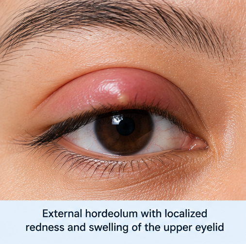
The patient initially noticed mild discomfort and localized redness near the eyelid margin. Over the following two days, the swelling gradually increased and became tender to touch. She also reported mild watering from the affected eye and discomfort while blinking.
There was no history of trauma, foreign-body exposure, chemical exposure, recent ocular surgery, or contact-lens use. She denied visual blurring, diplopia, photophobia, severe ocular pain, headache, fever, or purulent discharge from the eye.
The patient reported frequently touching and rubbing her eyes during prolonged computer use. She had experienced occasional mild eyelid irritation previously but had never required ophthalmological treatment.
There was no known history of diabetes mellitus, immunosuppression, chronic ocular disease, or recurrent skin infection.
On examination, the patient was afebrile and clinically stable.
Ocular examination demonstrated a small, localized, erythematous and tender swelling along the right upper eyelid margin. A focal pustular point was visible close to the base of an eyelash.
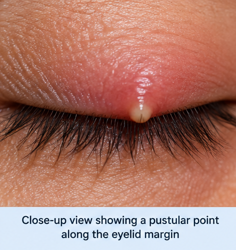
Mild surrounding eyelid edema was present, but there was no diffuse erythema extending beyond the immediate area of the lesion.
The conjunctiva showed minimal injection. There was no significant purulent conjunctival discharge.
Visual acuity was preserved in both eyes. Pupillary responses were normal, and extraocular movements were full and painless.
There was no proptosis, ophthalmoplegia, relative afferent pupillary defect, or evidence of orbital involvement.
The clinical appearance was consistent with an external hordeolum of the right upper eyelid.
Investigations
The diagnosis of an uncomplicated hordeolum is generally clinical, and extensive laboratory or imaging investigations are not routinely required.
In this patient, visual acuity assessment demonstrated no reduction in vision. External ocular examination showed a localized tender lesion involving the eyelid margin without evidence of deeper orbital disease.
No routine blood investigations were considered necessary because the patient was afebrile, systemically well, and had no clinical evidence of spreading infection.
Microbiological culture was not performed because there was no recurrent, unusually severe, or treatment-resistant infection.
Orbital imaging was also not indicated because there was no proptosis, painful or restricted ocular movement, visual impairment, systemic toxicity, or other clinical feature suggesting orbital cellulitis.
The characteristic localization of the lesion at the eyelid margin, associated tenderness, erythema, and focal pustular appearance supported the clinical diagnosis of an external hordeolum.
Differential Diagnosis
Chalazion
A chalazion is a chronic inflammatory lesion resulting from obstruction of a meibomian gland. It commonly presents as a firm, localized eyelid nodule and is typically less painful than an acute hordeolum.
In the present patient, the acute onset, marked tenderness, erythema, and pustular point at the eyelid margin favored a hordeolum rather than a chalazion.
Preseptal Cellulitis
Preseptal cellulitis may produce eyelid swelling, erythema, warmth, and tenderness. However, the inflammation is generally more diffuse rather than confined to a small focal lesion.
The absence of extensive eyelid involvement, fever, systemic symptoms, and progressive surrounding erythema made preseptal cellulitis less likely.
Orbital Cellulitis
Orbital cellulitis is a potentially serious infection involving tissues posterior to the orbital septum. Clinical features may include fever, proptosis, painful or restricted extraocular movements, reduced vision, diplopia, and systemic illness.
The absence of these findings effectively reduced clinical concern for orbital involvement in this patient.
Blepharitis
Blepharitis can cause eyelid-margin erythema, irritation, burning, crusting, and ocular discomfort. Although blepharitis may predispose to recurrent hordeola, the presence of a discrete, acutely painful pustular lesion was more characteristic of a hordeolum.
Eyelid Neoplasm
Persistent, recurrent, atypical, or non-healing eyelid lesions require evaluation for alternative diagnoses, including eyelid malignancy. Features such as lash loss, ulceration, abnormal vascularity, progressive enlargement, or repeated recurrence at the same location should prompt further ophthalmological assessment.
None of these features was observed in the present patient.
Management and Outcome
The patient was advised to apply a warm compress to the affected eyelid several times daily. Each application was continued for approximately 10–15 minutes to facilitate drainage and resolution of the obstructed gland.
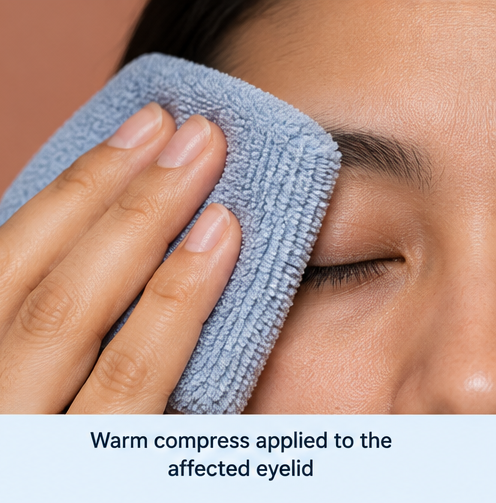
She was advised to maintain appropriate eyelid hygiene and to avoid rubbing, squeezing, puncturing, or attempting to manually drain the lesion.
The patient was also instructed to wash her hands before touching the periocular region and temporarily avoid eye makeup until the lesion had completely resolved.
Because the lesion was localized and there was no evidence of spreading infection or systemic involvement, systemic antibiotic therapy was not required.
The patient was advised to return promptly if she developed increasing eyelid swelling, fever, visual disturbance, severe ocular pain, painful or restricted eye movement, or rapidly progressive redness.
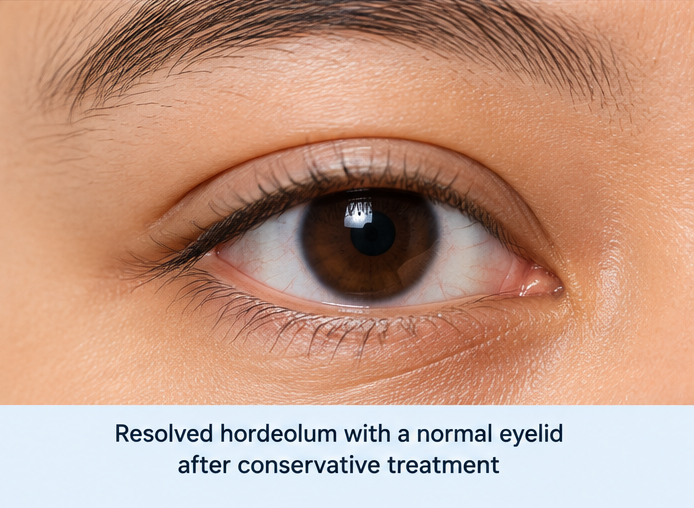
At follow-up, the pain and tenderness had substantially decreased. The localized swelling gradually reduced and the lesion resolved without complications.
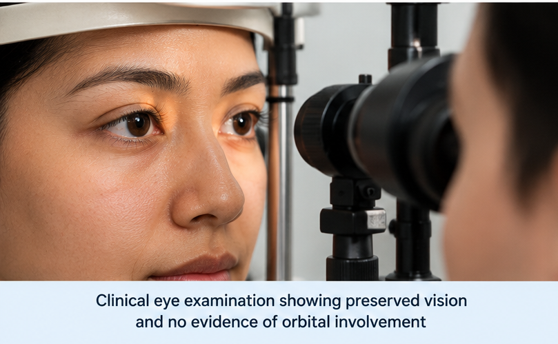
Visual acuity remained unaffected, and no evidence of preseptal or orbital extension developed.
Discussion
Hordeolum is one of the common acute inflammatory disorders affecting the eyelid. It occurs when an eyelid gland or associated follicular structure becomes obstructed and inflamed, frequently in association with bacterial infection.
The anatomical location determines whether the lesion is classified as external or internal. An external hordeolum develops near the eyelid margin and usually involves an eyelash follicle and its associated sebaceous or sweat glands. It commonly appears as a superficial tender pustule.
An internal hordeolum, in contrast, develops from infection involving a meibomian gland and may therefore produce deeper eyelid tenderness and swelling.
Typical clinical manifestations include localized eyelid pain, erythema, swelling, tenderness, and sometimes a visible pustular point. Mild tearing, foreign-body sensation, and discomfort while blinking may also occur.
The diagnosis is usually established through history and clinical examination. Additional testing is generally unnecessary in a straightforward presentation. Investigation becomes more relevant when the condition is recurrent, unusually severe, atypical, resistant to treatment, or associated with suspected cellulitis or systemic disease.
Conservative therapy remains central to the management of uncomplicated hordeola. Warm compresses are commonly recommended to promote drainage of glandular secretions and encourage spontaneous resolution.
Patients should be discouraged from squeezing or puncturing the lesion because manipulation may increase local inflammation and potentially facilitate spread of infection.
Antibiotic therapy is not necessarily required for every uncomplicated hordeolum. Clinical judgment should guide its use, particularly when there is associated blepharoconjunctivitis, secondary infection, or extension into surrounding eyelid tissues.
Persistent lesions may occasionally require incision and drainage by an ophthalmologist. A lesion that becomes chronic and relatively painless may evolve into or clinically resemble a chalazion.
An important component of clinical evaluation is recognition of complications and alternative diagnoses. Diffuse progressive eyelid erythema may suggest preseptal cellulitis, while visual impairment, proptosis, ophthalmoplegia, or painful eye movements should raise concern for orbital cellulitis and warrant urgent assessment.
Recurrent hordeola may be associated with underlying blepharitis or meibomian gland dysfunction. Addressing eyelid hygiene and associated ocular surface disease may therefore be important in patients experiencing repeated episodes.
In the present patient, the acute localized tender swelling and focal pustular lesion at the eyelid margin were characteristic of an external hordeolum. Preserved vision, normal ocular motility, absence of systemic illness, and lack of diffuse eyelid inflammation supported an uncomplicated presentation.
Conservative treatment with warm compresses and eyelid hygiene was followed by progressive clinical improvement and complete resolution without complications.
Conclusion
A stye or hordeolum is a common acute inflammatory condition of the eyelid that typically presents with localized pain, erythema, swelling, and tenderness.
External hordeola occur near the eyelid margin, whereas internal hordeola involve the deeper meibomian glands.
Diagnosis is usually clinical, and extensive investigations are generally unnecessary in uncomplicated cases.
Warm compresses, appropriate eyelid hygiene, avoidance of manipulation, and observation are central components of conservative management.
Clinicians should evaluate for progressive surrounding inflammation, visual disturbance, painful or restricted ocular movement, proptosis, fever, or other findings that could indicate preseptal or orbital extension.
Persistent, atypical, or recurrent lesions warrant further evaluation to identify underlying eyelid disease or an alternative diagnosis.
In this case, appropriate clinical recognition and conservative management resulted in complete resolution without visual impairment or infectious complications.
References
- Lindsley K, Nichols JJ, Dickersin K. Interventions for acute internal hordeolum. Cochrane Database Syst Rev. 2017;1(1). https://pmc.ncbi.nlm.nih.gov/articles/PMC5370090/
- Carlisle RT, Digiovanni J. Differential diagnosis of the swollen red eyelid. Am Fam Physician. 2015;92(2):106–112. https://www.aafp.org/pubs/afp/issues/2015/0715/p106.html
- Amescua G, Akpek EK, Farid M, et al. Blepharitis Preferred Practice Pattern®. Ophthalmology. 2019;126(1)–P93. https://pubmed.ncbi.nlm.nih.gov/30366797/
- Wladis EJ, Bradley EA, Bilyk JR, et al. Orbital cellulitis clinical statement. Ophthalmology. https://pubmed.ncbi.nlm.nih.gov/
- Willmann D, Guier CP, Patel BC, Melanson SW. Hordeolum (Stye). StatPearls. Treasure Island (FL): StatPearls Publishing. https://www.ncbi.nlm.nih.gov/books/NBK459349/
- Eberhardt M, Rammohan G. Blepharitis. StatPearls. Treasure Island (FL): StatPearls Publishing. https://www.ncbi.nlm.nih.gov/books/NBK459305/
Read more such content on @ Hidoc Dr | Medical Learning App for Doctors
Recommended News For You
Recommended Articles For You
Featured News
Featured Articles
Featured Events
Featured KOL Videos
1.
Tumor characteristics found to differ for melanomas in children, teens and young adults
2.
Unexplained CAR-T Death; Americans Spurn Sunscreen; Improving Diversity in Trials
3.
"A lot" of aspirin reduces the risk of ovarian cancer, regardless of genetic factors, according to the JAMA study.
4.
Approval in Endometrial Cancer Expanded; Masks at Cancer Centers; NPR Reporter Dies
5.
Is Lenalidomide Resistance in Myeloma Reversible with JAK Inhibition?
1.
Modern Oncology Unveiled with Treatment Innovations and Personalized Cancer Care
2.
Screening for Early Therapy-Related Cancer Susceptibility
3.
Unlocking the Potential of Bicalutamide 50 mg: A Revolutionary New Treatment for Prostate Cancer
4.
Skin-Sparing Mastectomy: Balancing Cancer Control with Cosmetic Results
5.
Reducing Modifiable Childhood Cancer Exposure Risks
1.
International Conference on Innovations in Critical Care for Oncology and Cardiology
2.
International Conference on Oncology, Cardiology and Critical Care Policy
3.
International Conference on Oncology, Cancer Prevention and Public Health
4.
International Conference on Cancer Nursing and Rehabilitation Strategies
5.
International Conference on Best Practices in Oncology, Cardiology and Critical Care
1.
Dacomitinib Case Presentation: Baseline Treatment and Current Status
2.
Targeting Oncologic Drivers with Dacomitinib: Further Discussion on Lung Cancer Treatment
3.
Should We Use DARA Up Front As First-Line Therapy in MM?
4.
Iron Deficiency Anemia: Ferric Maltol As a New Treatment Option- A New Perspective
5.
Importance of Cancer Screening and Early Detection

Address :
Hidoc Dr. Inc. | Delaware C Corp | 1309 Coffeen Ave. Suite 1200, Sheridan WY, 82801
Phone :
+1-415-463-3094
Email :
anishagadia@hidoc.co
© Copyright 2026 Hidoc Dr. Inc.
Terms & Conditions - LLP | Inc. | Privacy Policy - LLP | Inc. | Account Deactivation

To get started please enter your email ID
Welcome to Hidoc Dr.
Join to enhance your clinical skills and gain specialized in-depth medical knowledge