Choroiditis Presenting as Progressive Unilateral Blurred Vision with Floaters: A Case Report
OthersPage Navigation
Abstract
Choroiditis is an inflammatory disorder of the choroid that may occur as an isolated condition or as part of posterior uveitis. It may result from infectious or non-infectious etiologies and can lead to irreversible visual impairment if not diagnosed and treated promptly. Patients commonly present with blurred vision, floaters, photophobia, metamorphopsia, or visual field defects. Clinical examination and multimodal retinal imaging are essential for diagnosis, while laboratory investigations help identify the underlying cause. Treatment depends on the etiology and may include corticosteroids, immunosuppressive agents, or antimicrobial therapy. We report the case of a 34-year-old woman presenting with progressive unilateral blurred vision caused by non-infectious choroiditis. Early diagnosis and prompt corticosteroid therapy resulted in complete resolution of inflammation with preservation of visual acuity.
Introduction
Choroiditis is characterized by inflammation of the choroid and often involves adjacent structures including the retinal pigment epithelium and retina, resulting in chorioretinitis. The condition may be infectious, caused by organisms such as Toxoplasma gondii, Mycobacterium tuberculosis, herpes viruses, or fungi, or non-infectious, associated with autoimmune diseases including Vogt–Koyanagi–Harada disease, sarcoidosis, and Behçet disease.
Clinical manifestations vary depending on lesion location and severity but commonly include blurred vision, floaters, photophobia, decreased contrast sensitivity, and central or peripheral visual field defects. Prompt recognition is critical because untreated inflammation may result in permanent retinal damage, choroidal neovascularization, and irreversible vision loss.
Case Report
A 34-year-old woman presented to the ophthalmology clinic with gradually worsening blurred vision and multiple floaters in her left eye for two weeks. She also reported mild photophobia but denied ocular pain, redness, trauma, or previous ocular surgery.
Her medical history was unremarkable, with no known autoimmune disease, tuberculosis, or recent systemic infection. There was no history of similar ocular episodes or significant family history of inflammatory eye disease.
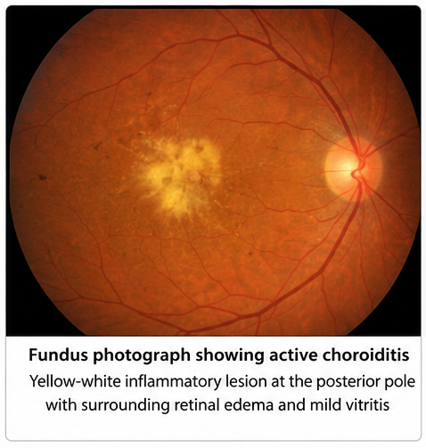
On examination, best-corrected visual acuity was 6/6 in the right eye and 6/18 in the left eye. Anterior segment examination was normal except for mild vitreous cells in the left eye. Fundus examination revealed a yellow-white inflammatory lesion involving the posterior pole with surrounding retinal edema and mild vitritis. The right eye appeared normal.
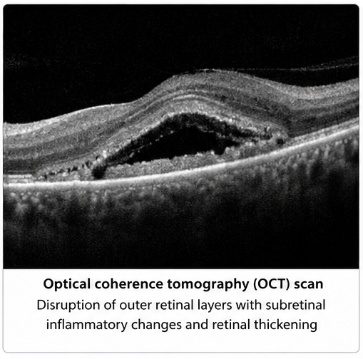
Optical coherence tomography demonstrated disruption of the outer retinal layers with subretinal inflammatory changes.
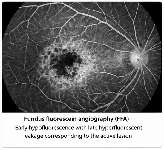
Fundus fluorescein angiography showed early hypofluorescence with late hyperfluorescent leakage corresponding to the active lesion.
Optical coherence tomography angiography excluded choroidal neovascularization.
Routine blood investigations were within normal limits. Serological testing for toxoplasmosis, syphilis, HIV, tuberculosis screening, and autoimmune markers were negative. Based on the clinical findings and exclusion of infectious causes, a diagnosis of unilateral non-infectious posterior choroiditis was established.
Management and Outcome
The patient was started on oral prednisolone at a dose of 1 mg/kg/day after infectious causes had been reasonably excluded through appropriate laboratory investigations. In addition, topical corticosteroid eye drops and cycloplegic agents were prescribed to control ocular inflammation, relieve ciliary muscle spasm, and improve patient comfort. The patient was advised regarding strict adherence to therapy and scheduled for regular ophthalmic follow-up with serial fundus examinations and optical coherence tomography (OCT).
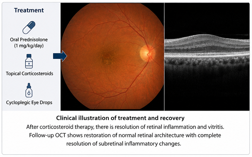
Oral corticosteroids were gradually tapered over an eight-week period according to the clinical response, with careful monitoring for disease recurrence and potential steroid-related adverse effects.
During follow-up, the patient demonstrated progressive improvement in visual acuity, accompanied by a marked reduction in vitreous inflammation and complete resolution of subjective symptoms, including floaters and photophobia. Serial OCT imaging showed restoration of the normal outer retinal architecture, resolution of subretinal inflammatory changes, and recovery of retinal morphology without evidence of macular edema or choroidal neovascularization. Fundus examination confirmed complete healing of the active choroidal lesion with minimal residual pigmentary alteration.
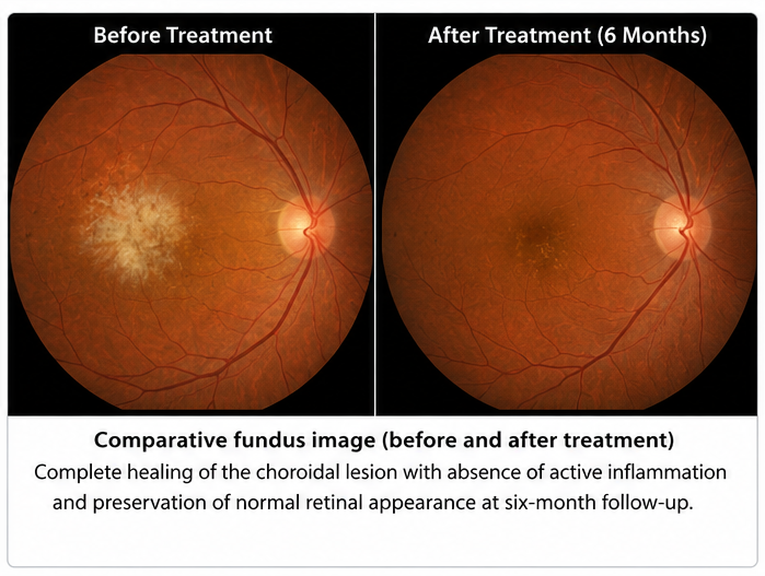
As the patient achieved sustained remission with corticosteroid therapy alone, additional immunosuppressive agents or biologic therapy were not required, and visual function remained stable throughout the follow-up period.
Follow-up
One Month
- Significant reduction in retinal inflammation.
- Visual acuity improved to 6/9.
- Floaters markedly reduced.
Three Months
- Complete resolution of active choroiditis.
- Visual acuity restored to 6/6.
- OCT demonstrated near-normal retinal anatomy.
Six Months
- No recurrence of inflammation.
- Stable retinal findings.
- Excellent visual outcome without complications.
Discussion
Choroiditis represents inflammation of the highly vascular choroidal tissue and may arise from infectious or immune-mediated mechanisms. Diagnosis relies on detailed ophthalmic examination supported by multimodal imaging, including optical coherence tomography, fluorescein angiography, indocyanine green angiography when indicated, and fundus autofluorescence.
A thorough systemic evaluation is essential to identify infectious causes before initiating corticosteroid therapy, as inappropriate immunosuppression may worsen underlying infections.
Differential diagnoses include central serous chorioretinopathy, age-related macular degeneration, posterior scleritis, retinal vasculitis, ocular lymphoma, and intraocular tumors.
Management depends on the underlying etiology. Non-infectious choroiditis is primarily treated with corticosteroids, while recurrent or severe disease may require steroid-sparing immunosuppressive agents or biologic therapy. Infectious choroiditis requires pathogen-specific antimicrobial treatment with corticosteroids introduced when appropriate.
Early intervention and regular follow-up with retinal imaging are essential to prevent permanent visual loss and detect complications such as choroidal neovascularization, macular edema, retinal scarring, and recurrence.
Prognosis
The visual prognosis depends on the underlying cause, lesion location, and promptness of treatment. Early diagnosis and appropriate therapy often result in good visual recovery. Delayed treatment may lead to irreversible retinal damage, recurrent inflammation, and permanent vision impairment. Long-term monitoring remains important because relapse may occur even after apparent clinical resolution.
Conclusion
Choroiditis should be considered in patients presenting with unilateral blurred vision, floaters, photophobia, or posterior segment inflammatory lesions, particularly when symptoms progress over a short period. A comprehensive ophthalmic examination supported by multimodal retinal imaging—including optical coherence tomography, fundus fluorescein angiography, and, when indicated, indocyanine green angiography—along with targeted laboratory investigations is essential for establishing the diagnosis, determining the underlying etiology, and excluding infectious causes before initiating immunosuppressive therapy. Early recognition and prompt treatment with corticosteroids for non-infectious choroiditis can effectively suppress inflammation, preserve retinal structure and visual function, reduce the risk of complications such as choroidal neovascularization and permanent retinal scarring, and improve long-term visual outcomes. Regular follow-up with clinical examination and retinal imaging remains crucial to monitor treatment response, detect recurrence, and ensure sustained disease control.
References
- Jabs DA, Nussenblatt RB, Rosenbaum JT. Standardization of Uveitis Nomenclature (SUN) Working Group. Standardization of Uveitis Nomenclature for Reporting Clinical Data. American Journal of Ophthalmology. 2005;140(3):509–516. https://pubmed.ncbi.nlm.nih.gov/16196117/
- Herbort CP Jr, Rao NA, Mochizuki M. International Criteria for the Diagnosis of Ocular Sarcoidosis. Ocular Immunology and Inflammation. 2009;17(3):160–169. https://pubmed.ncbi.nlm.nih.gov/19585358/
- Dick AD, Rosenbaum JT, Al-Dhibi HA, et al. Guidance on Non-Infectious Uveitis Management. Nature Reviews Disease Primers. 2018;4:18008. https://pubmed.ncbi.nlm.nih.gov/29545531/
- Pleyer U, Chee SP. Current Aspects on the Management of Infectious Posterior Uveitis. Clinical Ophthalmology. 2015;9:223–236. https://pubmed.ncbi.nlm.nih.gov/25673923/
Read more such content on @ Hidoc Dr | Medical Learning App for Doctors
Recommended News For You
Recommended Articles For You
Featured News
Featured Articles
Featured Events
Featured KOL Videos
1.
Innovative oncology therapeutics are being created in collaboration between Moderna and Immatics.
2.
A higher risk of chemotherapy-induced peripheral neuropathy is associated with a deficiency in vitamin D prior to treatment.
3.
New imaging guidelines for head and neck cancers, a step toward practice change
4.
How wildfires pose challenges to cancer care
5.
Annual whole-body, low-dose CT aids management of smoldering multiple myeloma
1.
Checkpoint Inhibitors in Oncology: Advances in PD-1/PD-L1 and CTLA-4 Targeted Therapy
2.
Mitoxantrone–Napabucasin Co-Nanoformulation Activates cGAS-STING in HCC Therapy
3.
Geriatric Medicine Through Long-Term Functional Survivorship After Cancer in Older Adults
4.
Omentum Cancer: Causes, Symptoms, and Treatment Options
5.
Advanced Directions in Oncology and Quality Improvement
1.
International Cancer Conference
2.
Asian Symposium on Advancement in Hematology and Oncology (ASAHO)
3.
International Cancer Conference
4.
Asian Symposium on Advancement in Hematology and Oncology (ASAHO)
5.
Asian Symposium on Advancement in Hematology and Oncology
1.
When Unexplained Bruising May Require Blood Testing
2.
Current Scenario of Cancer- The Incidence of Cancer in Men
3.
Untangling The Best Treatment Approaches For ALK Positive Lung Cancer - Part I
4.
Efficient Management of First line ALK-rearranged NSCLC - Part IV
5.
Recent Data Analysis for First-Line Treatment of ALK+ NSCLC: A Continuation

Address :
Hidoc Dr. Inc. | Delaware C Corp | 1309 Coffeen Ave. Suite 1200, Sheridan WY, 82801
Phone :
+1-415-463-3094
Email :
anishagadia@hidoc.co
© Copyright 2026 Hidoc Dr. Inc.
Terms & Conditions - LLP | Inc. | Privacy Policy - LLP | Inc. | Account Deactivation

To get started please enter your email ID
Welcome to Hidoc Dr.
Join to enhance your clinical skills and gain specialized in-depth medical knowledge