Dermoid Cyst Presenting as a Slowly Enlarging Painless Periorbital Swelling in a Young Adult: A Case Report
OthersPage Navigation
Abstract
Dermoid cysts are benign congenital choristomas that arise from ectodermal tissue trapped along embryonic fusion lines during development. They commonly occur around the orbit, particularly at the superotemporal aspect, and typically present as slowly enlarging, painless masses. Although generally benign, progressive enlargement may lead to cosmetic deformity, local pressure effects, bone remodeling, or rupture with secondary inflammation. Diagnosis is primarily based on clinical examination and imaging studies such as ultrasonography, computed tomography (CT), or magnetic resonance imaging (MRI). Complete surgical excision remains the treatment of choice and is usually curative. We report the case of a 25-year-old woman presenting with a gradually enlarging painless superotemporal orbital swelling diagnosed as a dermoid cyst. Complete surgical excision resulted in excellent cosmetic and functional outcomes without recurrence.
Introduction
Dermoid cysts are developmental inclusion cysts formed by sequestration of ectodermal elements during embryogenesis. They contain keratinized squamous epithelium with skin appendages including hair follicles, sebaceous glands, and sweat glands. Orbital dermoid cysts account for one of the most common benign orbital tumors in children and young adults.
Superficial dermoid cysts usually present as painless, slowly enlarging masses near the orbital rim, whereas deeper lesions may remain asymptomatic until adulthood and present with proptosis, diplopia, or visual disturbances due to mass effect. Imaging is essential to determine lesion extent, identify bony involvement, and assist surgical planning. Early surgical removal prevents enlargement, rupture, chronic inflammation, and cosmetic deformity.
Case Report
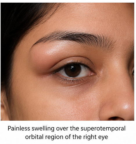
A 25-year-old woman presented to the ophthalmology clinic with a painless swelling over the outer aspect of her right upper eyelid that had gradually increased in size over the previous two years. She denied pain, redness, trauma, fever, diplopia, visual loss, or discharge from the lesion.
There was no history of previous orbital surgery, infection, or systemic illness. Family history was non-contributory.
On examination, best-corrected visual acuity was 6/6 in both eyes. A well-circumscribed, firm, non-tender, freely mobile mass measuring approximately 2.5 × 2 cm was palpable over the superotemporal orbital rim of the right eye. The overlying skin appeared normal without erythema or ulceration. Extraocular movements were full, pupillary reactions were normal, and there was no proptosis.
Anterior and posterior segment examinations were unremarkable in both eyes.
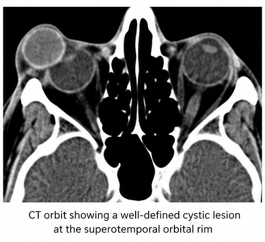
Orbital ultrasonography demonstrated a well-defined cystic lesion containing internal echogenic material. Computed tomography of the orbit revealed a sharply marginated, low-attenuation cystic lesion adjacent to the superotemporal orbital rim with mild pressure remodeling of the adjacent frontal bone but no intracranial extension.
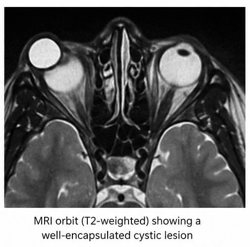
Magnetic resonance imaging confirmed a well-encapsulated lesion with imaging characteristics consistent with a dermoid cyst.
Based on the clinical and radiological findings, a diagnosis of superficial orbital dermoid cyst was established.
Management and Outcome
The patient underwent elective surgical excision through a superior eyelid crease incision under general anesthesia. Careful blunt dissection was performed to preserve the integrity of the cyst capsule and prevent intraoperative rupture. The lesion was completely excised without complications.
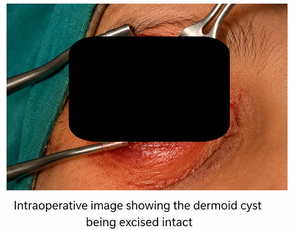
Gross examination revealed a cyst containing yellowish keratinous material and hair fragments. Histopathological examination demonstrated a cyst lined by keratinized stratified squamous epithelium containing sebaceous glands, sweat glands, and hair follicles, confirming the diagnosis of a dermoid cyst.
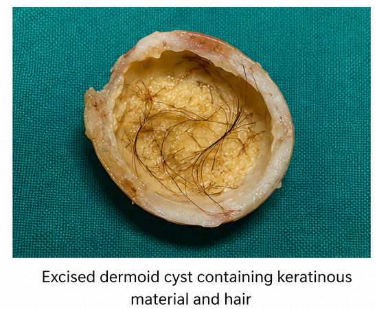
Postoperatively, topical antibiotic-steroid eye drops and oral analgesics were prescribed for one week. The surgical wound healed uneventfully with minimal scarring. At follow-up, the patient remained symptom-free with excellent cosmetic appearance and no evidence of recurrence.
Follow-up
One Month
-
Surgical wound healed well.
-
Complete resolution of swelling.
-
Excellent cosmetic outcome.
-
Visual acuity remained 6/6.
Three Months
-
No residual orbital mass.
-
Normal ocular movements.
-
No postoperative complications.
-
Histopathology confirmed dermoid cyst.
Six Months
-
No recurrence detected clinically.
-
Stable orbital examination.
-
Excellent cosmetic and functional outcome.
-
No visual impairment.
Discussion
Orbital dermoid cysts arise from congenital inclusion of ectodermal tissue during closure of embryonic suture lines. The frontozygomatic suture is the most common site, followed by the frontoethmoidal region.
Patients typically present with a painless, slowly enlarging orbital or periorbital mass. Superficial lesions are commonly diagnosed during childhood, whereas deeper orbital dermoid cysts may remain undetected until adulthood due to their concealed location.
Ultrasonography provides an initial assessment, while CT is particularly useful for identifying calcification, fat density, and bony remodeling. MRI offers superior evaluation of deep orbital extension and adjacent soft tissues.
Differential diagnoses include epidermoid cyst, lipoma, sebaceous cyst, lacrimal gland tumors, hemangioma, dacryocele, mucocele, and other orbital masses.
Complete surgical excision without rupture of the cyst capsule is the preferred treatment because leakage of keratinous material may induce a marked foreign-body granulomatous inflammatory reaction. Prognosis is excellent following complete removal, with recurrence being uncommon.
Prognosis
The prognosis following complete surgical excision is excellent. Most patients achieve complete resolution with minimal cosmetic deformity and preservation of normal visual function. Incomplete excision or intraoperative rupture may increase the risk of postoperative inflammation and recurrence. Long-term follow-up is recommended, particularly for deep orbital lesions or incompletely excised cysts.
Conclusion
Dermoid cyst should be considered in the differential diagnosis of painless, slowly enlarging orbital or periorbital masses, particularly those located near embryonic suture lines. A thorough ophthalmic examination supported by orbital imaging—including ultrasonography, computed tomography, and magnetic resonance imaging when indicated—is essential for confirming the diagnosis, assessing lesion extent, and planning appropriate surgical management. Complete surgical excision with preservation of the cyst capsule remains the definitive treatment and provides excellent functional and cosmetic outcomes while minimizing the risk of recurrence and postoperative inflammation. Early diagnosis and timely intervention can prevent progressive enlargement, bone remodeling, rupture, and secondary inflammatory complications.
References
-
Shields JA, Kaden IH, Eagle RC Jr, Shields CL. Orbital dermoid cysts: clinicopathologic correlations, classification, and management. Ophthalmic Plastic and Reconstructive Surgery. 1997;13(4):265–276. https://pubmed.ncbi.nlm.nih.gov/9430304/
-
Sherman RP, Rootman J, Lapointe JS. Orbital dermoids: clinical presentation and management. British Journal of Ophthalmology. 1984;68(9):642–652. https://pubmed.ncbi.nlm.nih.gov/6466593/
-
Lane CM, Ehrlich WW, Wright JE. Orbital dermoid cyst. Eye (London). 1987;1(Pt 4):504–511. https://pubmed.ncbi.nlm.nih.gov/3443205/
-
Pushker N, Meel R, Kumar A, et al. Orbital and periorbital dermoid/epidermoid cyst: a series of 280 cases and a brief review. Canadian Journal of Ophthalmology. 2020;55(2):167–171. https://pubmed.ncbi.nlm.nih.gov/31712044/
-
Dave TV, Gupta Rathi S, Kaliki S, Mishra D. Orbital and periorbital dermoid cysts: Comparison of clinical features and management outcomes in children and adults. European Journal of Ophthalmology. 2021;31(5):2631–2638. https://pubmed.ncbi.nlm.nih.gov/33198489/
Read more such content on @ Hidoc Dr | Medical Learning App for Doctors
Recommended News For You
Recommended Articles For You
Featured News
Featured Articles
Featured Events
Featured KOL Videos
1.
NPM1-Mutated AML: Not All Treatments Are Equal
2.
Paging Dr. AI; Myth Busting in Cancer; Oncologists Shine in Malpractice Survey
3.
MRI can save rectal cancer patients from surgery, study suggests
4.
Rectal Cancer 'Crisis'; Smells Like Cancer; Flushing Away Clues to Colon Cancer
5.
Rising rates of head and neck cancers in England
1.
A Closer Look at Breast Cancer: Examining the Ultrasound Images
2.
Exploring the Latest Advances in Hodgkin's Lymphoma Treatment
3.
Molecular Classification of Refractory Anemia: A Comprehensive Review
4.
Ferroptosis-Modulating Therapeutics in Precision Oncology
5.
Blood-Cell Morphology Foundation Models: Transforming Hematological Diagnostics and Clinical Practice
1.
International Cancer Conference
2.
Asian Symposium on Advancement in Hematology and Oncology (ASAHO)
3.
International Cancer Conference
4.
Asian Symposium on Advancement in Hematology and Oncology (ASAHO)
5.
Asian Symposium on Advancement in Hematology and Oncology
1.
CDK4/6 Inhibitors in Extending Overall Survival in HR+/HER2- aBC Patients in Clinical Trial and Real World
2.
Thromboprophylaxis In Medical Settings- An Update
3.
Nimotozumab Plus Chemo-radiotherapy v/s Placebo Plus Chemo-radiotherapy in Locally Advanced Nasopharyngeal Carcinoma
4.
How Platelet Disorders Can Affect the Body
5.
Treatment Sequencing Strategies in ALK + NSCLC Patients with CNS Diseases

Address :
Hidoc Dr. Inc. | Delaware C Corp | 1309 Coffeen Ave. Suite 1200, Sheridan WY, 82801
Phone :
+1-415-463-3094
Email :
anishagadia@hidoc.co
© Copyright 2026 Hidoc Dr. Inc.
Terms & Conditions - LLP | Inc. | Privacy Policy - LLP | Inc. | Account Deactivation

To get started please enter your email ID
Welcome to Hidoc Dr.
Join to enhance your clinical skills and gain specialized in-depth medical knowledge