Recalcitrant Periungual Warts Causing Nail Dystrophy
OthersPage Navigation
Introduction
Periungual warts are benign epidermal proliferations caused by infection with human papillomavirus. They arise around the proximal or lateral nail folds and may extend beneath the nail plate. Although common warts can occur at any age, periungual lesions are often difficult to eradicate because their location limits the intensity of treatment that can be delivered safely.
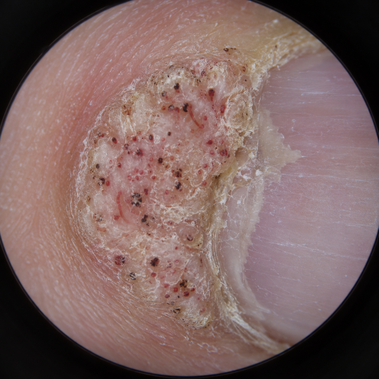
Typical lesions are rough, hyperkeratotic papules or plaques with an irregular surface. Disruption of dermatoglyphics and punctate black or red dots, representing thrombosed capillaries, support the clinical diagnosis. Nail biting, picking of the cuticle, repeated wet work, minor trauma, and impaired host immunity can promote inoculation and spread.
Case Report
A 29-year-old woman presented to the dermatology outpatient department with multiple rough growths around the nails of her right hand for approximately nine months. The first lesion appeared beside the right thumbnail and gradually increased in size. Similar smaller lesions subsequently developed around the index and middle fingernails.
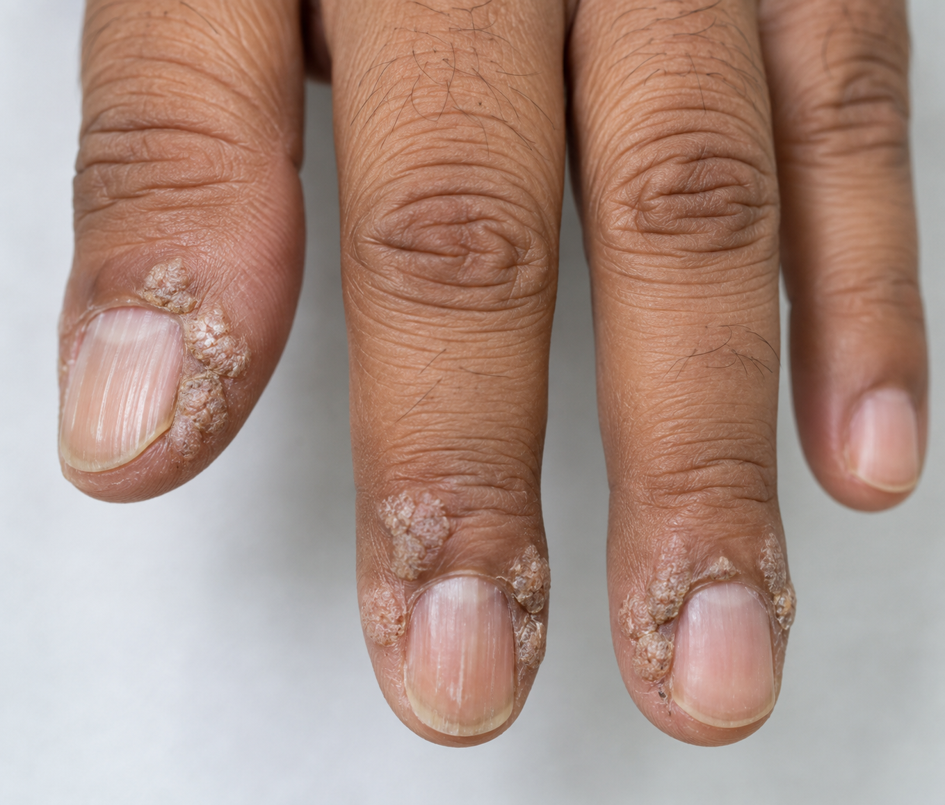
She reported mild pain while writing, opening containers, and performing household work. The thumbnail had become uneven and slightly elevated along its lateral margin. There was occasional bleeding after accidental trauma, but no spontaneous bleeding, purulent discharge, fever, or limitation of finger movement.
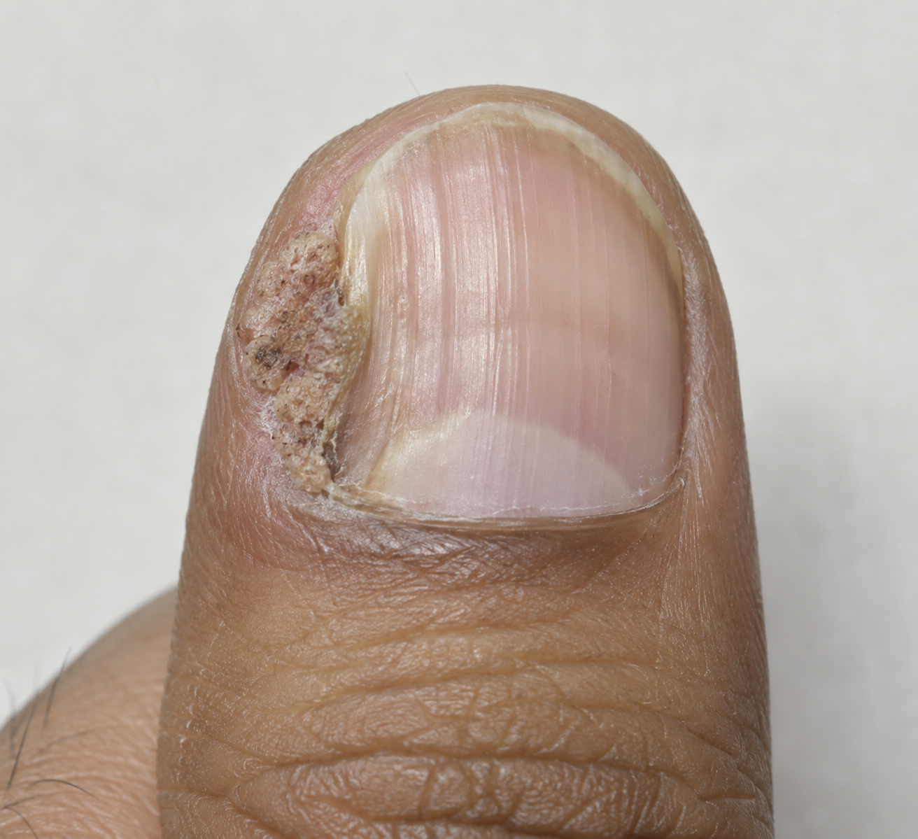
The patient frequently bit her nails and picked the surrounding cuticles during periods of stress. Her work involved repeated hand washing and prolonged exposure to water. She denied sharing manicure instruments and had not undergone professional nail procedures during the preceding year.
Cutaneous examination showed a 9 x 7 mm hyperkeratotic, verrucous plaque involving the lateral and proximal nail folds of the right thumb. Two discrete papules measuring 3-5 mm were present beside the index and middle fingernails. The surfaces were irregular, and interruption of normal skin lines was visible.
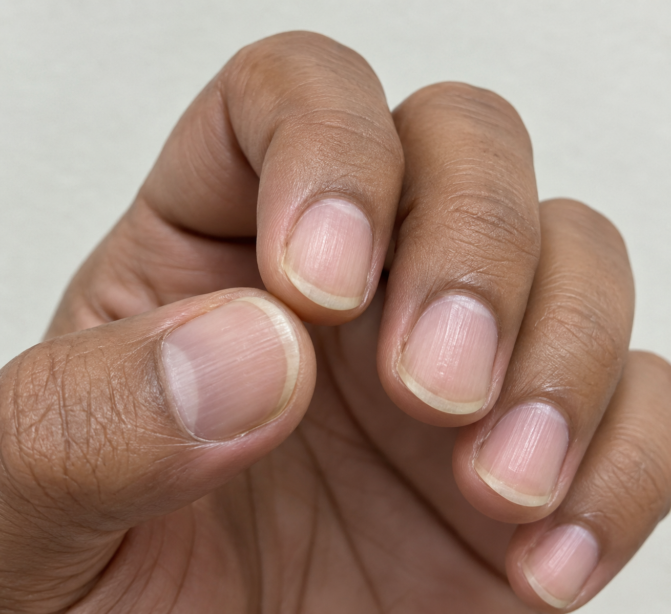
The right thumbnail showed mild lateral onycholysis and longitudinal ridging. The nail plate was not extensively destroyed, and there was no surrounding erythema, warmth, fluctuance, or discharge. The other fingernails and all toenails were normal. No palmar, plantar, facial, or anogenital warts were identified.
Investigations
Dermoscopy demonstrated a papillomatous surface with multiple red and black dots and short linear vessels on a whitish background. These findings, together with interruption of dermatoglyphics, were consistent with periungual warts.
Differential Diagnosis
Chronic Paronychia
Chronic paronychia commonly causes erythema, swelling, tenderness, and loss of the cuticle, particularly in people with repeated wet-work exposure. The absence of inflamed nail folds and the presence of discrete verrucous lesions made this diagnosis unlikely.
Onychomycosis
Fungal nail infection may cause nail thickening, subungual debris, and onycholysis. However, the primary abnormality in this patient was periungual verrucous growth, and microscopy of nail clippings did not show fungal elements.
Digital Squamous Cell Carcinoma
Squamous cell carcinoma of the nail unit can mimic a persistent periungual wart and should be considered in a solitary, enlarging, ulcerated, bleeding, painful, or treatment-resistant lesion. The patient's young age, multiplicity of typical lesions, and supportive dermoscopic findings favoured benign viral warts; biopsy would be required if the clinical behaviour became atypical.
Management and Outcome
The diagnosis, contagious nature, risk of autoinoculation, and possibility of recurrence were explained. The patient was advised to stop nail biting and cuticle picking, avoid trimming or manipulating the lesions, keep personal nail instruments separate, and use protective gloves for prolonged wet work.
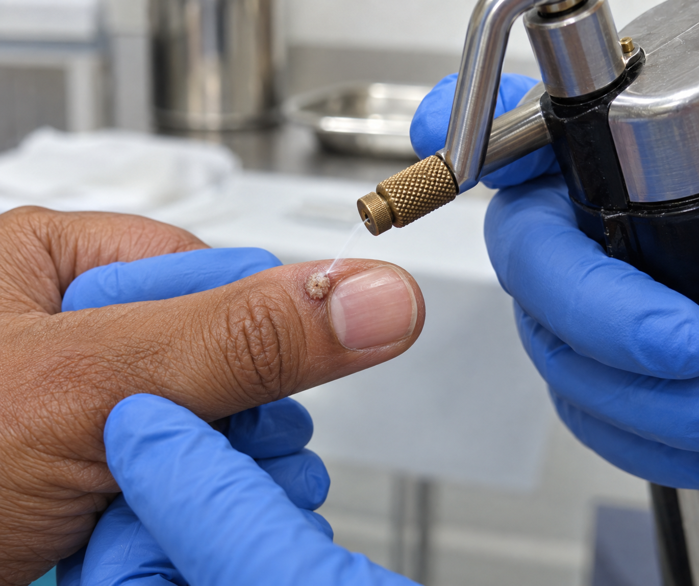
Because several lesions were symptomatic and the thumbnail was beginning to show dystrophy, active treatment was chosen. The thick keratin was gently pared by the dermatologist without entering the nail fold or causing bleeding. Liquid nitrogen cryotherapy was then applied conservatively to the visible wart tissue while protecting the surrounding nail fold and nail matrix. Two freeze-thaw cycles of approximately 10 seconds were used for the larger thumb lesion, and a shorter single cycle was used for each smaller lesion.
After re-epithelialisation, a keratolytic preparation containing salicylic acid was applied carefully to the residual wart surface each evening, with petroleum jelly used to protect adjacent normal skin. Treatment was paused if marked irritation occurred. Cryotherapy sessions were repeated at three-week intervals after reassessment of the nail folds.
At the second visit, the two smaller lesions had flattened substantially and the thumb lesion had reduced in thickness. Mild transient blistering had occurred after the first session, but there was no infection or worsening nail damage. A third conservative cryotherapy session was performed for the residual thumb lesion.
By week 12, all visible lesions had cleared, normal skin lines had begun to return, and no thrombosed capillary dots were seen on dermoscopy. The thumbnail remained mildly ridged but showed progressive distal growth of a smoother nail plate. The patient reported that pain during daily activities had resolved.
Discussion
Periungual warts can be therapeutically challenging. The nail folds create an irregular treatment surface, subungual extension may be difficult to reach, and aggressive destruction near the matrix can cause permanent nail dystrophy. Repeated trauma from nail biting and cuticle picking also facilitates autoinoculation and may reduce the likelihood of sustained clearance.
Topical salicylic acid is widely used because it gradually removes infected keratin and can be self-administered. Its effectiveness depends on regular application and protection of surrounding skin. Cryotherapy produces controlled tissue destruction and may be used for persistent or symptomatic common warts. In the nail unit, conservative application and adequate intervals between sessions are important to reduce pain and matrix injury.
No single treatment reliably clears every wart. Recalcitrant periungual lesions may be considered for intralesional antigen immunotherapy, bleomycin, local hyperthermia, laser-based treatment, or other dermatologist-supervised modalities. The choice depends on lesion number and location, previous treatment, patient age, immune status, pain tolerance, available expertise, and risk to the nail apparatus.
Atypical nail-unit lesions require particular caution because squamous cell carcinoma may be mistaken for a wart. Biopsy should be considered when a lesion is solitary and persistent, has destructive or ulcerative features, bleeds spontaneously, grows rapidly, causes disproportionate pain, or fails to respond as expected. Early histopathological assessment can prevent delay in the diagnosis of malignancy.
Conclusion
Periungual warts should be considered when rough hyperkeratotic papules or plaques disrupt the skin lines around a nail and show punctate vascular dots on dermoscopy.
Early treatment is important when lesions are painful, spreading, or beginning to distort the nail. In this patient, conservative cryotherapy combined with carefully applied topical keratolytic therapy achieved clearance while preserving the nail matrix.
Successful management also required correction of nail biting and cuticle picking, protection from repeated wet-work trauma, and follow-up for recurrence. Persistent or atypical nail-unit lesions should be biopsied to exclude squamous cell carcinoma or another mimicking disorder.
References
- Zhu P, Qi RQ, Yang Y, et al. Clinical guideline for the diagnosis and treatment of cutaneous warts (2022). Journal of Evidence-Based Medicine. 2022;15(3):284-301 https://pmc.ncbi.nlm.nih.gov/articles/PMC9825897/
- Lipke MM. An armamentarium of wart treatments. Clinical Medicine and Research. 2006;4(4):273-29. https://pmc.ncbi.nlm.nih.gov/articles/PMC1764803/
- Kwok CS, Gibbs S, Bennett C, Holland R, Abbott R. Topical treatments for cutaneous warts. Cochrane Database of Systematic Reviews. 2012;(9):CD001781 https://pmc.ncbi.nlm.nih.gov/articles/PMC8101088/
- Sterling JC, Gibbs S, Haque Hussain SS, Mohd Mustapa MF, Handfield-Jones SE. British Association of Dermatologists' guidelines for the management of cutaneous warts 2014. British Journal of Dermatology. 2014;171(4):696-712 https://pubmed.ncbi.nlm.nih.gov/25273231/
- Alghamdi KM, Khurram H. Successful treatment of periungual warts with diluted bleomycin using a translesional multipuncture technique: a pilot prospective study. Dermatologic Surgery. 2011;37(4):486-492 https://pubmed.ncbi.nlm.nih.gov/21352383/
- Tosti A, Piraccini BM. Warts of the nail unit: surgical and nonsurgical approaches. Dermatologic Surgery. 2001;27(3):235-239 https://pubmed.ncbi.nlm.nih.gov/11277886
Read more such content on @ Hidoc Dr | Medical Learning App for Doctors
Recommended News For You
Recommended Articles For You
Featured News
Featured Articles
Featured Events
Featured KOL Videos
1.
For MDS-Related Anemia, Telomerase Inhibitor Approved.
2.
Efficacy and safety of intravenous chemotherapy in children with intraocular retinoblastoma
3.
Admissions, medical schools, costs, and eligibility requirements information for FNB Onco-Anesthesia.
4.
Treating Depression: Crucial for Recovery From Fibromyalgia
5.
In postmenopausal women with hormone receptor-positive tumors, obesity increases the risk of breast cancer recurrence.
1.
Empowering Oncology with Data: Cloud Security, Real-World Evidence, and Clinical Insights
2.
Immune Regulation of Blood Cell Development
3.
Exploring the Effects of Radiation Therapy on Cystitis: A Journey to Better Health
4.
Transformative Frameworks in Oncology for Better Care
5.
Liposomal Doxorubicin and Mitomycin in Modern Cancer Treatment
1.
International Conference on Oncology, Cancer Prevention and Public Health
2.
International Conference on Cancer Nursing and Rehabilitation Strategies
3.
International Conference on Best Practices in Oncology, Cardiology and Critical Care
4.
International Conference on Innovations in Critical Care for Oncology and Cardiology
5.
International Symposium on Oncology, Cardiology and Critical Care Innovations
1.
Targeting Oncologic Drivers: A New Approach to Lung Cancer Treatment
2.
Newer Immunotherapies for Myeloma- A Comprehensive Overview
3.
Understanding the causes of anemia in adults beyond nutritional deficiencies
4.
Revolutionizing Treatment of ALK Rearranged NSCLC with Lorlatinib - Part III
5.
Guideline Recommendations of Lorlatinib as First-Line Treatment for ALK+ NSCLC

Address :
Hidoc Dr. Inc. | Delaware C Corp | 1309 Coffeen Ave. Suite 1200, Sheridan WY, 82801
Phone :
+1-415-463-3094
Email :
anishagadia@hidoc.co
© Copyright 2026 Hidoc Dr. Inc.
Terms & Conditions - LLP | Inc. | Privacy Policy - LLP | Inc. | Account Deactivation

To get started please enter your email ID
Welcome to Hidoc Dr.
Join to enhance your clinical skills and gain specialized in-depth medical knowledge