Acute Pyelonephritis: A Case Report of a Severe Clinical Presentation and Management Challenges
OthersPage Navigation
Abstract
Acute pyelonephritis is a bacterial infection involving the renal parenchyma and renal pelvis and represents an important form of upper urinary tract infection. The clinical presentation commonly includes fever, chills, flank pain, dysuria, urinary frequency, nausea, and vomiting. Prompt recognition and appropriate antimicrobial treatment are essential to prevent complications such as renal abscess, sepsis, and renal injury.
We report a hypothetical case of a 32-year-old woman who presented with high-grade fever, chills, right-sided flank pain, dysuria, and increased urinary frequency. Clinical examination revealed right costovertebral angle tenderness. Laboratory investigations demonstrated leukocytosis, elevated inflammatory markers, and urinalysis findings consistent with urinary tract infection. Urine culture isolated Escherichia coli. Renal ultrasonography demonstrated inflammatory changes involving the right kidney without evidence of urinary obstruction. The patient was diagnosed with acute uncomplicated pyelonephritis and treated with appropriate antibiotic therapy and supportive care.
This case highlights the importance of recognizing the characteristic clinical features of acute pyelonephritis and emphasizes the role of urinalysis, urine culture, laboratory investigations, and appropriate imaging in selected patients. Early diagnosis and timely antimicrobial therapy are essential for preventing progression to severe infection and other complications.
Introduction
Acute pyelonephritis is an infection involving the upper urinary tract, particularly the renal pelvis and renal parenchyma. It is commonly caused by ascending bacterial infection from the lower urinary tract, with Escherichia coli being the predominant causative organism.
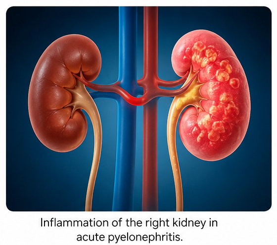
The condition may present with systemic and urinary symptoms, including fever, chills, flank pain, dysuria, urinary frequency, nausea, and vomiting. The combination of characteristic clinical findings and laboratory evidence of urinary infection is central to diagnosis. Urine culture with antimicrobial susceptibility testing is recommended to identify the causative organism and guide antimicrobial treatment.
Imaging is not routinely required in uncomplicated cases but may be useful when there is concern for urinary obstruction, anatomic abnormalities, treatment failure, or complications.
Management primarily involves appropriate antimicrobial therapy, with treatment selection influenced by the clinical severity, patient characteristics, and local antimicrobial susceptibility patterns. Supportive treatment is also important for controlling fever, pain, and maintaining adequate hydration.
We report a hypothetical case of acute pyelonephritis presenting with fever, flank pain, and urinary symptoms, followed by clinical, laboratory, microbiological, and imaging evaluation confirming the diagnosis.
Case Report
A 32-year-old woman presented with a 3-day history of high-grade fever associated with chills and right-sided flank pain. She also reported burning micturition, increased urinary frequency, and generalized weakness. The flank pain had progressively increased over the preceding 24 hours and was associated with reduced appetite.
There was no significant history of previous renal disease, recurrent urinary tract infections, urinary tract instrumentation, or known urinary tract abnormalities. She had no history of previous malignancy or major chronic illness.
On general examination, the patient was conscious and oriented but appeared acutely ill and mildly dehydrated. Her temperature was elevated, while other vital parameters were stable. Abdominal examination revealed mild tenderness over the right flank. Marked tenderness was elicited over the right costovertebral angle.
Investigations
Laboratory investigations revealed leukocytosis with neutrophilic predominance and elevated inflammatory markers. Renal function parameters were within acceptable limits.
Urinalysis demonstrated significant pyuria and bacteriuria, with positive leukocyte esterase and nitrite testing, supporting the diagnosis of urinary tract infection.
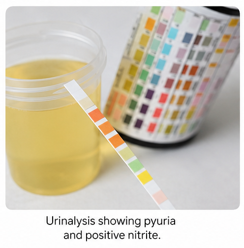
Urine culture grew Escherichia coli, and antimicrobial susceptibility testing was performed to guide antibiotic selection.

Urine culture and susceptibility testing are important components of the evaluation of acute pyelonephritis.

Renal ultrasonography demonstrated inflammatory changes involving the right kidney without evidence of hydronephrosis or an obstructing calculus. There was no imaging evidence of a renal abscess.
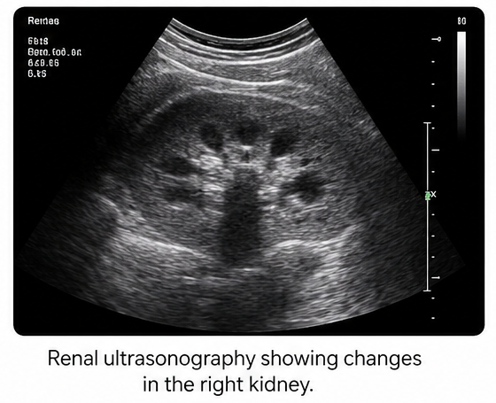
Based on the clinical presentation, laboratory findings, positive urine culture, and imaging assessment, the patient was diagnosed with acute right-sided pyelonephritis.
Diagnosis
The diagnosis was established based on:
- High-grade fever with chills
- Right-sided flank pain and costovertebral angle tenderness
- Dysuria and increased urinary frequency
- Urinalysis demonstrating pyuria and bacteriuria
- Urine culture positive for Escherichia coli
- Imaging demonstrating renal involvement without urinary obstruction
The overall findings were consistent with acute uncomplicated pyelonephritis.
Management and Outcome
After clinical assessment and review of the microbiological findings, antimicrobial therapy was initiated and subsequently tailored according to the urine culture and susceptibility results. Supportive treatment included adequate hydration, antipyretic therapy, and symptomatic management of flank pain.
The patient demonstrated progressive clinical improvement following initiation of treatment. Fever and chills subsided, urinary symptoms decreased, and the flank pain gradually resolved. Renal function remained stable during treatment.
The patient completed the prescribed antimicrobial course with regular clinical monitoring. No evidence of renal abscess, urinary obstruction, sepsis, or other acute complications was identified during the treatment period.
Follow-up
One Week
- Resolution of fever and chills
- Significant reduction in right-sided flank pain
- Improvement in dysuria and urinary frequency
- Improved appetite and general well-being
Four Weeks
- No recurrent fever
- Complete resolution of urinary symptoms
- No persistent flank tenderness
- Stable renal function
- Return to usual daily activities
Three Months
- No recurrence of urinary or systemic symptoms
- No further episodes of pyelonephritis
- Stable clinical condition
- Continued routine follow-up
Discussion
Acute pyelonephritis is a potentially serious upper urinary tract infection that can progress to systemic infection if not recognized and treated promptly. The clinical presentation is typically characterized by fever, flank pain, urinary symptoms, and laboratory evidence of urinary infection.
Escherichia coli is the most common causative organism. Identification of the responsible pathogen through urine culture allows antimicrobial treatment to be appropriately directed, particularly in the setting of increasing antimicrobial resistance.
Physical examination remains important in patients with suspected pyelonephritis. Costovertebral angle tenderness, when associated with fever and urinary symptoms, supports involvement of the upper urinary tract rather than an isolated lower urinary tract infection.
Urinalysis provides important evidence of urinary infection, while urine culture and antimicrobial susceptibility testing help identify the causative organism and guide treatment.
Imaging has a selective role in acute pyelonephritis. Uncomplicated cases can generally be diagnosed clinically and treated without routine imaging. However, imaging becomes particularly relevant in patients with risk factors for complications, atypical presentations, persistent symptoms, treatment failure, suspected obstruction, or possible renal abscess.
Ultrasonography can assist in identifying urinary obstruction and other structural abnormalities, while computed tomography can provide more detailed assessment of renal involvement and complications when clinically indicated.
Treatment requires appropriate antimicrobial therapy together with supportive management. In uncomplicated cases, patients who are clinically stable and able to tolerate oral treatment may be managed without hospitalization, whereas patients with severe disease or inability to tolerate oral therapy may require intravenous treatment and closer monitoring.
This hypothetical case demonstrates the importance of recognizing the combination of fever, flank pain, urinary symptoms, and laboratory evidence of infection. Prompt microbiological evaluation and appropriate treatment can lead to rapid clinical improvement and help prevent serious complications.
Prognosis
The prognosis of acute pyelonephritis is generally favorable when the condition is recognized early and treated appropriately. However, delayed treatment, urinary obstruction, diabetes, immunosuppression, resistant organisms, and other high-risk conditions may increase the likelihood of complications.
Potential complications include sepsis, renal abscess, obstruction-related infection, and renal injury. Continued clinical assessment is therefore important, particularly in patients who fail to respond to initial therapy or develop persistent systemic symptoms.
Conclusion
Acute pyelonephritis should be considered in patients presenting with fever, flank pain, and symptoms of urinary tract infection, particularly when accompanied by costovertebral angle tenderness and abnormal urinalysis findings.
This hypothetical case highlights the importance of clinical assessment, urinalysis, urine culture, antimicrobial susceptibility testing, and selective use of imaging in establishing the diagnosis and guiding treatment. Early recognition and appropriate antimicrobial therapy remain essential for achieving clinical recovery and preventing complications.
References
- Johnson JR, Russo TA. Acute pyelonephritis in adults: rapid evidence review. Am Fam Physician. 2020;102(3):173-180. https://pubmed.ncbi.nlm.nih.gov/32735433/
- Rollino C. Acute pyelonephritis in adults. G Ital Nefrol. 2007;24(2):121-131. https://pubmed.ncbi.nlm.nih.gov/17458827/
- Neumann I, Rojas MF, Moore P. Pyelonephritis (acute) in non-pregnant women. BMJ Clin Evid. 2008;2008:0807. https://pubmed.ncbi.nlm.nih.gov/19450332/
- Chitu MC, et al. Clinical characteristics, microbiological spectrum, biomarkers, and imaging insights in acute pyelonephritis and its complicated forms—a systematic review. Medicina (Kaunas). 2026. https://pubmed.ncbi.nlm.nih.gov/41597508/
Read more such content on @ Hidoc Dr | Medical Learning App for Doctors
Recommended News For You
Recommended Articles For You
Featured News
Featured Articles
Featured Events
Featured KOL Videos
1.
Research discovery halts childhood brain tumor before it forms
2.
Increased Data Support Active Monitoring for Low-Risk Prostate Cancer.
3.
'CDC Must Be Investigated'; David Lynch, Bob Uecker Die; Nasal Epinephrine Warning
4.
Increasing Access to Prostate Cancer Drugs; Reducing Toxic Emissions; FTC Files a 'Charity' Suit.
5.
Infections the Main Cause of Nonrelapse Mortality After CAR-T for Blood Cancers
1.
Beyond the Blinders: A Review of Targeted Therapeutic Strategies for Triple-Negative Breast Cancer in 2025
2.
AI-Based Cancer Follow-Up Monitoring: Transforming Survivorship Care through Intelligent Surveillance
3.
Preventing Sarcopenia During Cancer Treatment
4.
Guidance for Managing Complex Anticoagulation
5.
Unlocking the Potential of Sarclisa: A New Hope for Cancer Treatment
1.
International Cancer Conference
2.
Asian Symposium on Advancement in Hematology and Oncology (ASAHO)
3.
International Cancer Conference
4.
Asian Symposium on Advancement in Hematology and Oncology (ASAHO)
5.
Asian Symposium on Advancement in Hematology and Oncology
1.
Updates on Standard V/S High Risk Myeloma Treatment
2.
Navigating the Complexities of Ph Negative ALL - Part XIV
3.
Current Scenario of Blood Cancer- A Conclusion on Genomic Testing & Advancement in Diagnosis and Treatment
4.
Advances in Classification/ Risk Stratification of Plasma Cell Dyscrasias
5.
Pazopanib Takes Center Stage in Managing Renal Cell Carcinoma - Part I

Address :
Hidoc Dr. Inc. | Delaware C Corp | 1309 Coffeen Ave. Suite 1200, Sheridan WY, 82801
Phone :
+1-415-463-3094
Email :
anishagadia@hidoc.co
© Copyright 2026 Hidoc Dr. Inc.
Terms & Conditions - LLP | Inc. | Privacy Policy - LLP | Inc. | Account Deactivation

To get started please enter your email ID
Welcome to Hidoc Dr.
Join to enhance your clinical skills and gain specialized in-depth medical knowledge