Hepatic Coma Associated With Severe Hypoglycemia in Decompensated Cirrhosis
OthersPage Navigation
Introduction
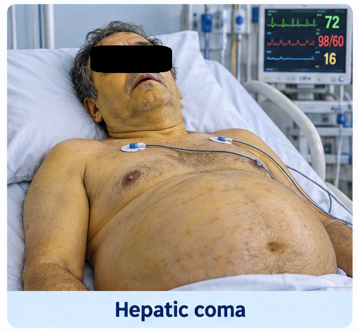
Hepatic encephalopathy is a potentially reversible neuropsychiatric complication of severe liver dysfunction or portosystemic shunting. Its manifestations range from impaired attention, disturbed sleep and confusion to stupor and coma. Hepatic coma generally refers to grade IV overt hepatic encephalopathy, in which the patient is unresponsive and requires urgent airway protection, metabolic correction and supportive management.
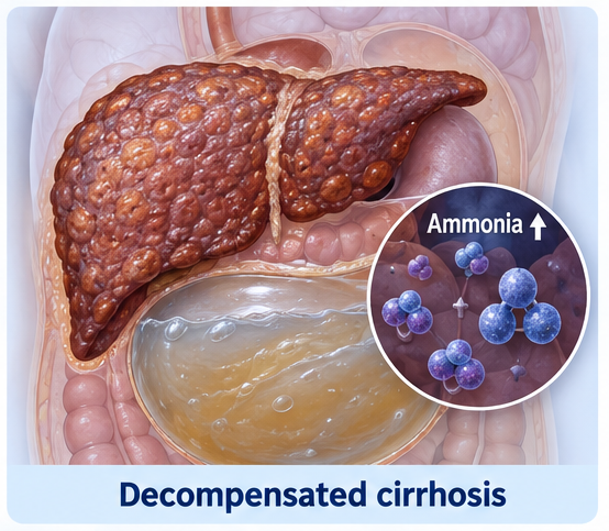
Hypoglycemia may develop in advanced liver disease because the damaged liver cannot adequately maintain glucose levels through glycogenolysis and gluconeogenesis. Poor oral intake, depleted glycogen stores, infection, alcohol consumption and renal dysfunction may further lower blood glucose. Severe hypoglycemia can independently cause seizures, neurological deficits and coma. Therefore, blood glucose should be checked immediately in every patient with liver disease who presents with altered consciousness.
Case Report
A 52-year-old man with alcohol-associated cirrhosis was brought to the emergency department with progressive drowsiness for one day. His family reported reduced food intake, increasing abdominal distension, constipation and disturbed sleep during the preceding four days. On the morning of admission, he became confused, stopped responding appropriately and subsequently lost consciousness.
There was no witnessed seizure, head injury, hematemesis or recent use of sedatives. He was not known to have diabetes and was not taking insulin or oral glucose-lowering medicines.
The patient had been diagnosed with cirrhosis two years earlier following an episode of ascites. His prescribed medicines included spironolactone, furosemide, propranolol and lactulose, but his adherence was irregular. He had consumed alcohol intermittently until approximately three weeks before admission.
On examination, the patient was deeply unconscious with a Glasgow Coma Scale score of 6/15. His temperature was 38.2°C, pulse rate was 108 beats/minute, blood pressure was 92/58 mmHg and respiratory rate was 24 breaths/minute. Oxygen saturation was 94% on room air.
He was icteric and pale, with bilateral pedal edema, tense ascites, splenomegaly and dilated abdominal veins. There was no neck stiffness or obvious focal neurological deficit. Asterixis could not be assessed because the patient was comatose.
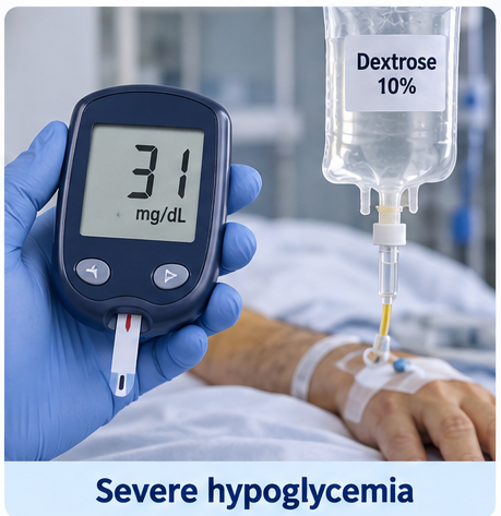
Immediate capillary blood glucose was 31 mg/dL. Intravenous access was secured, blood samples were collected and 25 g of intravenous dextrose was administered, followed by a continuous glucose infusion. His blood glucose increased to 112 mg/dL within 15 minutes. He became more responsive but remained markedly drowsy and disoriented. This partial response indicated that severe hypoglycemia had contributed to the coma but was not the only cause of his neurological impairment.
Investigations
Laboratory investigations showed hemoglobin of 9.4 g/dL, leukocyte count of 15,800 cells/mm³ and platelet count of 74,000 cells/mm³. Liver-function tests revealed total bilirubin of 7.8 mg/dL, serum albumin of 2.2 g/dL, aspartate aminotransferase of 126 U/L, alanine aminotransferase of 58 U/L and alkaline phosphatase of 188 U/L. The international normalized ratio was 2.1.
Serum creatinine was 1.6 mg/dL, sodium was 128 mmol/L, potassium was 3.4 mmol/L and venous lactate was 3.2 mmol/L. Plasma ammonia was elevated at 142 µmol/L. Repeated glucose measurements initially showed a tendency to fall whenever the glucose-infusion rate was reduced.
Diagnostic paracentesis produced cloudy ascitic fluid with a polymorphonuclear leukocyte count of 420 cells/mm³, supporting spontaneous bacterial peritonitis. Blood and ascitic-fluid cultures were collected before antibiotics were administered.
Non-contrast computed tomography of the head showed no intracranial hemorrhage, mass lesion or acute infarction. Abdominal ultrasonography demonstrated a coarse, nodular liver, splenomegaly, portal-vein dilatation and moderate ascites without biliary obstruction.
The clinical and laboratory findings supported grade IV overt hepatic encephalopathy in decompensated cirrhosis, precipitated by spontaneous bacterial peritonitis, constipation and poor oral intake. Severe hypoglycemia was considered an important additional factor that aggravated the coma.
Differential Diagnosis
Hypoglycemic Encephalopathy
Severe hypoglycemia was confirmed and required immediate treatment. Partial improvement following intravenous dextrose supported its contribution to the coma. However, persistent disorientation after normalization of glucose indicated concurrent hepatic encephalopathy.
Intracranial Event
Intracranial hemorrhage and ischemic stroke were considered because cirrhosis-associated coagulopathy and thrombocytopenia increase bleeding risk. The absence of a focal neurological deficit and a normal computed tomography scan made these conditions less likely.
Sepsis-Associated Encephalopathy
Fever, hypotension, leukocytosis and spontaneous bacterial peritonitis raised concern for sepsis-associated encephalopathy. Infection was considered a major precipitating factor that acted together with hepatic dysfunction and hypoglycemia.
Drug- or Alcohol-Related Coma
Sedative exposure, opioid toxicity, alcohol intoxication and accidental ingestion of glucose-lowering medication were considered. The history, medication review and laboratory findings did not support these diagnoses.
Management and Outcome
Airway protection, supplemental oxygen, continuous cardiac monitoring and frequent neurological assessment were initiated. Intravenous dextrose was continued, with bedside glucose monitoring every 30 minutes initially and then hourly. Once the glucose level remained stable, the infusion was gradually reduced while regular enteral nutrition was introduced.

Hepatic encephalopathy was treated with lactulose through a nasogastric tube. The dose was adjusted to produce two to three soft stools daily. Rifaximin 550 mg twice daily was added because of the severity of the encephalopathy.
Ceftriaxone and intravenous albumin were started for spontaneous bacterial peritonitis. Electrolyte abnormalities and intravascular depletion were corrected cautiously. Diuretics and propranolol were temporarily withheld because of hypotension and renal dysfunction.
No further blood glucose level below 70 mg/dL occurred after the first 12 hours. By 24 hours, the patient opened his eyes in response to speech and followed simple commands. By 48 hours, he was awake, oriented to person and place and able to tolerate oral feeding.
Ascitic-fluid culture subsequently grew Escherichia coli sensitive to ceftriaxone. Renal function improved, and the leukocyte count and ammonia level gradually decreased.
The patient was discharged after nine days with lactulose, rifaximin, nutritional counselling and alcohol-abstinence support. His family was educated to recognize confusion, sleep reversal, constipation, reduced food intake, fever and symptoms of recurrent hypoglycemia. Liver-transplant evaluation was recommended because of advanced decompensated cirrhosis.

Discussion
This case demonstrates that coma in a patient with cirrhosis should not automatically be attributed to hepatic encephalopathy alone. Hepatic encephalopathy is a clinical diagnosis that requires identification of liver dysfunction and exclusion of other causes of altered consciousness. Infection, gastrointestinal bleeding, constipation, dehydration, renal dysfunction, electrolyte abnormalities and sedative medicines are common precipitating factors.
The liver maintains fasting blood glucose through glycogen breakdown and gluconeogenesis. In advanced hepatic dysfunction, glycogen stores become depleted and the capacity to produce glucose from lactate, glycerol and amino acids is impaired. Poor food intake and sepsis further increase metabolic demand and may rapidly expose the reduced hepatic reserve.
Neurological improvement after dextrose may be incomplete when multiple mechanisms are responsible for coma. In this case, glucose correction produced an early partial response, whereas sustained recovery occurred only after treatment of infection, constipation, circulatory dysfunction and hepatic encephalopathy.
Plasma ammonia may support the diagnosis, but a single elevated value does not independently establish hepatic encephalopathy or accurately determine its clinical severity. Diagnosis should be based on clinical assessment, the presence of advanced liver disease and exclusion of other neurological and metabolic disorders.
Management requires immediate stabilization, correction of hypoglycemia and treatment of precipitating factors. Lactulose remains a primary treatment for overt hepatic encephalopathy, while rifaximin may be added in severe or recurrent cases. Frequent glucose monitoring is necessary because hypoglycemia may recur after an initial response to intravenous dextrose when hepatic glucose production remains impaired.
Conclusion
Severe hypoglycemia can precipitate or deepen coma in patients with advanced liver disease and may coexist with overt hepatic encephalopathy. In this patient, intravenous dextrose produced partial improvement, while full recovery required treatment of spontaneous bacterial peritonitis, constipation and hepatic encephalopathy.
Every patient with liver disease and altered consciousness should undergo immediate bedside glucose testing and systematic evaluation for additional precipitating factors. Rapid glucose correction, prevention of recurrence, treatment of infection and appropriate encephalopathy management can reverse coma and reduce the risk of permanent neurological injury.
References
-
Vilstrup H, Amodio P, Bajaj J, et al. Hepatic encephalopathy in chronic liver disease: 2014 practice guideline by AASLD and EASL. Hepatology. 2014;60(2):715–735. https://pubmed.ncbi.nlm.nih.gov/25042402/
- Wijdicks EFM. Hepatic encephalopathy. New England Journal of Medicine. 2016;375(17):1660–1670. https://pubmed.ncbi.nlm.nih.gov/27783916/
- Rose CF, Amodio P, Bajaj JS, et al. Hepatic encephalopathy: Novel insights into classification, pathophysiology and therapy. Journal of Hepatology. 2020;73(6):1526–1547. https://pubmed.ncbi.nlm.nih.gov/33097308/
- Stravitz RT, Lee WM. Acute liver failure. Lancet. 2019;394(10201):869–881. https://pubmed.ncbi.nlm.nih.gov/31498101/
- European Association for the Study of the Liver. EASL Clinical Practice Guidelines on the management of hepatic encephalopathy. Journal of Hepatology. 2022;77(3):807–824. https://pubmed.ncbi.nlm.nih.gov/35724930/
Read more such content on @ Hidoc Dr | Medical Learning App for Doctors
Recommended News For You
Recommended Articles For You
Featured News
Featured Articles
Featured Events
Featured KOL Videos
1.
Tumor characteristics found to differ for melanomas in children, teens and young adults
2.
Unexplained CAR-T Death; Americans Spurn Sunscreen; Improving Diversity in Trials
3.
"A lot" of aspirin reduces the risk of ovarian cancer, regardless of genetic factors, according to the JAMA study.
4.
Approval in Endometrial Cancer Expanded; Masks at Cancer Centers; NPR Reporter Dies
5.
Is Lenalidomide Resistance in Myeloma Reversible with JAK Inhibition?
1.
Modern Oncology Unveiled with Treatment Innovations and Personalized Cancer Care
2.
Screening for Early Therapy-Related Cancer Susceptibility
3.
Unlocking the Potential of Bicalutamide 50 mg: A Revolutionary New Treatment for Prostate Cancer
4.
Skin-Sparing Mastectomy: Balancing Cancer Control with Cosmetic Results
5.
Reducing Modifiable Childhood Cancer Exposure Risks
1.
International Conference on Innovations in Critical Care for Oncology and Cardiology
2.
International Conference on Oncology, Cardiology and Critical Care Policy
3.
International Conference on Oncology, Cancer Prevention and Public Health
4.
International Conference on Cancer Nursing and Rehabilitation Strategies
5.
International Conference on Best Practices in Oncology, Cardiology and Critical Care
1.
Dacomitinib Case Presentation: Baseline Treatment and Current Status
2.
Targeting Oncologic Drivers with Dacomitinib: Further Discussion on Lung Cancer Treatment
3.
Should We Use DARA Up Front As First-Line Therapy in MM?
4.
Iron Deficiency Anemia: Ferric Maltol As a New Treatment Option- A New Perspective
5.
Importance of Cancer Screening and Early Detection

Address :
Hidoc Dr. Inc. | Delaware C Corp | 1309 Coffeen Ave. Suite 1200, Sheridan WY, 82801
Phone :
+1-415-463-3094
Email :
anishagadia@hidoc.co
© Copyright 2026 Hidoc Dr. Inc.
Terms & Conditions - LLP | Inc. | Privacy Policy - LLP | Inc. | Account Deactivation

To get started please enter your email ID
Welcome to Hidoc Dr.
Join to enhance your clinical skills and gain specialized in-depth medical knowledge