Acute Exacerbation of Chronic Obstructive Pulmonary Disease in a Long Term Smoker
OthersPage Navigation
Introduction
Chronic obstructive pulmonary disease (COPD) is a common respiratory condition characterised by persistent respiratory symptoms and airflow obstruction resulting from abnormalities of the airways, alveoli, or both. Cigarette smoking is a major risk factor, although exposure to biomass fuel, occupational dust, fumes, and air pollution may also contribute.
Patients commonly experience progressive breathlessness, chronic cough, sputum production, wheezing, and reduced exercise tolerance. The diagnosis is confirmed using spirometry, which demonstrates persistent airflow obstruction following bronchodilator administration.
An acute exacerbation of COPD is an episode of worsening breathlessness, cough, or sputum production occurring over a short period. Respiratory infections and environmental pollutants are frequent triggers. Severe exacerbations may cause hypoxaemia, carbon dioxide retention, respiratory acidosis, hospitalisation, and respiratory failure.
Early recognition, controlled oxygen therapy, bronchodilators, systemic corticosteroids, appropriate antibiotics, and ventilatory support can reduce complications and improve clinical outcomes.
Case Report
A 64-year-old man presented to the emergency department with worsening breathlessness, productive cough, and increased sputum volume for four days. His sputum, which was normally scanty and white, had become thick and yellowish.
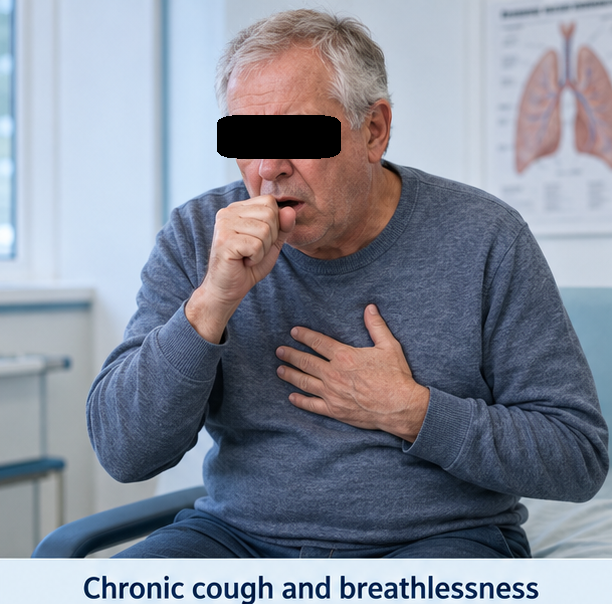
The patient had experienced gradually progressive breathlessness for approximately six years. Initially, breathlessness occurred only while climbing stairs or walking uphill. During the preceding year, he had started experiencing symptoms while walking on level ground and had reduced his daily activities.
He also reported a chronic morning cough with mucoid expectoration for several years. Intermittent wheezing was present, particularly during winter and following exposure to dust. He had experienced two similar episodes during the previous year but had not required hospitalisation.
The patient had smoked approximately 20 cigarettes daily for 35 years, corresponding to a smoking exposure of approximately 35 pack-years. He had stopped smoking six months before presentation. He had worked in a textile factory for 25 years and reported regular exposure to cotton dust.
He had previously been prescribed an inhaled bronchodilator but used it irregularly. His inhaler technique had never been formally assessed. He was not receiving long-term oxygen therapy and had not participated in pulmonary rehabilitation.
There was no history of high-grade fever, haemoptysis, chest trauma, recent surgery, prolonged immobilisation, or known tuberculosis. He denied orthopnoea, paroxysmal nocturnal dyspnoea, or swelling of the lower limbs. There was no previous diagnosis of bronchial asthma, bronchiectasis, interstitial lung disease, or heart failure.
On examination, the patient appeared anxious and breathless. He was sitting upright and using his accessory muscles of respiration. He could speak only in short sentences.
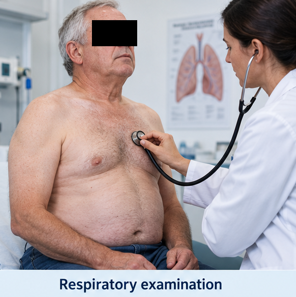
His temperature was 37.8°C, heart rate was 112 beats per minute, respiratory rate was 30 breaths per minute, blood pressure was 138/84 mmHg, and oxygen saturation was 84% while breathing room air.
Respiratory examination revealed a barrel-shaped chest with reduced bilateral chest expansion. The percussion note was hyper-resonant, and breath sounds were generally diminished. Prolonged expiration and widespread bilateral expiratory wheezing were present. Coarse crackles were heard over both lower lung fields.
Cardiovascular examination demonstrated tachycardia with normal heart sounds. There was no raised jugular venous pressure or peripheral oedema. Abdominal and neurological examinations were unremarkable.
The patient’s chronic progressive respiratory symptoms, substantial smoking exposure, occupational dust exposure, and acute increase in breathlessness with purulent sputum suggested an acute infective exacerbation of previously inadequately controlled COPD.
Investigations
Initial laboratory investigations demonstrated a total leucocyte count of 14,800 cells/mm³ with 82% neutrophils. Haemoglobin was 15.6 g/dL, and the platelet count was 248,000 cells/mm³. C-reactive protein was elevated at 68 mg/L.
Renal and liver function tests were within normal limits. Serum sodium was 137 mmol/L, and potassium was 4.2 mmol/L. Cardiac troponin was not elevated.
Arterial blood gas analysis performed before oxygen administration showed a pH of 7.31, partial pressure of carbon dioxide of 56 mmHg, partial pressure of oxygen of 49 mmHg, and bicarbonate concentration of 27 mmol/L. These findings indicated acute hypercapnic and hypoxaemic respiratory failure with respiratory acidosis.
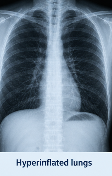
A chest radiograph demonstrated bilateral hyperinflation, flattened diaphragms, increased retrosternal airspace, and attenuation of peripheral vascular markings. No focal consolidation, pleural effusion, or pneumothorax was detected.
Electrocardiography showed sinus tachycardia without acute ischaemic changes. Echocardiography demonstrated preserved left ventricular systolic function and no significant valvular abnormality.
A sputum sample was obtained for Gram staining and culture. The culture subsequently demonstrated growth of Haemophilus influenzae, which was sensitive to amoxicillin–clavulanate. Blood cultures remained sterile.
After the patient’s condition stabilised, spirometry was performed before discharge. The post-bronchodilator forced expiratory volume in one second was 1.18 L, representing 44% of the predicted value. Forced vital capacity was 2.71 L, and the post-bronchodilator FEV₁/FVC ratio was 0.44. There was no significant bronchodilator reversibility.
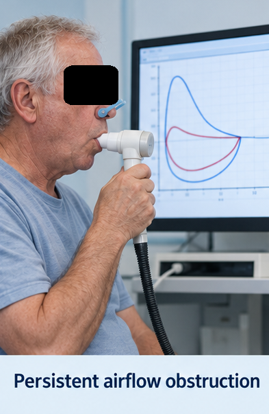
Persistent post-bronchodilator airflow obstruction, together with the patient’s symptoms and exposure history, confirmed COPD.
Differential Diagnosis
Bronchial Asthma
Asthma can cause episodic breathlessness, cough, and wheezing. However, this patient had progressive symptoms beginning later in life, substantial smoking exposure, persistent airflow obstruction, and no significant bronchodilator reversibility. These features favoured COPD.
Community Acquired Pneumonia
Pneumonia was considered because of the productive cough, purulent sputum, inflammatory marker elevation, and respiratory deterioration. However, the absence of focal consolidation on chest radiography made pneumonia less likely.
Acute Decompensated Heart Failure
Heart failure may present with acute breathlessness, hypoxaemia, and basal crackles. The absence of orthopnoea, peripheral oedema, cardiomegaly, or left ventricular dysfunction argued against this diagnosis.
Pulmonary Embolism
Pulmonary embolism may cause unexplained acute breathlessness, tachycardia, chest pain, and hypoxaemia. However, the patient had no thromboembolic risk factors or pleuritic chest pain, and the presence of increased purulent sputum and wheezing favoured an infective COPD exacerbation.
Bronchiectasis
Bronchiectasis may cause chronic productive cough and recurrent respiratory infections. However, the patient did not report large-volume sputum, recurrent haemoptysis, or frequent infections. The radiological findings were more consistent with emphysematous COPD.
Pneumothorax
Secondary spontaneous pneumothorax is an important cause of sudden deterioration in patients with COPD. It was excluded by the absence of unilateral examination findings and a normal pleural outline on chest radiography.
Management and Outcome
The patient was admitted to a monitored respiratory unit. Controlled oxygen was administered through a Venturi mask, with a target oxygen saturation of 88–92%. Oxygen flow was adjusted using repeated clinical assessment and arterial blood gas measurements.
Nebulised short-acting bronchodilator therapy with salbutamol and ipratropium bromide was initiated. Intravenous hydrocortisone was administered initially and subsequently changed to a short course of oral prednisolone.
Empirical antibiotic therapy with intravenous amoxicillin–clavulanate was started because the patient had increased breathlessness, sputum volume, and sputum purulence. The treatment was continued after the sputum culture demonstrated susceptible H. influenzae.
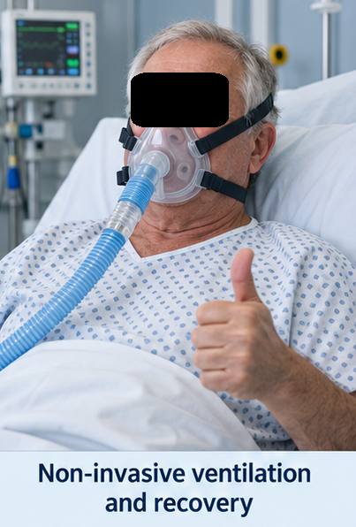
Despite initial treatment, the patient remained tachypnoeic and hypercapnic. Repeat arterial blood gas analysis showed persistent respiratory acidosis. Non-invasive ventilation using bilevel positive airway pressure was therefore initiated.
The patient tolerated non-invasive ventilation well. Over the following 24 hours, his respiratory rate decreased, accessory muscle use diminished, and arterial blood gas values improved. The pH increased to 7.37, while the partial pressure of carbon dioxide decreased to 47 mmHg.
Non-invasive ventilation was gradually discontinued after 36 hours. Oxygen supplementation was reduced as his respiratory condition improved. He remained haemodynamically stable and did not require invasive mechanical ventilation.
Before discharge, his inhaler technique was assessed and corrected. Maintenance treatment with a long-acting bronchodilator regimen was initiated based on his symptoms, exacerbation history, and clinical assessment. A short-acting inhaled bronchodilator was provided for rescue use.
The patient received counselling regarding complete smoking abstinence, avoidance of occupational and environmental respiratory irritants, regular medication adherence, physical activity, nutrition, vaccination, and early recognition of exacerbation symptoms.
He was referred for pulmonary rehabilitation and advised to attend respiratory follow-up. Assessment before discharge showed an oxygen saturation of 93% while breathing room air; therefore, long-term oxygen therapy was not prescribed.
At the four-week follow-up, the patient reported substantial improvement in breathlessness and exercise tolerance. His cough and sputum production had returned to baseline. He was using his inhalers regularly and demonstrated an appropriate inhalation technique.
At the three-month review, he had experienced no further exacerbations and had completed a pulmonary rehabilitation programme. He continued to abstain from smoking and reported improved confidence in managing his respiratory symptoms.
Discussion
This case illustrates an acute infective exacerbation leading to the diagnosis and comprehensive management of COPD in a patient with prolonged smoking and occupational dust exposure.
COPD should be considered in adults with chronic breathlessness, cough, sputum production, recurrent lower respiratory infections, or exposure to recognised respiratory risk factors. Clinical suspicion alone is insufficient; spirometry is required to demonstrate persistent airflow obstruction.
Exacerbations are clinically important because they can accelerate functional decline, impair quality of life, increase the likelihood of subsequent exacerbations, and result in hospitalisation or death. Increased breathlessness, sputum volume, and sputum purulence commonly indicate an exacerbation and may suggest a bacterial trigger.
Oxygen must be administered carefully in patients at risk of hypercapnic respiratory failure. Controlled oxygen therapy targeting a saturation of approximately 88–92% can correct dangerous hypoxaemia while limiting worsening carbon dioxide retention.
Short-acting inhaled bronchodilators are central to acute symptom relief. Systemic corticosteroids can shorten recovery and improve lung function, while antibiotics are appropriate when bacterial infection is suspected, particularly in patients with purulent sputum or severe exacerbations.
Non-invasive ventilation is particularly valuable in patients with acute hypercapnic respiratory failure and respiratory acidosis. It can reduce the work of breathing, improve gas exchange, decrease the need for endotracheal intubation, and improve outcomes when used in suitable patients.
Long-term management must extend beyond treatment of the acute episode. Smoking cessation is one of the most important interventions for limiting continued exposure-related lung injury. Correct inhaler selection and technique, medication adherence, vaccination, pulmonary rehabilitation, physical activity, and individualised follow-up contribute to improved symptom control and exacerbation prevention.
Conclusion
COPD should be suspected in patients with progressive breathlessness, chronic cough, sputum production, and substantial exposure to cigarette smoke or occupational pollutants.
In this patient, an acute infective exacerbation caused hypoxaemic and hypercapnic respiratory failure. Clinical assessment, arterial blood gas analysis, chest imaging, sputum culture, and post-stabilisation spirometry established the diagnosis and guided treatment.
Controlled oxygen therapy, short-acting bronchodilators, systemic corticosteroids, appropriate antibiotics, and non-invasive ventilation resulted in clinical recovery without the need for invasive mechanical ventilation.
Long-term improvement required smoking abstinence, maintenance inhaled therapy, correction of inhaler technique, pulmonary rehabilitation, vaccination, and structured respiratory follow-up.
References
- Global Initiative for Chronic Obstructive Lung Disease. Global Strategy for Prevention, Diagnosis and Management of COPD 2026 Report. https://goldcopd.org/2026-gold-report-and-pocket-guide/
- Cazzola M, Bajpai J, Calzetta L, et al. GOLD 2026: Transforming COPD management with early intervention, multidimensional assessment and personalised care. Drugs. 2026;86:581–597. https://pmc.ncbi.nlm.nih.gov/articles/PMC13109179/
- Khan KS, Nauriyal V, Rai SP, et al. Management of chronic obstructive pulmonary disease exacerbations in hospitalised patients. Cureus. 2023;15(8):e43625. https://pmc.ncbi.nlm.nih.gov/articles/PMC10505355/
- Bollmeier SG, Hartmann AP. Management of chronic obstructive pulmonary disease: A review focusing on exacerbations. American Journal of Health-System Pharmacy. 2020;77(4):259–268. https://pmc.ncbi.nlm.nih.gov/articles/PMC7005599/
- Perera B, Barton C, Osadnik C. General practice management of COPD patients following an acute exacerbation. Australian Journal of General Practice. 2023;52(3):117–123. https://pmc.ncbi.nlm.nih.gov/articles/PMC9975965/
- Mein SA, Ferrera MC, Mojica-Perez Y, et al. Management of asthma and COPD exacerbations in adults in the intensive care unit. Chest Critical Care. 2025;3(2):100109. https://pmc.ncbi.nlm.nih.gov/articles/PMC12054689/
Read more such content on @ Hidoc Dr | Medical Learning App for Doctors
Recommended News For You
Recommended Articles For You
Featured News
Featured Articles
Featured Events
Featured KOL Videos
1.
For the treatment of vestibular schwannomas in neurofibromatosis type 2, stereotactic radiosurgery has been found to be effective.
2.
FDA Advisors Recommend Galleri Multicancer Blood Test
3.
Women who miss their first mammogram face higher risk of breast cancer death, study finds
4.
Thriving while surviving: Understanding the social needs of cancer survivors
5.
Can Accelerated Salvage RT Improve Prostate Cancer Control?
1.
Fatigue and Work Participation in Blood Disease: A Comprehensive Review
2.
First-Line Immuno-Hematology Examinations: Essential Diagnostic Tools for Patient Care
3.
The benefits and risks of taking fludrocortisone for adrenal insufficiency
4.
The Algorithmic Revolution: How AI is Reshaping Precision Oncology from Bench to Bedside
5.
Childhood Cancer Prevention Through Modifiable Exposure Reduction
1.
International Conference on Oncology, Cancer Prevention and Public Health
2.
International Conference on Cancer Nursing and Rehabilitation Strategies
3.
International Conference on Best Practices in Oncology, Cardiology and Critical Care
4.
International Conference on Innovations in Critical Care for Oncology and Cardiology
5.
International Symposium on Oncology, Cardiology and Critical Care Innovations
1.
A Comprehensive Guide to First Line Management of ALK Positive Lung Cancer - Part VI
2.
Management of 1st line ALK+ mNSCLC (CROWN TRIAL Update) - Part III
3.
Understanding Common Causes of Abnormal Blood Counts
4.
Hematologic Fatigue and Work Function: Clinical Implications, Pathophysiology, and Management
5.
Treatment Paradigm for Patients with R/R Adult B-cell ALL- Expert Discussions

Address :
Hidoc Dr. Inc. | Delaware C Corp | 1309 Coffeen Ave. Suite 1200, Sheridan WY, 82801
Phone :
+1-415-463-3094
Email :
anishagadia@hidoc.co
© Copyright 2026 Hidoc Dr. Inc.
Terms & Conditions - LLP | Inc. | Privacy Policy - LLP | Inc. | Account Deactivation

To get started please enter your email ID
Welcome to Hidoc Dr.
Join to enhance your clinical skills and gain specialized in-depth medical knowledge