Hydatid Cyst of the Liver Presenting with Right Upper Quadrant Abdominal Pain – A Case Report
SurgeryPage Navigation
Abstract
Hydatid disease, also known as cystic echinococcosis, is a zoonotic parasitic infection caused primarily by the larval stage of Echinococcus granulosus. The liver is the most commonly affected organ, accounting for approximately 60–70% of cases, followed by the lungs. The disease remains endemic in many developing countries where livestock farming and close contact with dogs are common. Clinical manifestations vary depending on the size, location, and complications of the cyst. Many patients remain asymptomatic for years, while others may present with abdominal pain, hepatomegaly, jaundice, or cyst rupture.
We report the case of a 35-year-old woman who presented with intermittent right upper abdominal pain and abdominal fullness for six months. Ultrasonography and contrast-enhanced computed tomography revealed a large hydatid cyst in the right lobe of the liver. Serological testing supported the diagnosis. The patient underwent surgical management combined with antihelminthic therapy using albendazole, resulting in successful recovery without recurrence during follow-up. This case highlights the importance of early recognition, appropriate imaging, and timely intervention in the management of hepatic hydatid disease.
Introduction
Hydatid disease is a chronic parasitic infection caused by cestodes belonging to the genus Echinococcus. Humans serve as accidental intermediate hosts after ingesting parasite eggs shed in the feces of infected dogs and other canines.
Following ingestion, the embryos penetrate the intestinal mucosa and enter the portal circulation, where they are commonly trapped in the liver. Over time, the larvae develop into fluid-filled hydatid cysts containing daughter cysts and protoscolices.
Hydatid cyst disease remains a significant public health concern in endemic regions including parts of Asia, Africa, South America, the Middle East, and the Mediterranean region.
Risk factors include:
• Close contact with dogs
• Livestock farming
• Sheep and cattle rearing
• Poor sanitation
• Consumption of contaminated food or water
• Residence in endemic areas
Common clinical manifestations include:
• Right upper quadrant abdominal pain
• Abdominal fullness or mass
• Nausea and vomiting
• Hepatomegaly
• Jaundice (in complicated cases)
• Fever due to secondary infection
Early diagnosis and treatment are important to prevent complications such as cyst rupture, anaphylaxis, biliary obstruction, and secondary dissemination.
Case Report
Patient History
A 35-year-old woman presented to the general surgery outpatient department with complaints of:
• Dull aching pain in the right upper abdomen for six months
• Intermittent abdominal discomfort
• Sensation of fullness after meals
• Occasional nausea
The pain was non-radiating and gradually increasing in intensity over the preceding three months.
The patient reported living in a rural area and having frequent contact with domestic dogs and livestock.
Past medical history was unremarkable.
There was no history of:
• Tuberculosis
• Chronic liver disease
• Viral hepatitis
• Diabetes mellitus
• Previous abdominal surgery
Family history was non-contributory.
Clinical Examination
General examination revealed:
• Conscious and oriented patient
• Mild discomfort on abdominal palpation
• No pallor, cyanosis, or edema
Vital signs were stable:
• Pulse rate: 84 beats/minute
• Blood pressure: 124/78 mmHg
• Respiratory rate: 18 breaths/minute
• Temperature: Afebrile
Abdominal examination revealed:
• Mild hepatomegaly
• Tenderness in the right hypochondrium
• No guarding or rigidity
• No ascites
Other systemic examinations were within normal limits.
Clinical Evaluation
Differential Diagnosis
The following conditions were considered:
• Hydatid cyst of liver
• Simple hepatic cyst
• Liver abscess
• Hepatic hemangioma
• Biliary cystadenoma
• Hepatocellular carcinoma
Investigations
Radiological Evaluation
Ultrasonography
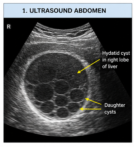
Abdominal ultrasonography demonstrated:
• A well-defined cystic lesion measuring approximately 10 × 8 cm in the right hepatic lobe
• Internal septations and daughter cysts
• Detached endocyst membrane appearance
These findings strongly suggested hepatic hydatid disease.
Contrast-Enhanced Computed Tomography (CECT)
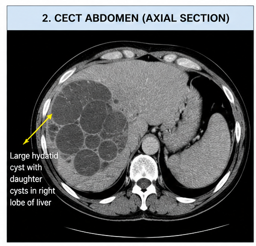
CECT abdomen revealed:
• Large cystic lesion in segment VII of the liver
• Multiple daughter cysts within the mother cyst
• Characteristic “wheel spoke” appearance
• No evidence of rupture
The imaging findings were consistent with a hepatic hydatid cyst.
Laboratory Evaluation
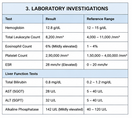
Routine investigations showed:
• Hemoglobin: 12.8 g/dL
• Total leukocyte count: 8,200/mm³
• Platelet count: 290,000/mm³
• Eosinophil count: Mildly elevated
• ESR: Elevated
Liver function tests revealed:
• Total bilirubin: Normal
• AST: Normal
• ALT: Normal
• Alkaline phosphatase: Mildly elevated
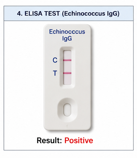
Serological testing:
• Enzyme-linked immunosorbent assay (ELISA) for Echinococcus antibodies: Positive
Diagnosis
Based on clinical presentation, imaging findings, and positive serological testing, a diagnosis of: Hydatid Cyst of the Liver (Cystic Echinococcosis) was established.
Management and Outcome
Medical Treatment
The patient was initiated on:
• Albendazole 400 mg twice daily
• Supportive therapy
• Liver function monitoring
Albendazole therapy was administered for four weeks before surgery to reduce cyst viability and minimize the risk of dissemination.
Surgical Management
Considering the large size of the cyst and symptomatic presentation, the patient underwent elective surgical intervention.
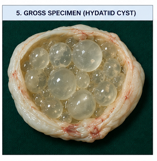
Intraoperative findings included:
• Large hydatid cyst in the right lobe of the liver
• Multiple daughter cysts within the cavity
• No communication with the biliary tree
The cyst contents were carefully evacuated and the residual cavity managed appropriately after instillation of a scolicidal agent.
The procedure was completed successfully without complications.
Follow-Up
At 1 Month
Clinical assessment revealed:
• Complete relief of abdominal pain
• Improved appetite
• No postoperative complications
At 3 Months
Follow-up ultrasonography showed:
• Reduction of residual cavity size
• No evidence of recurrence
At 6 Months
The patient remained asymptomatic with:
• Normal liver function tests
• No radiological evidence of recurrent disease
• Good quality of life
Discussion
Hydatid disease is caused by infection with the larval stage of Echinococcus granulosus. Humans become infected through accidental ingestion of parasite eggs present in contaminated food, water, or soil.
After entering the portal circulation, the larvae are most commonly filtered by the liver, explaining why hepatic involvement is observed in the majority of cases.
Clinical Features
The clinical manifestations depend largely on cyst size, location, and complications.
Common symptoms include:
• Right upper quadrant pain
• Abdominal swelling
• Nausea
• Hepatomegaly
• Loss of appetite
Complications may include:
• Cyst rupture
• Secondary bacterial infection
• Biliary obstruction
• Anaphylactic reactions
• Peritoneal dissemination
Many patients remain asymptomatic for years because hydatid cysts grow slowly.
Diagnostic Evaluation
Imaging plays a central role in diagnosis.
Ultrasonography is the preferred initial investigation because it can identify:
• Daughter cysts
• Floating membranes
• Hydatid sand
• Internal septations
CT scanning provides better anatomical definition and is particularly useful for surgical planning.
Serological tests such as ELISA assist in confirming the diagnosis, although sensitivity may vary depending on cyst location and stage.
Treatment
Management depends on cyst characteristics and patient factors.
Treatment options include:
• Albendazole therapy
• Percutaneous aspiration, injection, and re-aspiration (PAIR)
• Surgical excision
• Combined medical and surgical therapy
Albendazole remains the cornerstone of pharmacological treatment and is often used both preoperatively and postoperatively.
Surgery remains the preferred option for:
• Large cysts
• Symptomatic lesions
• Complicated cysts
• Cysts at risk of rupture
Successful treatment requires complete removal or inactivation of the parasite while preventing spillage of cyst contents.
Prevention
Hydatid disease can be prevented through:
• Regular deworming of dogs
• Proper disposal of animal carcasses
• Improved sanitation practices
• Health education in endemic communities
• Washing fruits and vegetables before consumption
• Avoiding feeding raw offal to dogs
Public health measures remain crucial for reducing disease transmission.
Prognosis
The prognosis of hepatic hydatid disease is generally favorable when diagnosed early and treated appropriately. Advances in imaging techniques, antihelminthic therapy, and surgical management have significantly improved patient outcomes. Most patients experience complete recovery following successful treatment, although recurrence may occur if daughter cysts are not completely eradicated or if intraoperative spillage takes place. Long-term follow-up with imaging studies is therefore recommended. Early diagnosis and timely intervention help prevent potentially life-threatening complications such as cyst rupture, secondary infection, biliary obstruction, and anaphylactic reactions. Patient education regarding preventive measures is equally important in reducing the risk of reinfection in endemic areas.
Conclusion
Hydatid cyst of the liver is an important parasitic disease that should be considered in patients presenting with chronic right upper quadrant abdominal pain, particularly in endemic regions. Characteristic imaging findings and positive serological tests facilitate diagnosis. Combined medical and surgical management can provide excellent outcomes and minimize recurrence. Early recognition and appropriate treatment remain essential for preventing complications and improving patient quality of life.
References
- Brunetti E, Kern P, Vuitton DA. Expert consensus for the diagnosis and treatment of cystic and alveolar echinococcosis. Acta Tropica. 2010;114(1):1–16. https://pubmed.ncbi.nlm.nih.gov/19931502/
- Wen H, Vuitton L, Tuxun T, Li J, Vuitton DA, Zhang W, McManus DP. Echinococcosis: Advances in the 21st Century. Clinical Microbiology Reviews. 2019;32(2):e00075-18. https://pubmed.ncbi.nlm.nih.gov/30700431/
- Moro P, Schantz PM. Echinococcosis: A Review. International Journal of Infectious Diseases. 2009;13(2):125–133. https://pubmed.ncbi.nlm.nih.gov/18938096/
- Dziri C, Haouet K, Fingerhut A. Management of cystic echinococcosis complications and dissemination. World Journal of Surgery. 2004;28(7):731–736. https://pubmed.ncbi.nlm.nih.gov/15383910/
- World Health Organization Informal Working Group on Echinococcosis (WHO-IWGE). International classification of ultrasound images in cystic echinococcosis. Acta Tropica. 2003;85(2):253–261. https://pubmed.ncbi.nlm.nih.gov/12606104/
Read more such content on @ Hidoc Dr | Medical Learning App for Doctors
Recommended News For You
Recommended Articles For You
Featured News
Featured Articles
Featured Events
Featured KOL Videos
1.
NPM1-Mutated AML: Not All Treatments Are Equal
2.
Paging Dr. AI; Myth Busting in Cancer; Oncologists Shine in Malpractice Survey
3.
MRI can save rectal cancer patients from surgery, study suggests
4.
Rectal Cancer 'Crisis'; Smells Like Cancer; Flushing Away Clues to Colon Cancer
5.
Rising rates of head and neck cancers in England
1.
A Closer Look at Breast Cancer: Examining the Ultrasound Images
2.
Exploring the Latest Advances in Hodgkin's Lymphoma Treatment
3.
Molecular Classification of Refractory Anemia: A Comprehensive Review
4.
Ferroptosis-Modulating Therapeutics in Precision Oncology
5.
Blood-Cell Morphology Foundation Models: Transforming Hematological Diagnostics and Clinical Practice
1.
International Cancer Conference
2.
Asian Symposium on Advancement in Hematology and Oncology (ASAHO)
3.
International Cancer Conference
4.
Asian Symposium on Advancement in Hematology and Oncology (ASAHO)
5.
Asian Symposium on Advancement in Hematology and Oncology
1.
CDK4/6 Inhibitors in Extending Overall Survival in HR+/HER2- aBC Patients in Clinical Trial and Real World
2.
Thromboprophylaxis In Medical Settings- An Update
3.
Nimotozumab Plus Chemo-radiotherapy v/s Placebo Plus Chemo-radiotherapy in Locally Advanced Nasopharyngeal Carcinoma
4.
How Platelet Disorders Can Affect the Body
5.
Treatment Sequencing Strategies in ALK + NSCLC Patients with CNS Diseases

Address :
Hidoc Dr. Inc. | Delaware C Corp | 1309 Coffeen Ave. Suite 1200, Sheridan WY, 82801
Phone :
+1-415-463-3094
Email :
anishagadia@hidoc.co
© Copyright 2026 Hidoc Dr. Inc.
Terms & Conditions - LLP | Inc. | Privacy Policy - LLP | Inc. | Account Deactivation

To get started please enter your email ID
Welcome to Hidoc Dr.
Join to enhance your clinical skills and gain specialized in-depth medical knowledge