Keloid Following Ear Piercing Presenting as a Progressive Painful Auricular Mass: A Case Report
OthersPage Navigation
Abstract
Keloids are benign fibroproliferative scars that develop due to excessive collagen deposition following cutaneous injury. Unlike hypertrophic scars, keloids extend beyond the boundaries of the original wound and rarely regress spontaneously. They commonly occur in genetically predisposed individuals following ear piercing, surgery, burns, acne, lacerations, or vaccination. Common sites include the earlobes, chest, shoulders, upper back, and jawline. Patients typically present with progressively enlarging, firm, raised lesions associated with pruritus, pain, cosmetic concerns, and occasionally functional impairment. Early diagnosis and multimodal treatment are important because recurrence following surgical excision alone is common. We report the case of a 28-year-old woman presenting with a progressively enlarging painful keloid over the right earlobe one year after ear piercing. Clinical examination confirmed the diagnosis. The patient underwent intralesional corticosteroid therapy followed by surgical excision with adjuvant steroid injections, resulting in satisfactory cosmetic and functional outcomes without recurrence during follow-up.
Introduction
Keloids are abnormal wound healing disorders characterized by excessive fibroblast proliferation and collagen deposition that extends beyond the margins of the initial skin injury. The exact pathogenesis remains incompletely understood but involves dysregulated inflammation, prolonged fibroblast activation, increased transforming growth factor-beta (TGF-β) signaling, and abnormal extracellular matrix remodeling.
Keloids are more common in younger individuals between 10 and 30 years of age and occur more frequently in patients with darker skin pigmentation and a positive family history. Ear piercing remains one of the most common precipitating factors for auricular keloids. Although benign, these lesions often cause significant cosmetic disfigurement, pain, itching, psychological distress, and high recurrence rates after treatment. Management typically requires a combination of surgery and adjuvant therapies to minimize recurrence.
Case Report
A 28-year-old woman presented to the dermatology outpatient department with a progressively enlarging swelling over the right earlobe for approximately one year. The lesion developed several months after cosmetic ear piercing and had gradually increased in size. She complained of intermittent pain, persistent itching, and occasional tenderness, particularly while sleeping on the affected side. The patient also expressed significant cosmetic concern.
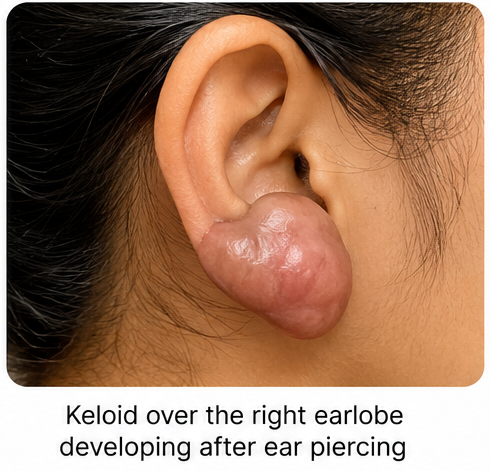
She had no history of diabetes mellitus, autoimmune disease, previous keloids, or connective tissue disorders. Family history revealed that her mother had developed keloids following surgical procedures. There was no history of trauma apart from the ear piercing, recurrent infection, or foreign body reaction.
On examination, a firm, smooth, shiny, hyperpigmented nodular lesion measuring approximately 2.8 × 2.5 cm was present over the posterior aspect of the right earlobe, extending beyond the original piercing site. The lesion was non-fluctuant, mildly tender on palpation, and firmly attached to the overlying skin but mobile over the underlying cartilage. No ulceration, discharge, secondary infection, or regional lymphadenopathy was observed. Examination of the contralateral ear and remaining skin was unremarkable.
Routine laboratory investigations, including complete blood count, blood glucose, renal function, and inflammatory markers, were within normal limits. Ultrasonography demonstrated a well-defined hypoechoic soft tissue lesion confined to the dermis and subcutaneous tissue without cartilage involvement. Based on the clinical appearance and imaging findings, a diagnosis of auricular keloid was established.
Management and Outcome
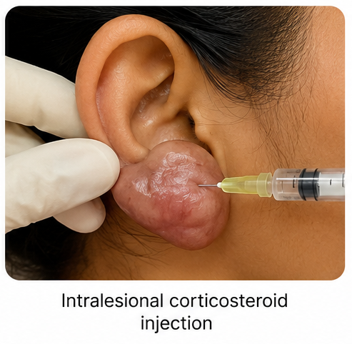
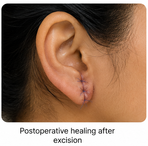
The patient initially received three sessions of intralesional triamcinolone acetonide injections at four-week intervals, resulting in partial reduction in lesion size, itching, and pain. She subsequently underwent complete surgical excision of the keloid with meticulous tension-free wound closure.
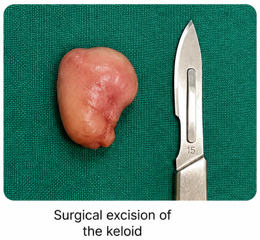
Postoperatively, intralesional corticosteroid injections were continued monthly for three additional sessions to reduce recurrence risk.
Silicone gel sheeting was initiated after complete wound healing and continued for three months.
The patient was advised to avoid further ear piercing and minimize local trauma.
The surgical wound healed uneventfully without infection or wound dehiscence. Progressive improvement in cosmetic appearance was observed, and symptoms resolved completely.
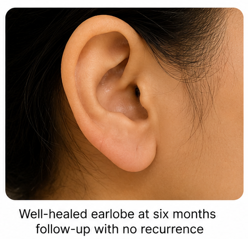
Follow-up
One Month
- Surgical wound healed completely.
- Significant reduction in itching and tenderness.
- Excellent cosmetic outcome.
Three Months
- No evidence of recurrent scar formation.
- Continued use of silicone gel sheeting.
- Patient satisfied with cosmetic appearance.
Six Months
- No clinical recurrence.
- Normal appearance of the earlobe.
- No pain, itching, or functional impairment.
Discussion
Keloids represent a pathological response to dermal injury resulting from excessive collagen synthesis and impaired wound remodeling. They differ from hypertrophic scars by extending beyond the original wound margins and demonstrating persistent growth. Increased fibroblast proliferation, elevated transforming growth factor-beta activity, vascular endothelial growth factor expression, and abnormal collagen type I and III production contribute to lesion formation.
The diagnosis is primarily clinical and based on a characteristic history of progressive scar enlargement beyond the original injury. Imaging is rarely necessary but may help assess lesion depth or exclude alternative soft tissue tumors in atypical cases. Histopathological examination demonstrates thick, hyalinized collagen bundles arranged in a haphazard pattern.
Differential diagnoses include hypertrophic scar, dermatofibroma, epidermoid cyst, dermatofibrosarcoma protuberans, pilomatricoma, and cutaneous sarcoidosis. Clinical history and lesion morphology generally distinguish these entities.
Management remains challenging because recurrence rates are high, particularly following surgical excision alone. Current evidence supports multimodal treatment combining intralesional corticosteroids, surgical excision, silicone gel therapy, pressure therapy, cryotherapy, laser therapy, radiotherapy in selected cases, and emerging biologic therapies. Intralesional corticosteroids remain the first-line treatment because they reduce fibroblast proliferation and collagen synthesis. Combined therapy significantly lowers recurrence compared with monotherapy.
Patients should receive counseling regarding recurrence risk, avoidance of unnecessary skin trauma, careful wound care, and early treatment of newly developing lesions.
Prognosis
Keloids are benign lesions with no malignant potential but frequently recur despite treatment. The prognosis improves considerably with early diagnosis and multimodal therapy. Combination treatment involving surgical excision and adjuvant corticosteroid therapy achieves lower recurrence rates than surgery alone. Long-term follow-up remains essential because recurrence may occur months or years after treatment.
Conclusion
Auricular keloids should be suspected in patients presenting with progressively enlarging raised scars following ear piercing or minor skin trauma. Clinical diagnosis is usually straightforward, and early intervention with multimodal therapy offers the best cosmetic and functional outcomes. Surgical excision combined with adjuvant intralesional corticosteroids and silicone therapy effectively reduces recurrence and improves patient satisfaction.
References
- Berman B, Maderal A, Raphael B. Keloids and Hypertrophic Scars: Pathophysiology, Classification, and Treatment. Dermatologic Surgery. 2017;43(Suppl 1):S3–S18. https://pubmed.ncbi.nlm.nih.gov/28609296/
- Ogawa R. Keloid and Hypertrophic Scars Are the Result of Chronic Inflammation in the Reticular Dermis. International Journal of Molecular Sciences. 2017;18(3):606. https://pubmed.ncbi.nlm.nih.gov/28300703/
- Gauglitz GG, Korting HC, Pavicic T, et al. Hypertrophic Scarring and Keloids: Pathomechanisms and Current and Emerging Treatment Strategies. Molecular Medicine. 2011;17(1-2):113–125. https://pubmed.ncbi.nlm.nih.gov/20927486/
- Limandjaja GC, Niessen FB, Scheper RJ, Gibbs S. The Keloid Disorder: Heterogeneity, Histopathology, Mechanisms and Models. Frontiers in Cell and Developmental Biology. 2020;8:360. https://pubmed.ncbi.nlm.nih.gov/32477995/
- Kelly AP. Medical and Surgical Therapies for Keloids. Dermatologic Therapy. 2004;17(2):212–218. https://pubmed.ncbi.nlm.nih.gov/15165204/
Read more such content on @ Hidoc Dr | Medical Learning App for Doctors
Recommended News For You
Recommended Articles For You
Featured News
Featured Articles
Featured Events
Featured KOL Videos
1.
NPM1-Mutated AML: Not All Treatments Are Equal
2.
Paging Dr. AI; Myth Busting in Cancer; Oncologists Shine in Malpractice Survey
3.
MRI can save rectal cancer patients from surgery, study suggests
4.
Rectal Cancer 'Crisis'; Smells Like Cancer; Flushing Away Clues to Colon Cancer
5.
Rising rates of head and neck cancers in England
1.
A Closer Look at Breast Cancer: Examining the Ultrasound Images
2.
Exploring the Latest Advances in Hodgkin's Lymphoma Treatment
3.
Molecular Classification of Refractory Anemia: A Comprehensive Review
4.
Ferroptosis-Modulating Therapeutics in Precision Oncology
5.
Blood-Cell Morphology Foundation Models: Transforming Hematological Diagnostics and Clinical Practice
1.
International Cancer Conference
2.
Asian Symposium on Advancement in Hematology and Oncology (ASAHO)
3.
International Cancer Conference
4.
Asian Symposium on Advancement in Hematology and Oncology (ASAHO)
5.
Asian Symposium on Advancement in Hematology and Oncology
1.
CDK4/6 Inhibitors in Extending Overall Survival in HR+/HER2- aBC Patients in Clinical Trial and Real World
2.
Thromboprophylaxis In Medical Settings- An Update
3.
Nimotozumab Plus Chemo-radiotherapy v/s Placebo Plus Chemo-radiotherapy in Locally Advanced Nasopharyngeal Carcinoma
4.
How Platelet Disorders Can Affect the Body
5.
Treatment Sequencing Strategies in ALK + NSCLC Patients with CNS Diseases

Address :
Hidoc Dr. Inc. | Delaware C Corp | 1309 Coffeen Ave. Suite 1200, Sheridan WY, 82801
Phone :
+1-415-463-3094
Email :
anishagadia@hidoc.co
© Copyright 2026 Hidoc Dr. Inc.
Terms & Conditions - LLP | Inc. | Privacy Policy - LLP | Inc. | Account Deactivation

To get started please enter your email ID
Welcome to Hidoc Dr.
Join to enhance your clinical skills and gain specialized in-depth medical knowledge