AI in Cancer Diagnostics and Imaging Interpretation
OncologyPage Navigation
Artificial intelligence (AI) has emerged as a transformative force in healthcare, and oncology is no exception. From enhancing diagnostic accuracy to optimizing imaging interpretation and enabling real-time treatment decisions, AI is redefining how cancer is detected, classified, and managed. For oncologists and pharmaceutical managers, the promise of AI in oncology diagnostics offers both clinical advantages and strategic opportunities across the care continuum.
In this blog, we explore the evolving role of AI in cancer diagnostics, the growing sophistication of oncology imaging interpretation training, and the emergence of AI-powered cancer diagnosis tools. We also analyze the implications for pharma stakeholders and highlight some of the most promising AI applications and platforms in oncology image analysis.
The Imperative for AI in Oncology Diagnostics
Cancer diagnostics have long depended on imaging, pathology, and biomarker analysis to detect, stage, and monitor disease progression. However, these traditional modalities often suffer from inter-observer variability, interpretation delays, and access disparities in low-resource settings. Additionally, the increasing volume and complexity of imaging and molecular data challenge even the most experienced clinicians.
This is where AI in oncology diagnostics offers a game-changing solution. AI algorithms, particularly those based on machine learning (ML) and deep learning (DL), can be trained to identify patterns in complex datasets with high speed and precision. These models support radiologists and pathologists in interpreting images, quantifying tumor features, and detecting subtle abnormalities that may escape the human eye.
For example, convolutional neural networks (CNNs) have demonstrated the ability to outperform human radiologists in certain image classification tasks, particularly in breast, lung, and prostate cancer imaging. The integration of AI tools into diagnostic workflows not only enhances accuracy but also reduces turnaround times, enabling faster clinical decisions.
AI-Powered Cancer Diagnosis: Capabilities and Benefits
AI-powered cancer diagnosis tools can process vast datasets from imaging modalities (CT, MRI, PET), electronic health records (EHRs), and molecular diagnostics. These systems generate diagnostic predictions, risk scores, and even treatment recommendations based on patient-specific features.
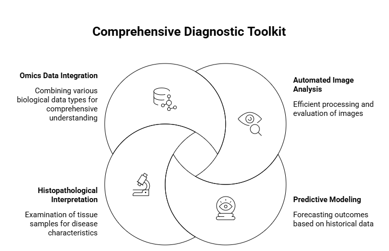
Key capabilities include:
-
Automated image analysis: AI models can detect and classify lesions, measure tumor volume, and evaluate progression or response to therapy.
-
Predictive modeling: AI can forecast disease trajectory, metastasis risk, or recurrence using historical patient data and biomarkers.
-
Histopathological interpretation: AI is increasingly used to analyze digital pathology slides for cancer subtyping, mitotic count, and immune infiltration, aiding in precision oncology.
-
Integration with omics data: By combining imaging and genomic information, AI supports multi-modal diagnostics and personalized treatment planning.
The benefits are substantial:
-
Improved diagnostic accuracy and consistency
-
Reduced diagnostic workload for clinicians
-
Early detection and intervention
-
Cost savings and operational efficiency
-
Better patient stratification for clinical trials
Oncology Imaging Interpretation Training: Preparing the Next Generation
As AI tools become more prevalent, oncologists and radiologists must adapt to new modes of diagnostic collaboration. This shift demands focused training in oncology imaging interpretation augmented by AI.
Leading medical institutions are incorporating AI modules into radiology and oncology fellowships, focusing on:

-
Understanding AI algorithms and their limitations
-
Validating AI outputs in clinical practice
-
Navigating medico-legal considerations and ethical implications
-
Collaborating with data scientists to refine diagnostic workflows
Additionally, industry stakeholders and professional societies are launching online courses and certifications in AI for medical imaging. These programs use case studies, real-world imaging datasets, and simulation platforms to help clinicians recognize AI’s role in enhancing; not replacing; clinical judgment.
Oncology Image Analysis AI Tools: What's Available?
A wide range of oncology image analysis AI tools are now available, many of which are FDA-approved or CE-marked for clinical use. These platforms cater to various oncological needs, including detection, segmentation, classification, and response assessment.
Some notable tools include:
-
PathAI: Specializes in digital pathology and histology analysis, assisting in tumor grading and immune response characterization.
-
Zebra Medical Vision: Offers automated cancer detection (e.g., breast, lung, liver) from imaging data using deep learning.
-
Aidoc: Supports radiologists with real-time triage and diagnostic alerts for suspected cancer findings.
-
Tempus: Integrates AI-based genomic profiling with imaging insights to drive precision oncology decisions.
-
Arterys: Provides cloud-based AI tools for lung and liver cancer diagnosis, with capabilities for tumor segmentation and tracking.
These tools not only streamline diagnostic workflows but also generate structured data outputs, enabling better integration with oncology clinical trials and drug development programs.
AI in Oncology Diagnostics and Clinical Decision Support
One of the most powerful applications of AI in cancer diagnostics is its role in real-time clinical decision support. By mining patient data, AI platforms can assist oncologists in:
-
Choosing the most appropriate diagnostic test
-
Recommending personalized treatment regimens
-
Predicting treatment efficacy based on tumor and patient characteristics
-
Identifying eligibility for biomarker-driven clinical trials
This synergy between diagnostics and therapeutics (Dx-Tx) has important implications for pharma managers, particularly those involved in companion diagnostics and targeted therapies. AI can help identify new biomarkers, stratify patient populations, and monitor therapeutic responses - key aspects of precision medicine strategies.
AI-Driven Insights in Cancer Imaging Biomarkers
The use of radiomics and AI-generated imaging biomarkers is a fast-growing area within oncology. Radiomics involves extracting high-dimensional features from standard imaging modalities, such as texture, shape, and intensity.
AI then analyzes these features to derive clinically relevant biomarkers, such as:
-
Tumor aggressiveness
-
Hypoxia levels
-
Immune infiltration status
-
Risk of metastasis
These non-invasive biomarkers are particularly valuable in contexts where biopsies are infeasible or risky. Pharma companies are increasingly interested in using AI-derived imaging biomarkers for drug response prediction, toxicity monitoring, and post-marketing surveillance.
Addressing Challenges in AI-Powered Cancer Diagnosis
Despite its potential, AI-powered cancer diagnosis is not without challenges. Key barriers to adoption include:
-
Data Quality and Bias
AI algorithms require large, diverse, and well-annotated datasets. Poor data quality or underrepresentation of certain populations can lead to biased or inaccurate results. -
Integration with Clinical Workflows
Adapting AI tools to existing hospital systems (PACS, EHRs) remains a technical and organizational hurdle. -
Regulatory and Ethical Concerns
Transparent algorithm validation, explainability, and accountability are essential to gain clinician trust and meet regulatory standards. -
Cost and Reimbursement
AI platforms can be costly to implement, and reimbursement models for AI-supported diagnostics are still evolving. -
Training and Cultural Shift
Clinicians must be trained not only to use AI tools but to critically evaluate their outputs; a major shift in diagnostic culture.
Pharma’s Role in Advancing AI in Oncology
Pharma managers are uniquely positioned to drive the adoption and co-development of AI tools in oncology. Strategic roles include:
-
Collaborating with AI companies to co-develop diagnostic tools that support drug pipelines
-
Integrating AI in clinical trials to improve patient recruitment, monitor outcomes, and analyze real-world evidence
-
Supporting AI research grants and academic partnerships to uncover novel diagnostic biomarkers
-
Driving regulatory frameworks by engaging with agencies to streamline approval of AI-based companion diagnostics
Furthermore, pharma companies can leverage AI to inform go-to-market strategies, optimize commercial forecasting, and support medical affairs teams with predictive analytics on disease burden and treatment gaps.
The Road Ahead: AI and the Future of Cancer Care
The future of AI in oncology diagnostics lies in its seamless integration across the cancer care continuum from early detection and diagnosis to therapy selection and survivorship monitoring. We can expect increasing use of multimodal AI platforms that combine:
-
Radiological imaging
-
Histopathology
-
Genomic data
-
Clinical records
-
Wearable health data
Such holistic approaches will enable oncologists to make faster, more informed, and personalized decisions.
In the next decade, AI is also expected to:
-
Facilitate virtual tumor boards with real-time decision support
-
Automate trial matching based on AI-evaluated eligibility
-
Empower low-resource settings with cloud-based diagnostic platforms
-
Enhance patient engagement through AI-driven symptom monitoring apps
For both clinicians and pharmaceutical leaders, staying ahead of this technological curve is not optional; it is critical to delivering value-based, precision oncology.
Conclusion
The integration of AI in oncology diagnostics marks a paradigm shift in cancer care. With tools that enhance imaging interpretation, enable faster cancer detection, and support personalized treatment decisions, AI is not just a technological advancement; it is a clinical necessity.
For oncologists, AI offers decision-making support, reduced diagnostic burden, and improved outcomes. For pharma managers, it opens new avenues for biomarker discovery, trial optimization, and product differentiation in an increasingly competitive market.
Whether it's through AI-powered cancer diagnosis, oncology imaging interpretation training, or cutting-edge oncology image analysis AI tools, the path forward is clear: AI is not just shaping the future of cancer diagnostics; it is the future.
Read more such content on @ Hidoc Dr | Medical Learning App for Doctors
Recommended News For You
Recommended Articles For You
Featured News
Featured Articles
Featured Events
Featured KOL Videos
1.
Underprescribed for Alcohol Use Disorder is pharmacotherapy.
2.
Providing essential cancer care to rural communities
3.
Study finds 10% of pediatric blood cancers may stem from medical imaging radiation
4.
Limits to Anti-Nausea Pill Coverage Wear on Cancer Patients and Doctors
5.
AI Breath Test IDs Cancers; More Grief for Prior Auth; Imatinib Discoverer Resigns
1.
Everything You Need to Know About Normal Potassium Levels
2.
A Review of Cancer Treatment Evolution Towards Stem Cells and Gene Therapy
3.
Unmasking the Subtle Symptoms of Colon Cancer
4.
Real-World Evidence and Patient Voices: Redefining Outcomes in Modern Oncology
5.
Strategic Deals and FDA Approvals Transforming U.S. Oncology Drug Development
1.
International Lung Cancer Congress®
2.
Genito-Urinary Oncology Summit 2026
3.
Future NRG Oncology Meeting
4.
ISMB 2026 (Intelligent Systems for Molecular Biology)
5.
Annual International Congress on the Future of Breast Cancer East
1.
An Eagles View - Evidence-based discussion on Iron Deficiency Anemia
2.
ESMO Breast Cancer 2022: P Reality X- A Restrospective Analysis
3.
Untangling The Best Treatment Approaches For ALK Positive Lung Cancer - Part VIII
4.
Revolutionizing Treatment of ALK Rearranged NSCLC with Lorlatinib - Part I
5.
Evolving Space of First-Line Treatment for Urothelial Carcinoma- Case Discussion

Address :
Hidoc Dr. Inc. | Delaware C Corp | 1309 Coffeen Ave. Suite 1200, Sheridan WY, 82801
Phone :
+1-415-463-3094
Email :
anishagadia@hidoc.co
© Copyright 2026 Hidoc Dr. Inc.
Terms & Conditions - LLP | Inc. | Privacy Policy - LLP | Inc. | Account Deactivation

To get started please enter your email ID
Welcome to Hidoc Dr.
Join to enhance your clinical skills and gain specialized in-depth medical knowledge