Immune Thrombocytopenia Presenting With Petechiae, Mucosal Bleeding and Severe Isolated Thrombocytopenia
OthersPage Navigation
Introduction
Immune thrombocytopenia (ITP) is an acquired autoimmune disorder characterised by a platelet count below 100 × 10⁹/L in the absence of another clear cause of thrombocytopenia. It is mediated by accelerated immune destruction of platelets together with impaired platelet production.
Clinical presentation is variable. Some patients are asymptomatic, whereas others develop petechiae, purpura, easy bruising, epistaxis, gingival bleeding, heavy menstrual bleeding, or, rarely, life-threatening internal haemorrhage. Bleeding severity does not correlate perfectly with the platelet count and must be assessed individually.
Primary ITP is diagnosed when no underlying disorder is identified. Secondary immune thrombocytopenia may occur with autoimmune disease, chronic viral infection, lymphoproliferative disorders, immune deficiency, certain medications, or other conditions.
No single laboratory test confirms primary ITP. Diagnosis therefore depends on clinical assessment, confirmation of isolated thrombocytopenia, examination of the peripheral blood film, and exclusion of alternative causes.
Early recognition is important because severe thrombocytopenia accompanied by active mucosal or internal bleeding may require urgent treatment. The immediate objective is to achieve a haemostatically safe platelet count rather than a normal platelet count.
Case Report
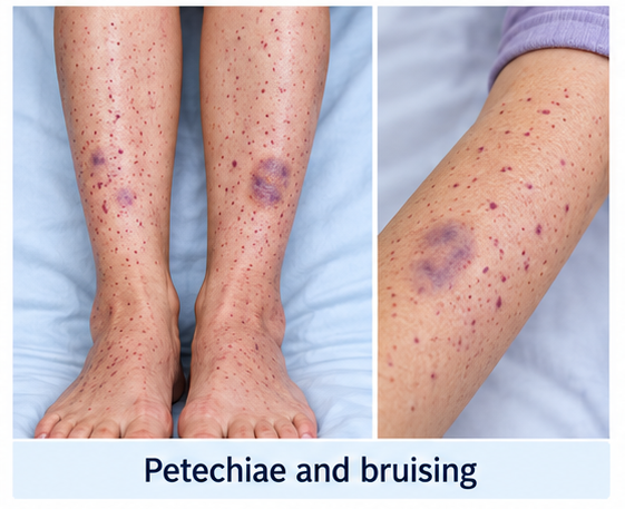
A 29-year-old woman presented with spontaneous bruising over both lower limbs and multiple pinpoint red spots over the ankles and forearms for approximately two weeks.
During the preceding four days, she had experienced intermittent gum bleeding while brushing and two episodes of spontaneous epistaxis, each lasting approximately ten minutes. She also reported that her most recent menstrual period had been unusually heavy and had continued for eight days.
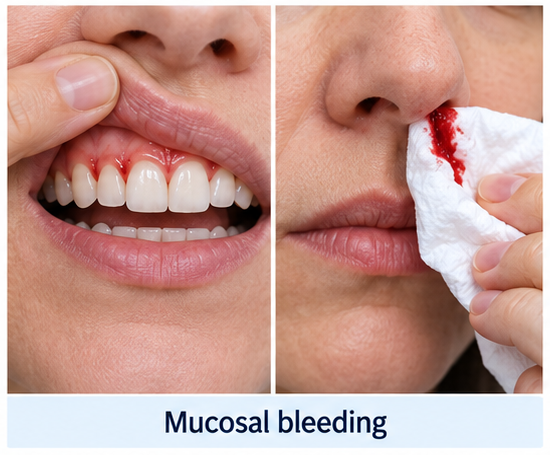
She denied haematemesis, melaena, haematuria, severe headache, confusion, visual disturbance, focal weakness, chest pain, breathlessness, or abdominal pain. There was no recent trauma or surgical procedure.
She had experienced a self-limiting upper respiratory illness approximately three weeks earlier. There was no history of persistent fever, weight loss, night sweats, recurrent infection, joint pain, photosensitivity, oral ulceration, or skin rash apart from the bleeding manifestations.
The patient had no known liver, renal, thyroid, autoimmune, or haematological disease. She was not pregnant and had no previous history of thrombocytopenia, excessive bleeding after dental procedures, or postpartum haemorrhage.
She was not taking heparin, quinine, sulfonamides, antiepileptic drugs, antiplatelet agents, anticoagulants, or herbal preparations. She reported occasional paracetamol use but denied recent use of aspirin or nonsteroidal anti-inflammatory drugs.
There was no history of alcohol misuse, recent blood transfusion, or exposure to industrial chemicals. No family member had thrombocytopenia or an inherited bleeding disorder.
On examination, the patient was alert, haemodynamically stable, and afebrile. Her pulse was 88 beats per minute, blood pressure was 112/72 mmHg, respiratory rate was 16 breaths per minute, and oxygen saturation was 99% on room air.
Multiple non-blanching petechiae were present over both ankles, lower legs, and forearms. Several ecchymoses of varying size were visible over the thighs and arms without a corresponding history of trauma.
Small haemorrhagic areas were observed along the gingival margin, but there was no active epistaxis at the time of examination. No large oral blood blisters were present.
There was no pallor, icterus, lymphadenopathy, hepatomegaly, or splenomegaly. Cardiovascular, respiratory, abdominal, and neurological examinations were otherwise unremarkable.
The combination of petechiae, ecchymoses, mucosal bleeding, heavy menstrual bleeding, and an otherwise normal examination suggested a platelet disorder rather than a coagulation-factor deficiency.
Investigations
Initial laboratory investigations showed:
- Platelet count: 8 × 10⁹/L
- Haemoglobin: 11.2 g/dL
- Total leucocyte count: 7.4 × 10⁹/L
- Absolute neutrophil count: 4.3 × 10⁹/L
- Mean corpuscular volume: 82 fL
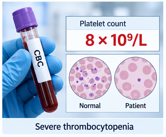
Repeat platelet counting in a citrate-anticoagulated sample confirmed severe thrombocytopenia and excluded EDTA-dependent platelet clumping.
Peripheral blood-film examination showed markedly reduced platelets with occasional large platelets. Red-cell and white-cell morphology was normal. No platelet clumps, schistocytes, blasts, dysplastic cells, or malarial parasites were identified.
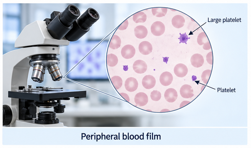
Prothrombin time, activated partial thromboplastin time, fibrinogen, and D-dimer were within reference ranges. These findings did not support disseminated intravascular coagulation or a primary coagulation-factor disorder.
The reticulocyte count, lactate dehydrogenase, bilirubin, haptoglobin, serum creatinine, and urinalysis were normal. The direct antiglobulin test was negative. There was no biochemical or morphological evidence of haemolysis or thrombotic microangiopathy.
Liver-function tests, thyroid-stimulating hormone, serum vitamin B12, folate, and quantitative immunoglobulin levels were within reference ranges.
Serological testing for human immunodeficiency virus, hepatitis B virus, and hepatitis C virus was negative. Helicobacter pylori stool-antigen testing was negative.
Antinuclear antibody testing was negative, and there were no clinical features of systemic lupus erythematosus or antiphospholipid syndrome. A urine pregnancy test was negative.
Abdominal ultrasonography showed normal liver and spleen size without portal hypertension or abdominal lymphadenopathy.
Bone-marrow examination was not performed because the patient had typical isolated thrombocytopenia, a compatible peripheral smear, no organomegaly or systemic warning features, and no abnormality in the other blood-cell lines.
Based on severe isolated thrombocytopenia, typical mucocutaneous bleeding, exclusion of pseudothrombocytopenia and secondary causes, and an otherwise normal blood film, newly diagnosed primary immune thrombocytopenia was considered the most likely diagnosis.
Differential Diagnosis
Pseudothrombocytopenia
EDTA-dependent platelet aggregation can produce a falsely low automated platelet count. The absence of platelet clumps on the blood film and confirmation of thrombocytopenia in a citrate sample excluded this laboratory artefact.
Thrombotic Thrombocytopenic Purpura
Thrombotic thrombocytopenic purpura may present with severe thrombocytopenia and requires urgent plasma-exchange therapy. However, the patient had no neurological or renal abnormality, fever, microangiopathic haemolytic anaemia, elevated lactate dehydrogenase, or schistocytes.
Disseminated Intravascular Coagulation
Disseminated intravascular coagulation can cause thrombocytopenia and bleeding, usually in association with severe infection, trauma, malignancy, or obstetric complications. Normal coagulation tests, fibrinogen, and D-dimer, together with clinical stability, argued strongly against this diagnosis.
Drug-Induced Immune Thrombocytopenia
Several medications may trigger abrupt and profound immune thrombocytopenia. A detailed medication history did not identify a plausible culprit drug, and the patient had not recently received heparin.
Bone-Marrow Failure or Haematological Malignancy
Aplastic anaemia, acute leukaemia, myelodysplastic syndromes, marrow infiltration, and megaloblastic anaemia may cause thrombocytopenia. Normal leucocyte and red-cell morphology, absence of blasts or pancytopenia, and lack of constitutional symptoms, lymphadenopathy, or organomegaly made these diagnoses unlikely.
Secondary Immune Thrombocytopenia
Secondary ITP associated with systemic lupus erythematosus, chronic viral infection, immune deficiency, lymphoproliferative disease, or Helicobacter pylori infection was considered. Clinical evaluation and targeted investigations did not identify an underlying disorder.
Management and Outcome
Because the platelet count was 8 × 10⁹/L and the patient had gingival bleeding, recurrent epistaxis, and heavy menstrual bleeding, she was admitted for treatment and close clinical observation.
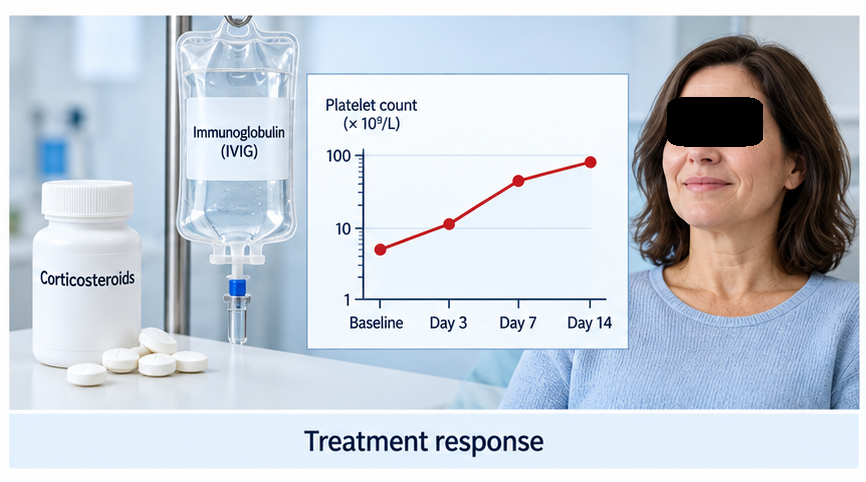
She received oral dexamethasone 40 mg once daily for four days. Intravenous immunoglobulin at 1 g/kg was administered as a single dose to obtain a more rapid platelet response because of active mucosal bleeding.
Local measures were used for epistaxis and gingival bleeding. Antifibrinolytic therapy was considered for mucosal bleeding after assessment of thrombotic risk and contraindications. Platelet transfusion was not given because there was no critical or life-threatening haemorrhage.
She was advised to avoid aspirin, nonsteroidal anti-inflammatory drugs, intramuscular injections, contact sports, and other activities associated with trauma. Venepuncture and blood-pressure cuff use were minimised when possible.
The platelet count increased to 24 × 10⁹/L after 24 hours and 68 × 10⁹/L by day three. Epistaxis and gingival bleeding stopped, and no new petechiae or ecchymoses developed.
She was discharged after completion of dexamethasone with a platelet count of 91 × 10⁹/L. Clear instructions were provided to return immediately for persistent bleeding, haematemesis, melaena, haematuria, severe headache, vomiting, confusion, visual symptoms, or neurological weakness.
At the one-week review, the platelet count was 164 × 10⁹/L and all mucosal bleeding had resolved. Mild bruising remained but was fading.
At four weeks, the platelet count was 138 × 10⁹/L without further treatment. Her next menstrual period was normal in duration and flow.
At three months, she remained clinically well with a platelet count of 112 × 10⁹/L. She continued haematology follow-up because relapse may occur and the subsequent disease course cannot be predicted from the initial response alone.
Discussion
This case illustrates a typical presentation of newly diagnosed primary ITP in an adult. Mucocutaneous bleeding, including petechiae, purpura, bruising, epistaxis, gingival bleeding, and heavy menstrual bleeding, reflects defective primary haemostasis caused by severe thrombocytopenia.
ITP is an immune-mediated disorder involving both increased platelet clearance and impaired platelet production. Autoantibodies may bind platelet glycoproteins and promote clearance by macrophages, while T-cell abnormalities and immune effects on megakaryocytes may further reduce platelet survival and production.
The diagnosis remains one of exclusion. A complete history must examine bleeding severity, previous blood counts, infection, pregnancy, medications, alcohol exposure, transfusion, autoimmune symptoms, malignancy-associated symptoms, and family history.
Physical examination should assess the pattern and extent of bleeding and look for fever, pallor, jaundice, lymphadenopathy, hepatomegaly, splenomegaly, neurological abnormalities, and signs of chronic liver or systemic autoimmune disease. Substantial splenomegaly is atypical for primary ITP and should prompt investigation for another cause.
The complete blood count usually shows isolated thrombocytopenia. Peripheral blood-film review is essential to exclude platelet clumping, schistocytes, blasts, dysplasia, and features of inherited thrombocytopenia.
Bone-marrow examination is not routinely required in an otherwise typical presentation but may be appropriate when blood-count or smear abnormalities, systemic features, treatment failure, relapse after remission, or diagnostic uncertainty are present.
The decision to treat should not be based on the platelet count alone. Active bleeding, age, comorbidities, anticoagulant or antiplatelet therapy, fall risk, lifestyle, access to emergency care, and planned procedures all influence management.
For newly diagnosed adults requiring therapy, a short course of corticosteroids is generally preferred to prolonged corticosteroid exposure. Intravenous immunoglobulin can produce a faster but often temporary platelet increase and is useful when a rapid response is needed, including clinically important bleeding or preparation for an urgent procedure.
Treatment aims to stop bleeding and maintain a safe platelet count while minimising toxicity. It is not necessary to normalise the platelet count. Platelet transfusion is generally reserved for critical bleeding and is used together with therapies that reduce immune-mediated platelet destruction.
Patients who develop persistent or chronic ITP and require further therapy may be considered for thrombopoietin-receptor agonists, rituximab, fostamatinib, splenectomy, or other individualised options. Treatment choice depends on disease duration, bleeding risk, comorbidity, expected durability, adverse effects, cost, availability, and patient preference.
Education is central to long-term care. Patients should understand which medicines increase bleeding risk, when activity modification is appropriate, how often blood counts are required, and which symptoms demand urgent assessment. Women should be asked directly about menstrual blood loss because it may be clinically important but underreported.
Conclusion
Immune thrombocytopenia should be considered in patients with unexplained petechiae, purpura, easy bruising, epistaxis, gingival bleeding, or heavy menstrual bleeding, particularly when the complete blood count shows isolated thrombocytopenia.
In this illustrative case, severe isolated thrombocytopenia, a compatible peripheral blood film, normal coagulation and haemolysis studies, and exclusion of secondary causes supported the diagnosis of newly diagnosed primary ITP.
A short course of high-dose dexamethasone combined with intravenous immunoglobulin produced a rapid clinical and platelet response, allowing safe discharge and outpatient monitoring.
Careful exclusion of urgent alternative diagnoses, treatment based on bleeding risk, avoidance of unnecessary prolonged corticosteroid exposure, patient education, and structured follow-up are essential for safe management.
References
- Provan D, Arnold DM, Bussel JB, et al. Updated international consensus report on the investigation and management of primary immune thrombocytopenia. Blood Advances. 2019;3(22):3780–3817 https://pmc.ncbi.nlm.nih.gov/articles/PMC6880896/
- Neunert C, Terrell DR, Arnold DM, et al. American Society of Hematology 2019 guidelines for immune thrombocytopenia. Blood Advances. 2019;3(23):3829–3866 https://pmc.ncbi.nlm.nih.gov/articles/PMC6963252/
- Lambert MP, Gernsheimer TB. Clinical updates in adult immune thrombocytopenia. Blood. 2017;129(21):2829–2835 https://pmc.ncbi.nlm.nih.gov/articles/PMC5813736/
- Bussel JB, Garcia CA. Diagnosis of immune thrombocytopenia, including secondary forms, and selection of second-line treatment. Haematologica. 2022;107(9):2018–2036 https://haematologica.org/article/view/haematol.2021.279513
- Mititelu A, Onisâi MC, Roșca A, Vlădăreanu AM. Current understanding of immune thrombocytopenia: A review of pathogenesis and treatment options. International Journal of Molecular Sciences. 2024;25(4):2163 https://pmc.ncbi.nlm.nih.gov/articles/PMC10889445/
Read more such content on @ Hidoc Dr | Medical Learning App for Doctors
Recommended News For You
Recommended Articles For You
Featured News
Featured Articles
Featured Events
Featured KOL Videos
1.
Tumor characteristics found to differ for melanomas in children, teens and young adults
2.
Unexplained CAR-T Death; Americans Spurn Sunscreen; Improving Diversity in Trials
3.
"A lot" of aspirin reduces the risk of ovarian cancer, regardless of genetic factors, according to the JAMA study.
4.
Approval in Endometrial Cancer Expanded; Masks at Cancer Centers; NPR Reporter Dies
5.
Is Lenalidomide Resistance in Myeloma Reversible with JAK Inhibition?
1.
Modern Oncology Unveiled with Treatment Innovations and Personalized Cancer Care
2.
Screening for Early Therapy-Related Cancer Susceptibility
3.
Unlocking the Potential of Bicalutamide 50 mg: A Revolutionary New Treatment for Prostate Cancer
4.
Skin-Sparing Mastectomy: Balancing Cancer Control with Cosmetic Results
5.
Reducing Modifiable Childhood Cancer Exposure Risks
1.
International Conference on Innovations in Critical Care for Oncology and Cardiology
2.
International Conference on Oncology, Cardiology and Critical Care Policy
3.
International Conference on Oncology, Cancer Prevention and Public Health
4.
International Conference on Cancer Nursing and Rehabilitation Strategies
5.
International Conference on Best Practices in Oncology, Cardiology and Critical Care
1.
Dacomitinib Case Presentation: Baseline Treatment and Current Status
2.
Targeting Oncologic Drivers with Dacomitinib: Further Discussion on Lung Cancer Treatment
3.
Should We Use DARA Up Front As First-Line Therapy in MM?
4.
Iron Deficiency Anemia: Ferric Maltol As a New Treatment Option- A New Perspective
5.
Importance of Cancer Screening and Early Detection

Address :
Hidoc Dr. Inc. | Delaware C Corp | 1309 Coffeen Ave. Suite 1200, Sheridan WY, 82801
Phone :
+1-415-463-3094
Email :
anishagadia@hidoc.co
© Copyright 2026 Hidoc Dr. Inc.
Terms & Conditions - LLP | Inc. | Privacy Policy - LLP | Inc. | Account Deactivation

To get started please enter your email ID
Welcome to Hidoc Dr.
Join to enhance your clinical skills and gain specialized in-depth medical knowledge