Kaposi Sarcoma Presenting with Violaceous Cutaneous Lesions: A Case Report
DermatologyPage Navigation
Abstract
Kaposi sarcoma (KS) is a vascular neoplasm associated with infection by human herpesvirus 8 (HHV-8), also known as Kaposi sarcoma-associated herpesvirus. It occurs in several epidemiological forms, including classic, endemic, HIV-associated, and iatrogenic disease. The clinical presentation ranges from localized cutaneous lesions to extensive mucosal, lymphatic, and visceral involvement. We report the case of a 45-year-old man who presented with gradually progressive, painless violaceous lesions over the lower extremities. Clinical examination revealed multiple violaceous macules, plaques, and nodules involving both lower limbs, with mild pedal edema. A skin biopsy demonstrated spindle-cell proliferation with slit-like vascular spaces and extravasated erythrocytes. Immunohistochemical staining was positive for CD34 and HHV-8, supporting the diagnosis of Kaposi sarcoma. Further evaluation revealed HIV infection with a reduced CD4 count. The patient was initiated on antiretroviral therapy and referred for oncology and dermatology management. This case highlights the importance of recognizing characteristic violaceous skin lesions, confirming the diagnosis histologically, assessing for HHV-8 and underlying immunosuppression, and evaluating patients for mucosal or visceral disease.
Introduction
Kaposi sarcoma is an angioproliferative neoplasm involving endothelial-lineage cells and is strongly associated with HHV-8 infection. HHV-8 is considered the causative viral agent across the recognized epidemiological forms of KS.
Four major clinical forms are generally recognized: classic KS, endemic African KS, epidemic or HIV-associated KS, and iatrogenic KS, particularly in patients receiving immunosuppressive therapy after organ transplantation.
Cutaneous disease commonly manifests as violaceous, reddish-brown, or bluish macules, plaques, and nodules. Lesions may progress from an early patch stage to plaque and tumor stages. KS can also involve mucosal surfaces, lymph nodes, and visceral organs, particularly in more advanced disease.
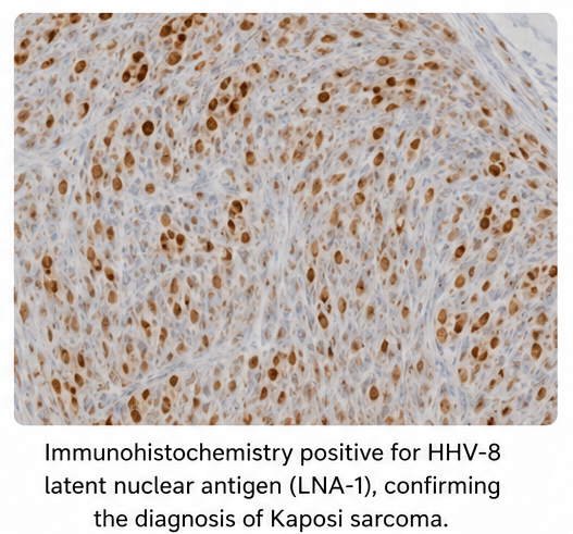
The diagnosis is primarily clinical but requires histopathological confirmation. Histology typically demonstrates spindle-cell proliferation, irregular slit-like vascular spaces, and extravasated red blood cells. Immunohistochemical detection of HHV-8 latent nuclear antigen is particularly useful in confirming the diagnosis and distinguishing KS from its clinical and histological mimics.
Management depends on the clinical subtype, extent of disease, symptoms, immune status, and presence of visceral involvement. In HIV-associated KS, effective antiretroviral therapy is an important component of management, while systemic therapy may be required for extensive, symptomatic, or progressive disease.
We report a case of HIV-associated Kaposi sarcoma presenting with characteristic violaceous cutaneous lesions.
Case Report
A 45-year-old man presented to the dermatology outpatient department with a 3-month history of gradually progressive, painless skin lesions over both lower limbs. The lesions had initially appeared as small reddish-purple macules and progressively increased in number and size.

During the preceding few weeks, the patient noticed mild swelling of both ankles. There was no history of significant trauma, fever, or recent allergic reaction. He reported no previous diagnosis of malignancy or chronic dermatological disease.
On examination, multiple violaceous macules, plaques, and nodular lesions were noted over both lower extremities, predominantly involving the feet and distal legs. Mild bilateral pedal edema was present. The lesions were non-tender and showed no significant discharge or ulceration.
There were no obvious lesions over the trunk. Oral examination was performed to assess for mucosal involvement. The patient was subsequently evaluated for possible immunosuppression and systemic involvement.
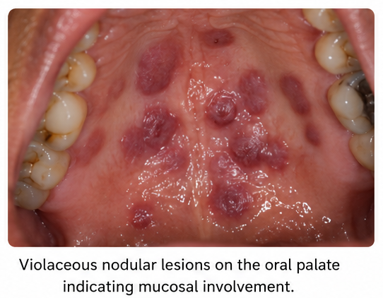
A punch biopsy was obtained from one of the representative lesions for histopathological examination.

Investigations
Histopathological examination demonstrated:
- Spindle-cell proliferation in the dermis
- Irregular slit-like vascular spaces
- Extravasation of erythrocytes
- Hemosiderin deposition
- Mild inflammatory cell infiltration
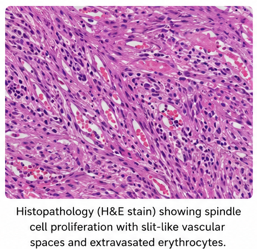
Immunohistochemical evaluation demonstrated HHV-8 positivity, supporting the diagnosis of Kaposi sarcoma. CD34 staining highlighted the vascular component of the lesion.
Further laboratory evaluation revealed previously undiagnosed HIV infection with a reduced CD4 T-cell count. HIV viral load assessment was performed, and additional investigations were undertaken to evaluate the extent of disease.
Imaging and systemic assessment did not demonstrate clinically significant visceral involvement at the time of initial evaluation.
The overall clinical, histopathological, immunohistochemical, and laboratory findings were consistent with HIV-associated Kaposi sarcoma.
Management and Outcome
The patient was referred for multidisciplinary management involving dermatology, infectious disease, and oncology teams.
Management included:
- Initiation of appropriate antiretroviral therapy
- Assessment and monitoring of CD4 count and HIV viral load
- Evaluation for mucosal and visceral involvement
- Regular dermatological assessment of cutaneous lesions
- Monitoring for progression or development of new lesions
- Consideration of local or systemic KS-directed therapy depending on treatment response and disease extent
Antiretroviral therapy was continued with close monitoring. During follow-up, the patient demonstrated gradual improvement in the cutaneous lesions, with reduction in lesion size and intensity of pigmentation.
No significant progression to visceral disease was identified during the documented follow-up period.
Follow-up
One Month
- No significant increase in the number of lesions
- Reduction in the intensity of violaceous discoloration
- Pedal edema improved
- Antiretroviral therapy was tolerated
- Continued dermatological and infectious disease follow-up
Three Months
- Further regression of cutaneous lesions
- No new significant lesions
- Improvement in lower-limb edema
- Continued monitoring of immune status and HIV viral load
- No evidence of clinically apparent visceral involvement
Six Months
- Stable clinical condition
- Marked improvement in existing skin lesions
- No significant new cutaneous lesions
- Continued antiretroviral therapy
- Ongoing surveillance for disease recurrence or progression
Discussion
Kaposi sarcoma is a distinctive vascular neoplasm whose development is closely associated with HHV-8 infection and immune dysregulation. Although HHV-8 is necessary for the development of KS, additional factors, particularly immune suppression and inflammatory signaling, influence disease development and progression.
The clinical appearance of KS varies according to disease stage and subtype. Early lesions may appear as flat macules, which can progressively evolve into plaques and nodules. The typical violaceous or reddish-brown coloration results from the vascular nature of the tumor and associated extravasation of erythrocytes.
In the present case, the presence of multiple progressive violaceous lesions over the lower extremities prompted biopsy. Histopathology demonstrated the characteristic spindle-cell and slit-like vascular pattern. HHV-8 immunohistochemistry further supported the diagnosis. Histological confirmation is particularly important because several conditions can clinically or microscopically mimic KS.
The association between KS and HIV is well established. Before the widespread availability of effective antiretroviral therapy, KS was a common AIDS-associated malignancy. The incidence and severity of HIV-associated KS have declined substantially with improved HIV treatment and immune restoration.
Assessment of a patient with suspected KS should extend beyond the skin. Depending on the clinical presentation, evaluation may include examination of the oral cavity and lymph nodes and assessment for gastrointestinal, pulmonary, or other visceral involvement. Visceral disease may occur even when cutaneous findings are limited.
Treatment is individualized according to disease extent, symptoms, rate of progression, immune status, and clinical subtype. Localized lesions may be treated using approaches such as radiotherapy, intralesional therapy, cryotherapy, or other local modalities. Systemic therapy is generally considered for extensive, progressive, symptomatic, or visceral disease. European consensus recommendations identify pegylated liposomal doxorubicin and paclitaxel among the principal systemic options for advanced disease.
For HIV-associated KS, effective antiretroviral therapy is fundamental because immune restoration may produce significant regression of KS lesions. Patients with aggressive or extensive disease may require additional systemic treatment.
This case emphasizes the importance of considering Kaposi sarcoma when patients present with unexplained violaceous cutaneous lesions, particularly when immunosuppression or HIV infection is suspected. Early biopsy, HHV-8 confirmation, assessment of immune status, and evaluation for systemic involvement are essential for appropriate management.
Prognosis
The prognosis of Kaposi sarcoma varies according to the clinical subtype, immune status, extent of disease, visceral involvement, and response to treatment.
Localized cutaneous disease may follow a relatively indolent course, whereas HIV-associated, iatrogenic, or extensive disease may progress more rapidly. Restoration of immune function with effective antiretroviral therapy has substantially improved outcomes in HIV-associated KS.
Patients require continued follow-up to monitor for new cutaneous lesions, recurrence, mucosal involvement, visceral disease, and treatment-related complications.
Conclusion
Kaposi sarcoma is an HHV-8-associated vascular neoplasm that can present with characteristic violaceous macules, plaques, and nodules, particularly in patients with impaired immunity. Diagnosis requires clinical recognition followed by histopathological examination and confirmation with HHV-8 immunohistochemistry when appropriate.
This case highlights the importance of considering Kaposi sarcoma in patients with progressive violaceous skin lesions and evaluating for underlying immunosuppression, particularly HIV infection. Early diagnosis, assessment of disease extent, appropriate antiretroviral therapy in HIV-associated disease, and individualized local or systemic treatment can help achieve effective disease control.
References
- Cesarman E, Damania B, Krown SE, Martin J, Bower M, Whitby D. Kaposi sarcoma. Nat Rev Dis Primers. 2019;5(1):9. https://pubmed.ncbi.nlm.nih.gov/30705286/
- Lebbé C, Garbe C, Stratigos AJ, et al. Diagnosis and treatment of Kaposi's sarcoma: European consensus-based interdisciplinary guidelines (EDF/EADO/EORTC). Eur J Cancer. 2019;114:117-127.https://pubmed.ncbi.nlm.nih.gov/31096150/
- Iftode N, Rădulescu MA, Aramă SS, Aramă V. Update on Kaposi sarcoma-associated herpesvirus (KSHV or HHV8) - review. Rom J Intern Med. 2020;58(4):199-208. https://pubmed.ncbi.nlm.nih.gov/32681788/
- Dupin N. Update on oncogenesis and therapy for Kaposi sarcoma. Curr Opin Oncol. 2020;32(2):122-128.https://pubmed.ncbi.nlm.nih.gov/31815777/
- Liew YCC, Tam YCS, Oh CC. Treatments for AIDS/HIV-related Kaposi sarcoma: A systematic review of the literature. Int J Dermatol. 2022;61(11):1311-1324.https://pubmed.ncbi.nlm.nih.gov/35775738/
- O'Neill M, et al. Changing therapeutic landscape in advanced Kaposi sarcoma: Current state and future directions. Curr Opin Oncol. 2023.https://pubmed.ncbi.nlm.nih.gov/36718515/
- WHO. WHO guidelines on the management of advanced HIV disease. 2025. The updated guideline includes evidence-informed recommendations for management of Kaposi sarcoma in advanced HIV disease. https://www.who.int/publications/i/item/9789240118164
Read more such content on @ Hidoc Dr | Medical Learning App for Doctors
Recommended News For You
Recommended Articles For You
Featured News
Featured Articles
Featured Events
Featured KOL Videos
1.
A three-quarters reduction in the risk of prostate cancer is associated with an annual increase in cardiorespiratory fitness of more than three percent.
2.
Study Homes In on Simpler Lung Cancer Screening Criteria
3.
Novel analysis identifies markers that may predict failure of metastatic prostate cancer treatment
4.
No Survival Bump in Metastatic NSCLC With Addition of ADC to Immunotherapy
5.
More Positive Survival Data in Lung Cancer With Perioperative Therapy
1.
Bone Marrow Microenvironment Biomarkers in Hematologic Health
2.
Emerging Insights in Hematology for Better Care
3.
Biologic Therapies for Cutaneous Immune-Related Adverse Events in the Era of Immune Checkpoint Inhibitors
4.
Radioligand Therapy Advances: PSMA, Neuroendocrine, and Solid Tumor Clinical Insights
5.
Comprehensive Breakthroughs in Oncology for Better Care
1.
International Conference on Oncology, Cardiology and Critical Care Policy
2.
International Conference on Innovations in Critical Care for Oncology and Cardiology
3.
International Conference on Oncology, Cancer Prevention and Public Health
4.
International Conference on Cancer Nursing and Rehabilitation Strategies
5.
International Conference on Cancer Nursing and Hematology Support
1.
Expert Perspectives on Hematology
2.
Untangling The Best Treatment Approaches For ALK Positive Lung Cancer - Part VI
3.
How Multidisciplinary Teams Support Modern Cancer Care
4.
Revolutionizing Treatment of ALK Rearranged NSCLC with Lorlatinib - Part IV
5.
Redefining Treatment Pathways in Relapsed/Refractory Adult B-Cell ALL

Address :
Hidoc Dr. Inc. | Delaware C Corp | 1309 Coffeen Ave. Suite 1200, Sheridan WY, 82801
Phone :
+1-415-463-3094
Email :
anishagadia@hidoc.co
© Copyright 2026 Hidoc Dr. Inc.
Terms & Conditions - LLP | Inc. | Privacy Policy - LLP | Inc. | Account Deactivation

To get started please enter your email ID
Welcome to Hidoc Dr.
Join to enhance your clinical skills and gain specialized in-depth medical knowledge