Hyperparathyroidism Presenting as Recurrent Nephrolithiasis and Persistent Hypercalcemia: A Case Report
OthersPage Navigation
Abstract
Hyperparathyroidism is an endocrine disorder characterized by excessive secretion of parathyroid hormone (PTH), resulting in hypercalcemia and disturbances in calcium and phosphate metabolism. Primary hyperparathyroidism is most commonly caused by a solitary parathyroid adenoma, whereas secondary and tertiary forms are associated with chronic kidney disease and prolonged hypocalcemia. Clinical manifestations range from asymptomatic hypercalcemia to nephrolithiasis, osteoporosis, bone pain, gastrointestinal disturbances, neuropsychiatric symptoms, and cardiovascular complications. Diagnosis relies on biochemical evaluation, including elevated serum calcium and PTH levels, followed by localization studies such as neck ultrasonography and technetium-99m sestamibi scintigraphy. Surgical parathyroidectomy remains the definitive treatment for symptomatic primary hyperparathyroidism. We report the case of a 48-year-old woman presenting with recurrent renal stones and persistent hypercalcemia caused by a parathyroid adenoma. Early diagnosis and successful parathyroidectomy resulted in normalization of biochemical parameters and complete symptom resolution.
Introduction
Hyperparathyroidism is a disorder of excessive parathyroid hormone secretion leading to dysregulation of calcium homeostasis. Primary hyperparathyroidism is usually caused by a solitary parathyroid adenoma, accounting for nearly 80–85% of cases, while parathyroid hyperplasia and carcinoma are less common causes.
Patients may present with classic manifestations summarized as "bones, stones, abdominal groans, and psychiatric overtones," including osteoporosis, nephrolithiasis, constipation, abdominal pain, fatigue, depression, muscle weakness, and cognitive impairment. Many patients are diagnosed incidentally following routine biochemical testing demonstrating hypercalcemia.
Accurate biochemical diagnosis combined with localization imaging is essential for appropriate management. Early surgical intervention prevents progressive skeletal, renal, and cardiovascular complications.
Case Report
A 48-year-old woman presented to the endocrinology clinic with recurrent episodes of flank pain and two documented episodes of renal stone passage over the previous eighteen months. She also complained of generalized fatigue, diffuse bone pain, constipation, and increased thirst for approximately six months.
She had no history of chronic kidney disease, malignancy, or previous neck surgery. There was no significant family history of endocrine disorders.
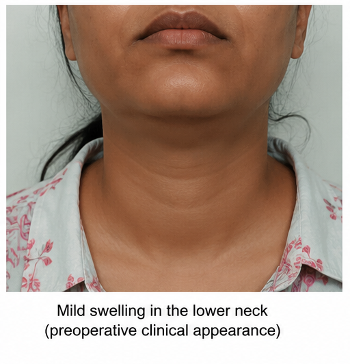
On examination, her vital signs were stable. Physical examination was unremarkable except for mild generalized proximal muscle weakness. A palpable neck swelling was identified.
Laboratory investigations demonstrated:
- Serum calcium: 11.8 mg/dL
- Serum phosphate: 2.1 mg/dL
- Intact parathyroid hormone (PTH): 185 pg/mL
- Serum creatinine: Normal
- 25-hydroxyvitamin D: Mild insufficiency
Twenty-four-hour urinary calcium excretion was elevated.
Dual-energy X-ray absorptiometry (DEXA) demonstrated reduced bone mineral density consistent with osteopenia.
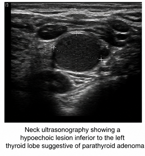
Neck ultrasonography revealed a well-defined hypoechoic lesion measuring 1.8 cm inferior to the left thyroid lobe.
Technetium-99m sestamibi scintigraphy demonstrated focal radiotracer uptake corresponding to the lesion, confirming a left inferior parathyroid adenoma.
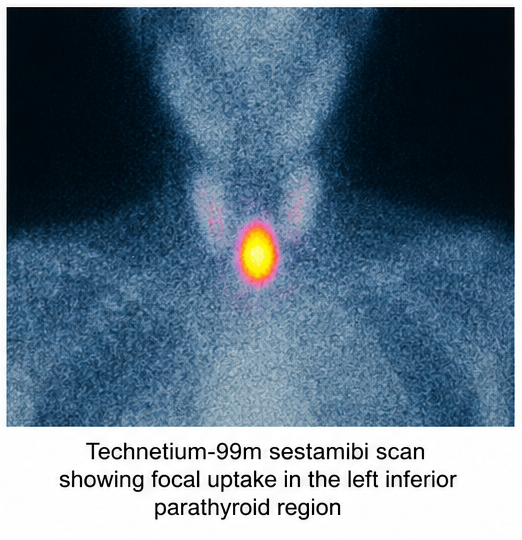
Based on the biochemical profile and imaging findings, a diagnosis of symptomatic primary hyperparathyroidism secondary to parathyroid adenoma was established.
Management and Outcome
The patient underwent minimally invasive left inferior parathyroidectomy under general anesthesia. Intraoperative parathyroid hormone monitoring demonstrated a greater than 50% reduction in PTH levels within ten minutes following adenoma excision, confirming successful removal.
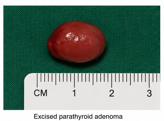
Histopathological examination confirmed a benign parathyroid adenoma.
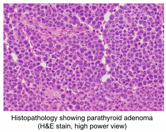
Postoperatively, serum calcium levels were monitored closely. Oral calcium supplementation and vitamin D were initiated temporarily to prevent hungry bone syndrome. The patient's symptoms gradually improved, and biochemical parameters normalized within several weeks.
At follow-up, renal symptoms had resolved completely, bone pain diminished significantly, and serum calcium and PTH levels remained within normal limits.
Follow-up
One Month
- Serum calcium normalized.
- PTH levels significantly reduced.
- Constipation and fatigue resolved.
- No postoperative complications.
Three Months
- Bone pain markedly improved.
- Renal function remained normal.
- No further renal stone episodes.
- Improved quality of life.
Six Months
- Stable calcium and PTH levels.
- Bone mineral density showed improvement.
- No disease recurrence.
- Excellent clinical outcome.
Discussion
Primary hyperparathyroidism is one of the most common causes of persistent hypercalcemia. Excessive secretion of parathyroid hormone increases bone resorption, renal calcium reabsorption, and intestinal calcium absorption through activation of vitamin D.
Although many patients are identified incidentally, symptomatic individuals commonly present with nephrolithiasis, osteoporosis, fractures, muscle weakness, neurocognitive symptoms, gastrointestinal complaints, and cardiovascular manifestations.
Biochemical diagnosis is established by elevated serum calcium with inappropriately elevated PTH concentrations. Neck ultrasonography and technetium-99m sestamibi scintigraphy remain the primary localization studies before surgery. Four-dimensional CT may be considered when first-line imaging is inconclusive.
Differential diagnoses include familial hypocalciuric hypercalcemia, malignancy-associated hypercalcemia, vitamin D intoxication, granulomatous disease, multiple myeloma, and medication-induced hypercalcemia.
Parathyroidectomy is the definitive treatment for symptomatic primary hyperparathyroidism and significantly reduces the risk of recurrent nephrolithiasis, fractures, and progressive renal dysfunction. Medical therapy with cinacalcet or bisphosphonates may be considered in selected patients who are not surgical candidates.
Prognosis
The prognosis following successful parathyroidectomy is excellent. Most patients experience normalization of calcium metabolism, significant improvement in symptoms, reduced recurrence of nephrolithiasis, and stabilization or improvement of bone mineral density. Long-term follow-up with periodic biochemical monitoring is recommended to detect recurrent or persistent disease.
Conclusion
Hyperparathyroidism should be considered in patients presenting with recurrent nephrolithiasis, persistent or unexplained hypercalcemia, osteoporosis, fragility fractures, bone pain, proximal muscle weakness, neurocognitive symptoms, or nonspecific complaints such as fatigue, constipation, depression, and excessive thirst. Because the clinical presentation may be subtle or nonspecific, a high index of suspicion is essential, particularly in patients with recurrent renal stones or incidental hypercalcemia identified during routine laboratory evaluation.
A comprehensive diagnostic workup should include measurement of serum calcium, phosphate, intact parathyroid hormone (PTH), vitamin D levels, renal function, and 24-hour urinary calcium excretion. Localization studies, including neck ultrasonography and technetium-99m sestamibi scintigraphy, play a crucial role in identifying abnormal parathyroid glands and facilitating surgical planning. Additional imaging modalities such as four-dimensional computed tomography (4D-CT) or magnetic resonance imaging (MRI) may be considered when conventional imaging is inconclusive or in cases of persistent or recurrent disease.
Early recognition and timely diagnosis are essential to prevent long-term complications, including recurrent nephrolithiasis, nephrocalcinosis, chronic kidney disease, osteoporosis, pathological fractures, cardiovascular abnormalities, and deterioration in quality of life. Surgical parathyroidectomy remains the definitive treatment for symptomatic primary hyperparathyroidism and offers excellent long-term outcomes, with normalization of serum calcium and PTH levels, improvement in bone mineral density, reduction in renal stone recurrence, and resolution of many systemic symptoms.
Careful postoperative monitoring is equally important to detect hypocalcemia, hungry bone syndrome, or persistent and recurrent hyperparathyroidism. Long-term follow-up with periodic biochemical assessment and, when appropriate, bone mineral density evaluation helps ensure sustained disease remission, preservation of skeletal and renal health, and continued improvement in overall patient outcomes. Early intervention combined with regular surveillance remains the cornerstone of successful management in patients with primary hyperparathyroidism.
References
- Bilezikian JP, Brandi ML, Eastell R, et al. Evaluation and Management of Primary Hyperparathyroidism: Summary Statement and Guidelines. Journal of Bone and Mineral Research. 2022. https://pubmed.ncbi.nlm.nih.gov/35718831/
- Wilhelm SM, Wang TS, Ruan DT, et al. The American Association of Endocrine Surgeons Guidelines for Definitive Management of Primary Hyperparathyroidism. JAMA Surgery. 2016. https://pubmed.ncbi.nlm.nih.gov/27532368/
- Khan AA, Hanley DA, Rizzoli R, et al. Primary Hyperparathyroidism: Review and Recommendations on Evaluation, Diagnosis, and Management. Osteoporosis International. 2017. https://pubmed.ncbi.nlm.nih.gov/27812881/
- Silverberg SJ, Bilezikian JP. Primary Hyperparathyroidism: Clinical Presentation and Management. Journal of Clinical Endocrinology & Metabolism. 1996. https://pubmed.ncbi.nlm.nih.gov/8855804/
- Fraser WD. Hyperparathyroidism. The Lancet. 2009;374(9684):145–158. https://pubmed.ncbi.nlm.nih.gov/19595349/
Read more such content on @ Hidoc Dr | Medical Learning App for Doctors
Recommended News For You
Recommended Articles For You
Featured News
Featured Articles
Featured Events
Featured KOL Videos
1.
NPM1-Mutated AML: Not All Treatments Are Equal
2.
Paging Dr. AI; Myth Busting in Cancer; Oncologists Shine in Malpractice Survey
3.
MRI can save rectal cancer patients from surgery, study suggests
4.
Rectal Cancer 'Crisis'; Smells Like Cancer; Flushing Away Clues to Colon Cancer
5.
Rising rates of head and neck cancers in England
1.
A Closer Look at Breast Cancer: Examining the Ultrasound Images
2.
Exploring the Latest Advances in Hodgkin's Lymphoma Treatment
3.
Molecular Classification of Refractory Anemia: A Comprehensive Review
4.
Ferroptosis-Modulating Therapeutics in Precision Oncology
5.
Blood-Cell Morphology Foundation Models: Transforming Hematological Diagnostics and Clinical Practice
1.
International Cancer Conference
2.
Asian Symposium on Advancement in Hematology and Oncology (ASAHO)
3.
International Cancer Conference
4.
Asian Symposium on Advancement in Hematology and Oncology (ASAHO)
5.
Asian Symposium on Advancement in Hematology and Oncology
1.
CDK4/6 Inhibitors in Extending Overall Survival in HR+/HER2- aBC Patients in Clinical Trial and Real World
2.
Thromboprophylaxis In Medical Settings- An Update
3.
Nimotozumab Plus Chemo-radiotherapy v/s Placebo Plus Chemo-radiotherapy in Locally Advanced Nasopharyngeal Carcinoma
4.
How Platelet Disorders Can Affect the Body
5.
Treatment Sequencing Strategies in ALK + NSCLC Patients with CNS Diseases

Address :
Hidoc Dr. Inc. | Delaware C Corp | 1309 Coffeen Ave. Suite 1200, Sheridan WY, 82801
Phone :
+1-415-463-3094
Email :
anishagadia@hidoc.co
© Copyright 2026 Hidoc Dr. Inc.
Terms & Conditions - LLP | Inc. | Privacy Policy - LLP | Inc. | Account Deactivation

To get started please enter your email ID
Welcome to Hidoc Dr.
Join to enhance your clinical skills and gain specialized in-depth medical knowledge