Melasma Presenting as Bilateral Facial Hyperpigmentation: A Case Report
OthersPage Navigation
Abstract
Melasma is a common acquired disorder of hyperpigmentation characterized by symmetrical, hyperpigmented macules and patches, predominantly affecting sun-exposed areas of the face. It is frequently associated with ultraviolet (UV) exposure, hormonal influences, and genetic predisposition. We report the case of a 34-year-old woman who presented with gradually progressive, bilateral brownish facial pigmentation over the cheeks. Clinical evaluation and pattern recognition were consistent with melasma. Differential diagnoses including photodermatitis, rosacea, and cutaneous manifestations of systemic lupus erythematosus were considered and excluded based on clinical features. This case highlights the importance of careful clinical examination and pattern-based diagnosis in dermatology, particularly in differentiating melasma from other facial dermatoses with overlapping presentations.
Introduction
Facial hyperpigmentation is a frequent dermatological concern and may result from a wide spectrum of conditions ranging from benign pigmentary disorders to inflammatory or autoimmune diseases. Among these, melasma is one of the most commonly encountered acquired hypermelanoses, particularly in women of reproductive age [1]. It is characterized by symmetrical, brown to gray-brown macules and patches, most commonly involving the cheeks, forehead, upper lip, and chin.
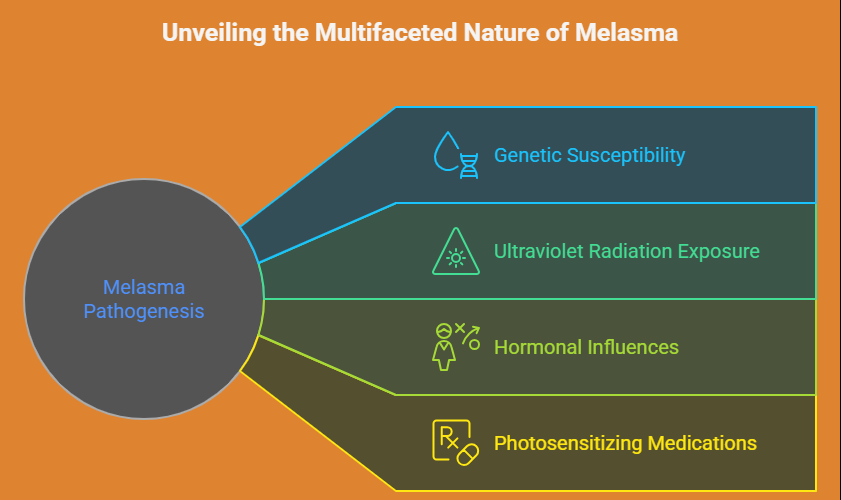
The pathogenesis of melasma is multifactorial and includes genetic susceptibility, ultraviolet radiation exposure, hormonal influences such as pregnancy and oral contraceptive use, and photosensitizing medications [2].
Although diagnosis is primarily clinical, distinguishing melasma from other causes of facial erythema and pigmentation—such as photodermatitis, rosacea, or connective tissue diseases—is essential for appropriate management.
This case report describes a typical presentation of melasma with classical clinical features and discusses key differential diagnoses, emphasizing the value of detailed clinical assessment in routine dermatology practice.
Case Report
Patient History
A 34-year-old woman presented to the dermatology outpatient clinic with complaints of gradually progressive discoloration over both cheeks for the past eight months. The pigmentation was asymptomatic and had darkened over time, particularly following sun exposure.
There was no history of itching, burning sensation, vesiculation, or scaling. The patient reported regular outdoor exposure and inconsistent use of sunscreen. She had no known photosensitivity disorders.
There was a history of oral contraceptive use for the past one year. No similar lesions were noted elsewhere on the body.
There was no personal or family history of autoimmune disease, connective tissue disorders, or chronic dermatological conditions.
Clinical Findings
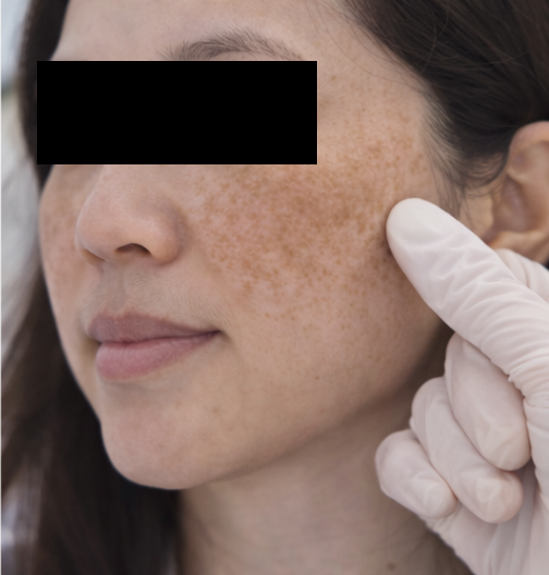
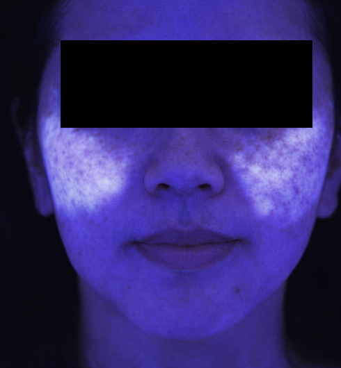
Cutaneous examination revealed bilateral, symmetrical, brownish hyperpigmented patches over the malar regions of the face.
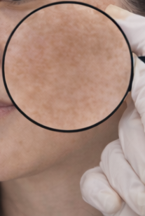
The lesions were sharply demarcated, non-scaly, and non-erythematous. No telangiectasia, papules, pustules, or vesicles were observed.
The forehead, nasolabial folds, and perioral region were spared. There was no evidence of scarring, atrophy, or induration. Examination of the scalp, oral mucosa, and nails was unremarkable. Systemic examination revealed no abnormalities.
Differential Diagnosis
Based on the clinical presentation, the following differential diagnoses were considered:
- Melasma – suggested by bilateral symmetrical hyperpigmented patches on sun-exposed facial areas, absence of inflammation, and association with hormonal factors.
- Photodermatitis – typically presents with acute erythema, itching, vesiculation, or scaling following sun exposure, which were absent in this case.
- Systemic lupus erythematosus (malar rash) – usually erythematous rather than hyperpigmented, may show photosensitivity and systemic features, and often spares the nasolabial folds.
- Rosacea – characterized by central facial erythema, papules, pustules, and telangiectasia, none of which were present.
Based on clinical correlation, melasma was considered the most likely diagnosis.
Investigations
As the diagnosis of melasma is primarily clinical, based on characteristic distribution and morphology of the lesions, no invasive investigations were required. In the absence of systemic symptoms or atypical clinical features, routine laboratory investigations were not indicated. There was no clinical suspicion of underlying endocrine, autoimmune, or metabolic disorders that would necessitate further testing.
The patient was counseled in detail regarding the benign and chronic nature of melasma, its multifactorial etiology, and common contributing factors such as ultraviolet exposure, hormonal influences, and genetic predisposition. Emphasis was placed on the relapsing course of the condition, the importance of adherence to photoprotective measures, and realistic expectations from treatment, including the need for long-term maintenance and follow-up.
Management and Outcome
The patient was advised strict photoprotection, including the regular use of a broad-spectrum sunscreen with high SPF, avoidance of peak sunlight exposure, and use of physical protective measures such as hats and umbrellas.
Topical depigmenting therapy was initiated, consisting of a combination regimen targeting melanogenesis. The patient was also counseled regarding the role of hormonal factors and the chronic, relapsing nature of melasma.
At follow-up after six weeks, mild lightening of the pigmentation was noted, with no adverse effects. The patient reported improved adherence to sun-protective measures and was advised to continue long-term maintenance therapy.
Discussion
Melasma is a chronic acquired pigmentary disorder with a considerable psychosocial and cosmetic impact, particularly among women of reproductive age. The condition arises from increased melanin synthesis and abnormal deposition within the epidermis, dermis, or a combination of both, leading to the characteristic hyperpigmented appearance. Key etiological factors include prolonged ultraviolet exposure, hormonal influences such as pregnancy and oral contraceptive use, genetic predisposition, and, in some cases, photosensitizing medications [2,3]. These factors act synergistically to stimulate melanocyte activity and disrupt normal pigment regulation.
The symmetrical distribution of lesions, predilection for sun-exposed facial areas, and absence of overt inflammatory features serve as important clinical clues that help distinguish melasma from other facial dermatoses. In contrast, conditions such as photodermatitis and rosacea commonly present with erythema, pruritus, burning sensations, or inflammatory lesions including papules and pustules. Autoimmune disorders, particularly lupus erythematosus, are often associated with photosensitivity, systemic symptoms, and distinctive rash morphology, features that are typically absent in melasma and aid in clinical differentiation [4].
Early recognition of melasma is essential, as it allows timely initiation of preventive and therapeutic strategies aimed at limiting disease progression and reducing recurrence. Emphasis on strict photoprotection, appropriate topical or procedural therapies, and modification of contributing factors forms the cornerstone of management. Comprehensive patient education regarding sun avoidance, consistent use of sunscreen, realistic treatment expectations, and adherence to long-term maintenance therapy is critical for achieving sustained improvement and successful long-term disease control.
Conclusion
Melasma is a common and important cause of bilateral facial hyperpigmentation and is primarily diagnosed based on its characteristic clinical features, including symmetrical distribution, well-defined hyperpigmented patches, and predilection for sun-exposed areas of the face. This case underscores the importance of careful visual assessment, thorough clinical examination, and thoughtful consideration of relevant differential diagnoses when evaluating patients presenting with facial pigmentation, as several dermatological and systemic conditions may present with overlapping features.
Accurate identification of melasma allows for timely and appropriate patient counseling, initiation of effective management strategies, and avoidance of unnecessary laboratory investigations or invasive diagnostic procedures. Furthermore, integration of sound clinical expertise with patient education regarding disease chronicity, contributing factors, photoprotection, and treatment expectations remains central to optimizing long-term outcomes and improving quality of life in individuals affected by this chronic dermatological condition.
References
- Grimes PE. Melasma: Etiologic and therapeutic considerations. Arch Dermatol. 1995;131(12):1453–1457.
- Kang HY, Ortonne JP. What should be considered in the treatment of melasma. Ann Dermatol. 2010;22(4):373–378.
- Sanchez NP, et al. Melasma: A clinical, light microscopic, ultrastructural, and immunofluorescence study. J Am Acad Dermatol. 1981;4(6):698–710.
- James WD, Elston DM, Treat JR, Rosenbach MA. Andrews’ Diseases of the Skin: Clinical Dermatology. 13th ed. Elsevier; 2020.
Read more such content on @ Hidoc Dr | Medical Learning App for Doctors
Recommended News For You
Recommended Articles For You
Featured News
Featured Articles
Featured Events
Featured KOL Videos
1.
Does pollution cause cancer?
2.
AI is equally capable of reading breast cancer scans as human radiologists.
3.
EVP Beats Cisplatin for Resectable MIBC
4.
New research points out a promising strategy for treating metastatic medulloblastoma
5.
Academics + Pharma = Big Bucks; New CAR-T Warnings; Patients Seek Cancer Tests.
1.
A Closer Look at Breast Cancer: Examining the Ultrasound Images
2.
Unlocking the Secrets of Oral Cancer Staging: A New Approach to Early Detection
3.
Impact of Hormone Therapy Cessation on Tumor Growth: Case Study of Ki-67 Reduction
4.
Unraveling the Mysteries of Lymphoma: A Journey into the Unknown
5.
Refining AML Survival: Prognostic Factors, Therapies, and Stem Cell Strategies Reviewed
1.
International Lung Cancer Congress®
2.
Genito-Urinary Oncology Summit 2026
3.
Future NRG Oncology Meeting
4.
ISMB 2026 (Intelligent Systems for Molecular Biology)
5.
Annual International Congress on the Future of Breast Cancer East
1.
Navigating the Complexities of Ph Negative ALL - Part III
2.
A Comprehensive Guide to First Line Management of ALK Positive Lung Cancer - Part VIII
3.
Management of 1st line ALK+ mNSCLC (CROWN TRIAL Update)
4.
Expert Group meeting with the management of EGFR mutation positive NSCLC - Part III
5.
Virtual Case Study on Pedal Edema and Triple Vessel Disease - An Initiative by Hidoc Dr.

Address :
Hidoc Dr. Inc. | Delaware C Corp | 1309 Coffeen Ave. Suite 1200, Sheridan WY, 82801
Phone :
+1-415-463-3094
Email :
anishagadia@hidoc.co
© Copyright 2026 Hidoc Dr. Inc.
Terms & Conditions - LLP | Inc. | Privacy Policy - LLP | Inc. | Account Deactivation

To get started please enter your email ID
Welcome to Hidoc Dr.
Join to enhance your clinical skills and gain specialized in-depth medical knowledge