Helicobacter pylori Associated Peptic Ulcer Disease Presenting With Upper Gastrointestinal Bleeding
OthersPage Navigation
Introduction
Peptic ulcer disease is characterized by a break in the gastric or duodenal mucosa extending through the muscularis mucosae. Helicobacter pylori infection and the use of nonsteroidal anti-inflammatory drugs are the principal causes. Other contributing factors include smoking, severe physiological stress, corticosteroid use with nonsteroidal anti-inflammatory drugs, and hypersecretory conditions.
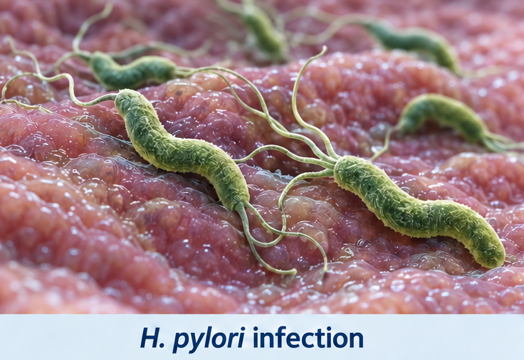
Clinical manifestations range from mild epigastric discomfort to life-threatening bleeding or perforation. Upper gastrointestinal bleeding is the most frequent complication and may present with hematemesis, coffee-ground vomiting, melena, dizziness, or circulatory instability. Prompt resuscitation, early endoscopy, endoscopic hemostasis, acid suppression, and treatment of the underlying cause are essential for a favourable outcome.
Case Report
A 48-year-old man presented to the emergency department with two episodes of black, tarry stools and one episode of coffee-ground vomiting during the preceding 12 hours. He also reported dizziness, generalized weakness, and a burning pain in the upper abdomen.

The epigastric pain had been present intermittently for approximately three months. It usually developed two to three hours after meals and occasionally awakened him at night. The discomfort was temporarily relieved by food or over-the-counter antacids. During the previous week, the pain had increased in intensity and was associated with nausea and reduced appetite.
The patient had osteoarthritis of both knees and had been taking diclofenac 50 mg twice daily without medical supervision for approximately six weeks. He was not using a proton-pump inhibitor or other gastroprotective medicine. He smoked approximately 10 cigarettes daily and consumed alcohol occasionally.
There was no previous history of peptic ulcer disease, gastrointestinal bleeding, chronic liver disease, renal disease, or bleeding disorder. He was not taking aspirin, anticoagulants, corticosteroids, or antiplatelet medicines. There was no history of significant weight loss, progressive difficulty swallowing, persistent vomiting, or gastrointestinal malignancy in a first-degree relative.
On examination, the patient appeared pale and anxious. His pulse rate was 112 beats/minute, blood pressure was 94/60 mmHg, respiratory rate was 22 breaths/minute, and oxygen saturation was 97% on room air. He was afebrile.
The abdomen was soft, with moderate tenderness in the epigastric region. There was no guarding, rigidity, rebound tenderness, palpable mass, or organ enlargement. Bowel sounds were present. Digital rectal examination confirmed black, tarry stool. No clinical features of chronic liver disease or portal hypertension were observed.

Investigations
Initial laboratory investigations showed a hemoglobin level of 8.2 g/dL, hematocrit of 25%, leukocyte count of 11,600 cells/mm³, and platelet count of 238,000 cells/mm³. The blood urea nitrogen level was elevated at 38 mg/dL, while serum creatinine was 1.1 mg/dL. The disproportionate elevation in blood urea nitrogen supported an upper gastrointestinal source of bleeding.
Serum sodium was 136 mmol/L and potassium was 3.8 mmol/L. Liver-function tests, serum amylase, lipase, prothrombin time, and international normalized ratio were within normal limits. Electrocardiography showed sinus tachycardia without ischemic changes.
After initial stabilization, oesophagogastroduodenoscopy was performed. It revealed altered blood within the stomach and a solitary 1.2-cm ulcer on the anterior wall of the duodenal bulb. The ulcer had an actively oozing visible vessel. The surrounding mucosa was edematous and inflamed. There were no oesophageal varices, gastric masses, or features of portal hypertensive gastropathy.
Endoscopic hemostasis was achieved using diluted epinephrine injection followed by thermal coagulation. Gastric biopsies obtained from the antrum and body were positive for H. pylori on histopathological examination. A rapid urease test was also positive.
The final diagnosis was H. pylori-associated bleeding duodenal ulcer, with unsupervised diclofenac use considered an important additional mucosal risk factor.
Differential Diagnosis
Erosive Gastritis
Nonsteroidal anti-inflammatory drug use may cause gastric erosions and upper gastrointestinal bleeding. However, endoscopy demonstrated a discrete duodenal ulcer with a visible bleeding vessel rather than diffuse gastric erosions.
Gastric Malignancy
Gastric cancer may present with epigastric pain, anemia, reduced appetite, weight loss, or gastrointestinal bleeding. The absence of a gastric mass on endoscopy and the duodenal location of the lesion made malignancy unlikely.
Oesophageal Variceal Bleeding
Variceal bleeding was considered because of the presentation with hematemesis and melena. Nevertheless, the patient had no clinical or laboratory evidence of chronic liver disease, and endoscopy showed no oesophageal or gastric varices.
Mallory Weiss Tear
A mucosal tear at the gastro-oesophageal junction may follow repeated retching or vomiting. In this patient, bleeding occurred without preceding forceful vomiting, and no tear was identified during endoscopy.
Acute Pancreatitis
Acute pancreatitis can produce severe epigastric pain, nausea, and vomiting. Normal serum amylase and lipase levels, the absence of pain radiating to the back, and the endoscopic findings excluded this diagnosis.
Management and Outcome
Two large-bore intravenous cannulas were inserted immediately. The patient received intravenous crystalloid fluids, supplemental oxygen, and continuous monitoring of blood pressure, pulse rate, urine output, and mental status. Blood samples were obtained for complete blood count, coagulation testing, and cross-matching.
Intravenous pantoprazole was initiated before endoscopy. Because the patient had symptomatic anemia, hemodynamic instability, and ongoing gastrointestinal bleeding, packed red blood cells were transfused. Diclofenac was discontinued, and the patient was advised to avoid nonsteroidal anti-inflammatory drugs unless specifically prescribed.
Following successful endoscopic hemostasis, high-dose intravenous proton-pump inhibitor therapy was continued for 72 hours. The patient was initially kept fasting and subsequently progressed from clear fluids to a soft diet after remaining hemodynamically stable without recurrent bleeding.

His pulse rate and blood pressure normalized during the first 24 hours. No further hematemesis or melena occurred, and serial hemoglobin measurements remained stable after transfusion.
After stabilization, he was switched to oral pantoprazole. H. pylori eradication therapy was initiated with a 14-day bismuth quadruple regimen consisting of a proton-pump inhibitor, bismuth, tetracycline, and metronidazole. The importance of completing the entire treatment course was explained.
The patient was discharged after four days. He was advised to stop smoking, avoid self-medication with analgesics, and seek urgent medical attention if he developed hematemesis, black stools, severe abdominal pain, fainting, or progressive weakness.
At review six weeks later, he was asymptomatic and his hemoglobin had increased to 11.6 g/dL. Eradication testing performed after completion of treatment and an appropriate interval without proton-pump inhibitor therapy was negative, confirming successful eradication of H. pylori.
Discussion
This case illustrates the interaction between two major causes of peptic ulcer disease: H. pylori infection and nonsteroidal anti-inflammatory drug exposure. Both factors disrupt the normal balance between gastroduodenal mucosal defence and damaging influences such as acid and pepsin.
H. pylori causes chronic gastric inflammation and alters acid regulation. Depending on the distribution of gastritis, infection may increase acid delivery to the duodenum, promote gastric metaplasia, weaken mucosal protection, and contribute to duodenal ulcer formation. Eradication of the organism promotes ulcer healing and substantially decreases recurrence.
Nonsteroidal anti-inflammatory drugs inhibit cyclooxygenase enzymes and reduce prostaglandin synthesis. This decreases mucus and bicarbonate secretion, impairs mucosal blood flow, and limits epithelial repair. The direct topical effect of certain agents may produce additional injury. Concurrent H. pylori infection can further increase the risk of ulceration and gastrointestinal bleeding.
The clinical pattern of duodenal ulcer pain commonly includes epigastric discomfort occurring several hours after meals or during the night, with temporary relief after eating or taking antacids. Nevertheless, symptoms alone cannot reliably distinguish peptic ulcer disease from functional dyspepsia, gastritis, gastro-oesophageal reflux disease, pancreatobiliary disorders, or malignancy.
Upper gastrointestinal endoscopy is important in patients with bleeding, anemia, hemodynamic instability, persistent vomiting, progressive weight loss, or other alarm features. It identifies the source of bleeding, enables risk assessment, and permits endoscopic treatment. Combination endoscopic therapy is generally preferred for actively bleeding ulcers or ulcers with high-risk stigmata.
High-dose proton-pump inhibitor therapy after endoscopic treatment reduces gastric acidity, stabilizes clot formation, and lowers the risk of recurrent bleeding. Continued monitoring is necessary because rebleeding may present with renewed hematemesis, melena, tachycardia, hypotension, or a decrease in hemoglobin.
Eradication therapy should be selected according to local antibiotic resistance patterns, previous antibiotic exposure, allergies, and current treatment guidelines. Confirmation of eradication is essential because symptom resolution does not establish microbiological cure. Testing is generally performed with a urea breath test, stool antigen test, or biopsy-based method after an appropriate post-treatment interval.

Conclusion
Peptic ulcer disease may progress from intermittent epigastric discomfort to potentially life-threatening gastrointestinal bleeding. In this patient, H. pylori infection and unsupervised diclofenac use contributed to the development of a bleeding duodenal ulcer.
Rapid hemodynamic stabilization, early endoscopy, endoscopic hemostasis, proton-pump inhibitor therapy, withdrawal of the offending medicine, and eradication of H. pylori resulted in complete clinical recovery. Patients should also receive counselling regarding nonsteroidal anti-inflammatory drug use, smoking cessation, treatment adherence, warning symptoms, and confirmation of H. pylori eradication.
References
- Lanas A, Chan FKL. Peptic ulcer disease. Lancet. 2017;390(10094):613–624. https://pubmed.ncbi.nlm.nih.gov/28242110/
- Chey WD, Leontiadis GI, Howden CW, Moss SF. ACG clinical guideline: Treatment of Helicobacter pylori infection. Am J Gastroenterol. 2017;112(2):212–239. https://pubmed.ncbi.nlm.nih.gov/28071659/
- Laine L, Barkun AN, Saltzman JR, Martel M, Leontiadis GI. ACG clinical guideline: Upper gastrointestinal and ulcer bleeding. Am J Gastroenterol. 2021;116(5):899–917. https://pubmed.ncbi.nlm.nih.gov/33929377/
- Malfertheiner P, Megraud F, Rokkas T, et al. Management of Helicobacter pylori infection: The Maastricht VI Florence Consensus Report. Gut. 2022;71(9):1724–1762. https://pubmed.ncbi.nlm.nih.gov/35944925/
- Sung JJY, Tsoi KKF, Ma TKW, Yung MY, Lau JYW, Chiu PWY. Causes of mortality in patients with peptic ulcer bleeding: A prospective cohort study of 10,428 cases. Am J Gastroenterol. 2010;105(1):84–89. https://pubmed.ncbi.nlm.nih.gov/19755976/
Read more such content on @ Hidoc Dr | Medical Learning App for Doctors
Recommended News For You
Recommended Articles For You
Featured News
Featured Articles
Featured Events
Featured KOL Videos
1.
Tumor characteristics found to differ for melanomas in children, teens and young adults
2.
Unexplained CAR-T Death; Americans Spurn Sunscreen; Improving Diversity in Trials
3.
"A lot" of aspirin reduces the risk of ovarian cancer, regardless of genetic factors, according to the JAMA study.
4.
Approval in Endometrial Cancer Expanded; Masks at Cancer Centers; NPR Reporter Dies
5.
Is Lenalidomide Resistance in Myeloma Reversible with JAK Inhibition?
1.
Modern Oncology Unveiled with Treatment Innovations and Personalized Cancer Care
2.
Screening for Early Therapy-Related Cancer Susceptibility
3.
Unlocking the Potential of Bicalutamide 50 mg: A Revolutionary New Treatment for Prostate Cancer
4.
Skin-Sparing Mastectomy: Balancing Cancer Control with Cosmetic Results
5.
Reducing Modifiable Childhood Cancer Exposure Risks
1.
International Conference on Innovations in Critical Care for Oncology and Cardiology
2.
International Conference on Oncology, Cardiology and Critical Care Policy
3.
International Conference on Oncology, Cancer Prevention and Public Health
4.
International Conference on Cancer Nursing and Rehabilitation Strategies
5.
International Conference on Best Practices in Oncology, Cardiology and Critical Care
1.
Dacomitinib Case Presentation: Baseline Treatment and Current Status
2.
Targeting Oncologic Drivers with Dacomitinib: Further Discussion on Lung Cancer Treatment
3.
Should We Use DARA Up Front As First-Line Therapy in MM?
4.
Iron Deficiency Anemia: Ferric Maltol As a New Treatment Option- A New Perspective
5.
Importance of Cancer Screening and Early Detection

Address :
Hidoc Dr. Inc. | Delaware C Corp | 1309 Coffeen Ave. Suite 1200, Sheridan WY, 82801
Phone :
+1-415-463-3094
Email :
anishagadia@hidoc.co
© Copyright 2026 Hidoc Dr. Inc.
Terms & Conditions - LLP | Inc. | Privacy Policy - LLP | Inc. | Account Deactivation

To get started please enter your email ID
Welcome to Hidoc Dr.
Join to enhance your clinical skills and gain specialized in-depth medical knowledge