Androgenetic Alopecia Presenting With Progressive Frontotemporal and Vertex Hair Thinning
OthersPage Navigation
Introduction
Androgenetic alopecia (AGA), also called pattern hair loss, is a common, progressive, non-scarring alopecia influenced by genetic susceptibility and androgen signalling. In predisposed follicles, dihydrotestosterone contributes to gradual follicular miniaturisation, shortening of the anagen phase, and replacement of terminal hairs by finer, shorter hairs.
Men usually develop bitemporal recession and thinning over the frontal and vertex scalp, while the occipital fringe is relatively preserved. Diagnosis is generally clinical and may be supported by trichoscopy. Early recognition is important because established miniaturisation may become difficult to reverse, whereas treatment can slow progression and improve scalp coverage.
Case Report
A 28-year-old man presented with progressive thinning of scalp hair for approximately three years. He first noticed recession at both temples, followed by reduced density over the frontal scalp and vertex. The change had become more visible during the preceding eight months and caused considerable concern about his appearance.
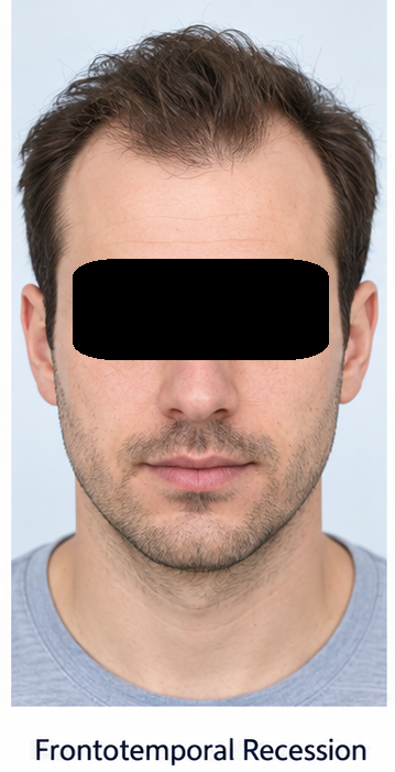
He described mild daily shedding but no sudden loss of handfuls of hair. There were no discrete bald patches, scalp pain, burning, marked itching, pustules, or scaling. He denied recent high fever, major surgery, rapid weight loss, severe emotional stress, or use of bodybuilding hormones. His diet was mixed, and he reported no symptoms suggestive of anaemia or thyroid dysfunction.
His father and elder brother had developed patterned hair loss before 35 years of age. There was no family history of scarring alopecia or autoimmune disease. He had no significant medical illness and was not taking retinoids, anticoagulants, anticonvulsants, or other medicines associated with hair shedding.
Examination showed symmetrical bitemporal recession with diffuse reduction in hair density over the frontal scalp and vertex. The frontal hairline had assumed an M-shaped contour. The occipital and lateral scalp were relatively preserved. The pattern corresponded approximately to Hamilton–Norwood grade III vertex.
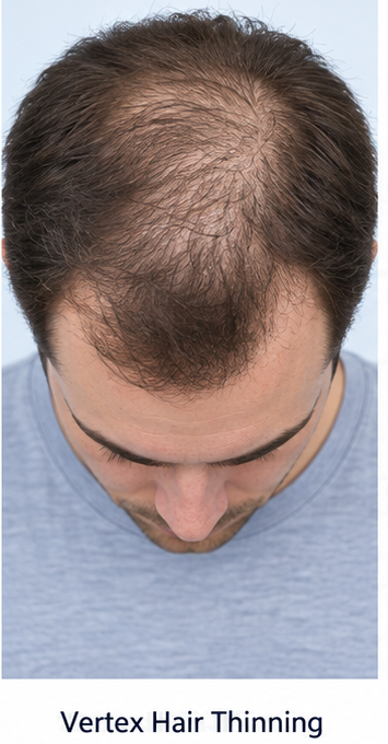
The scalp skin appeared normal without erythema, perifollicular scaling, pustules, loss of follicular openings, or shiny atrophic areas. The hair-pull test was negative. Eyebrows, eyelashes, beard, body hair, nails, and oral mucosa were normal. There were no clinical features of systemic disease or nutritional deficiency.
Investigations
Trichoscopy of the frontal and vertex scalp showed hair-shaft diameter diversity, an increased proportion of thin and miniaturised hairs, perifollicular brown discoloration, and an increased number of single-hair follicular units. Follicular openings were preserved, supporting a non-scarring process. The occipital scalp showed relatively uniform terminal hairs.

Because the presentation was typical, extensive investigation was not required. A complete blood count, serum ferritin, thyroid-stimulating hormone, liver-function tests, renal-function tests, and serum vitamin B12 were within reference ranges. There was no laboratory evidence of anaemia, iron deficiency, thyroid disease, or systemic illness contributing to diffuse shedding.
The gradual course, characteristic distribution, positive family history, preserved follicular openings, and trichoscopic miniaturisation supported the diagnosis of male androgenetic alopecia. Scalp biopsy was not performed because the clinical and trichoscopic findings were concordant and there were no signs of inflammation or scarring.
Differential Diagnosis
Telogen Effluvium
Telogen effluvium usually causes abrupt, diffuse shedding two to three months after a physiological or psychological trigger. The patterned distribution, negative hair-pull test, three-year progression, and absence of a relevant trigger made this unlikely.
Alopecia Areata
Alopecia areata commonly produces sharply defined patches, exclamation-mark hairs, black dots, or yellow dots. None of these features was present, and the distribution was typical of androgenetic alopecia.
Scarring Alopecia
Frontal fibrosing alopecia and lichen planopilaris may cause hairline recession with perifollicular erythema, scaling, symptoms, and loss of follicular openings. The absence of these findings argued against cicatricial alopecia.
Traction Alopecia and Hair-Shaft Damage
There was no history of sustained traction, chemical processing, or heat-related breakage. Examination did not show broken hairs or a marginal traction pattern.
Management and Outcome
The patient was counselled that androgenetic alopecia is a chronic condition and that treatment aims to maintain existing hair, slow progression, and obtain partial regrowth rather than restore the original adolescent hairline. Standardised baseline photographs were obtained.
Topical minoxidil 5% solution was started at 1 mL twice daily on the affected dry scalp. He was advised that temporary increased shedding might occur during the first few weeks, improvement usually requires several months, and the benefit is gradually lost after discontinuation. Possible irritation and unwanted facial hair growth were discussed.

After an informed discussion of the benefits, limitations, and potential adverse effects, oral finasteride 1 mg once daily was added. Counselling included possible sexual adverse effects, mood changes, breast symptoms, and the need to seek medical review if these occurred. He was advised not to alter or discontinue treatment without consultation.
At eight weeks, the mild early shedding had settled, and no adverse effects were reported. At three months, standardised photographs showed stabilisation with reduced hair shedding.
At six months, the patient and clinician observed improved vertex coverage and modest thickening over the frontal scalp. Trichoscopy showed fewer markedly miniaturised hairs in the monitored area. He elected to continue both treatments, with follow-up planned every six months.

Discussion
This case demonstrates a typical young-adult presentation of male androgenetic alopecia. The central pathological process is progressive miniaturisation of genetically susceptible hair follicles. Testosterone is converted to dihydrotestosterone by 5-alpha-reductase. Androgen-receptor activity within susceptible scalp follicles contributes to shorter growth cycles and reduced hair calibre.
Pattern recognition is central to diagnosis. Trichoscopy improves diagnostic confidence by demonstrating variability in hair-shaft diameter, follicular miniaturisation, altered follicular-unit composition, and peripilar signs.
Laboratory testing should be targeted rather than automatic. It is most useful when hair shedding is diffuse, the pattern is atypical, or the history and examination suggest iron deficiency, endocrine disease, nutritional deficiency, or systemic illness.
Topical minoxidil and oral finasteride remain the principal evidence-based medical treatments for male androgenetic alopecia. The response is gradual and variable; therefore, baseline photographs, consistent application, and assessment after at least six to twelve months improve evaluation. Adherence is essential because treatment-related gains generally diminish when effective therapy is discontinued.
Management should also address patient expectations and the psychological effects of hair loss. Patients may be vulnerable to expensive products with limited supporting evidence. Clear discussion of realistic goals, adverse effects, treatment duration, and available alternatives supports shared decision-making.
Hair transplantation may be considered for selected patients with stable hair loss and adequate donor density. However, medical treatment is often continued to protect non-transplanted hair and prevent further progression.
Conclusion
Androgenetic alopecia should be considered when a patient develops gradual, patterned frontotemporal and vertex thinning with relative preservation of the occipital scalp. In this case, the positive family history, typical clinical appearance, preserved follicular openings, and trichoscopic evidence of miniaturisation established the diagnosis without requiring scalp biopsy.
Early diagnosis, realistic counselling, standardised monitoring, and sustained evidence-based therapy can reduce disease progression and improve scalp coverage. Treatment must be individualised, with careful discussion of its benefits, adverse effects, adherence requirements, and need for long-term follow-up.
References
- Kaiser M, Abdin R, Gaumond SI, Issa NT, Jimenez JJ. Treatment of androgenetic alopecia: Current guidance and unmet needs. Clinical, Cosmetic and Investigational Dermatology. 2023;16:1387–1406. https://pmc.ncbi.nlm.nih.gov/articles/PMC10239632/
- Ntshingila S, Oputu O, Arowolo AT, Khumalo NP. Androgenetic alopecia: An update. JAAD International. 2023;13:150–158. https://pmc.ncbi.nlm.nih.gov/articles/PMC10562178/
- Ho CH, Sood T, Zito PM. Androgenetic Alopecia. StatPearls. Updated 2024. https://www.ncbi.nlm.nih.gov/books/NBK430924/
- Lolli F, Pallotti F, Rossi A, et al. Androgenetic alopecia: A review. Endocrine. 2017;57(1):9–17. https://pmc.ncbi.nlm.nih.gov/articles/PMC9298335/
- Piraccini BM, Alessandrini A. Androgenetic alopecia. Giornale Italiano di Dermatologia e Venereologia. 2014;149(1):15–24. https://pubmed.ncbi.nlm.nih.gov/24566563/
Read more such content on @ Hidoc Dr | Medical Learning App for Doctors
Recommended News For You
Recommended Articles For You
Featured News
Featured Articles
Featured Events
Featured KOL Videos
1.
Tumor characteristics found to differ for melanomas in children, teens and young adults
2.
Unexplained CAR-T Death; Americans Spurn Sunscreen; Improving Diversity in Trials
3.
"A lot" of aspirin reduces the risk of ovarian cancer, regardless of genetic factors, according to the JAMA study.
4.
Approval in Endometrial Cancer Expanded; Masks at Cancer Centers; NPR Reporter Dies
5.
Is Lenalidomide Resistance in Myeloma Reversible with JAK Inhibition?
1.
Modern Oncology Unveiled with Treatment Innovations and Personalized Cancer Care
2.
Screening for Early Therapy-Related Cancer Susceptibility
3.
Unlocking the Potential of Bicalutamide 50 mg: A Revolutionary New Treatment for Prostate Cancer
4.
Skin-Sparing Mastectomy: Balancing Cancer Control with Cosmetic Results
5.
Reducing Modifiable Childhood Cancer Exposure Risks
1.
International Conference on Innovations in Critical Care for Oncology and Cardiology
2.
International Conference on Oncology, Cardiology and Critical Care Policy
3.
International Conference on Oncology, Cancer Prevention and Public Health
4.
International Conference on Cancer Nursing and Rehabilitation Strategies
5.
International Conference on Best Practices in Oncology, Cardiology and Critical Care
1.
Dacomitinib Case Presentation: Baseline Treatment and Current Status
2.
Targeting Oncologic Drivers with Dacomitinib: Further Discussion on Lung Cancer Treatment
3.
Should We Use DARA Up Front As First-Line Therapy in MM?
4.
Iron Deficiency Anemia: Ferric Maltol As a New Treatment Option- A New Perspective
5.
Importance of Cancer Screening and Early Detection

Address :
Hidoc Dr. Inc. | Delaware C Corp | 1309 Coffeen Ave. Suite 1200, Sheridan WY, 82801
Phone :
+1-415-463-3094
Email :
anishagadia@hidoc.co
© Copyright 2026 Hidoc Dr. Inc.
Terms & Conditions - LLP | Inc. | Privacy Policy - LLP | Inc. | Account Deactivation

To get started please enter your email ID
Welcome to Hidoc Dr.
Join to enhance your clinical skills and gain specialized in-depth medical knowledge