Acute Lower-Limb Cellulitis Presenting With Painful Erythema and Swelling: A Case Report
OthersPage Navigation
Introduction
Cellulitis is an acute bacterial infection involving the dermis and subcutaneous tissues. It commonly presents with localized erythema, warmth, swelling, tenderness, and pain and may occasionally be accompanied by fever and other systemic manifestations. The lower extremities are among the most frequently affected sites in adults.
The infection usually develops when bacteria enter through a disruption in the normal skin barrier. Minor trauma, abrasions, ulcers, eczema, fissures, and fungal infections may provide a portal of entry. Other recognized risk factors include edema, lymphedema, venous insufficiency, obesity, and previous episodes of cellulitis.
Diagnosis is primarily clinical. However, careful evaluation is required to determine the severity of infection and exclude conditions such as deep-vein thrombosis, venous stasis dermatitis, abscess, and necrotizing soft-tissue infection. Early recognition and appropriate antimicrobial treatment are important for preventing disease progression and complications.
Case Report
A 46-year-old man presented to the outpatient department with a three-day history of progressively increasing redness, swelling, warmth, and pain involving the left lower leg.
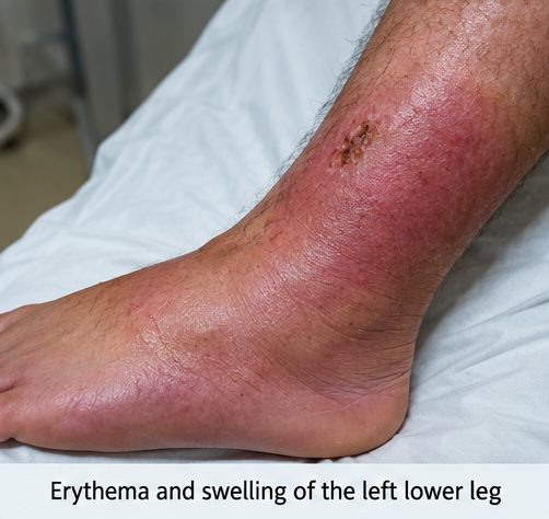
The symptoms initially began as a small area of redness over the lower shin. During the following two days, the erythema gradually extended to the surrounding skin and was accompanied by increasing swelling and tenderness. Walking became uncomfortable because of pain and a sensation of tightness in the affected leg.

The patient also reported low-grade fever, mild chills, generalized weakness, and reduced activity. There was no history of vomiting, breathlessness, altered consciousness, or other significant systemic symptoms.
On further questioning, he recalled sustaining a small superficial abrasion over the lower leg approximately five days before presentation. He had cleaned the wound at home and had not sought medical attention because the injury initially appeared minor.
There was no history of animal bite, prolonged water exposure, recent surgery, intravenous drug use, recurrent cellulitis, or chronic lower-limb ulceration.
On examination, the patient was conscious, oriented, and hemodynamically stable. Examination of the left lower limb demonstrated diffuse erythema and edema involving the lower leg. The affected skin was warm and tender on palpation.
The margins of erythema were relatively indistinct. A small healing abrasion was visible over the lower shin and was considered the probable portal of bacterial entry. There was no purulent discharge, fluctuant swelling, skin necrosis, crepitus, or hemorrhagic bullae.
Peripheral pulses were palpable, and distal perfusion was preserved. There was no evidence of neurovascular compromise. The overall clinical presentation was consistent with acute non-purulent cellulitis of the left lower limb.
Investigations
Initial laboratory investigations were performed to evaluate the inflammatory response and severity of infection.
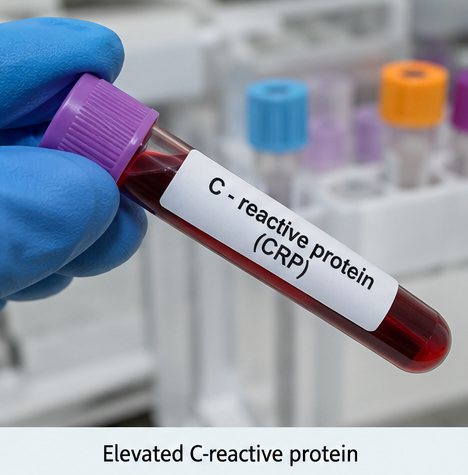
Complete blood count demonstrated mild leukocytosis with neutrophilic predominance. C-reactive protein was elevated, supporting the presence of an acute inflammatory process. Renal and liver function parameters were within acceptable limits, with no significant electrolyte abnormalities.
As the patient had typical non-purulent cellulitis without severe systemic toxicity or immunosuppression, extensive microbiological investigations were not considered necessary.
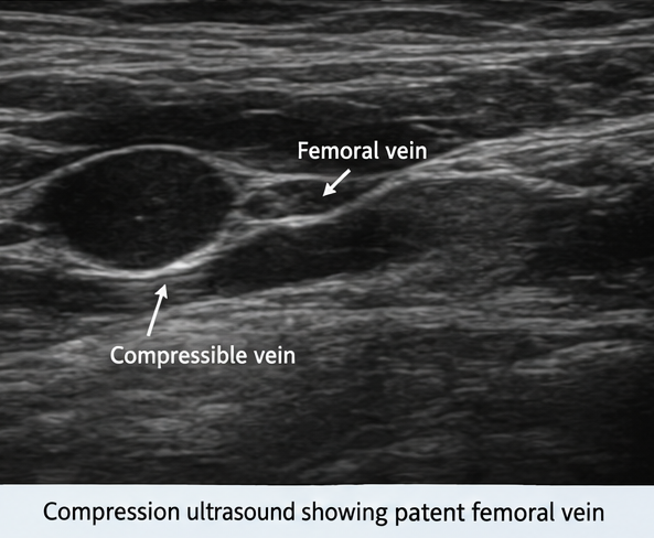
There was no clinical evidence of a localized abscess requiring drainage. Imaging was therefore not routinely required. Further imaging would have been considered if there was suspicion of an underlying abscess, foreign body, osteomyelitis, gas-forming infection, or deeper soft-tissue involvement.
Differential Diagnosis
Deep-Vein Thrombosis
Deep-vein thrombosis may cause unilateral lower-limb swelling, pain, warmth, and erythema and is therefore an important differential diagnosis. In this patient, prominent superficial inflammation together with a preceding skin abrasion favored cellulitis.
Venous Stasis Dermatitis
Venous stasis dermatitis may produce lower-limb erythema, edema, scaling, and discomfort. However, it usually follows a more chronic course and may involve both lower limbs. The acute onset and progressive localized inflammatory changes in this patient were more suggestive of cellulitis.
Erysipelas
Erysipelas is a more superficial bacterial infection involving the upper dermis and superficial lymphatics. It typically produces bright erythema with raised, sharply demarcated margins. The relatively diffuse inflammation and indistinct margins observed in this patient favored cellulitis.
Cutaneous Abscess
An abscess may present with pain, erythema, warmth, and swelling. However, a localized fluctuant or purulent collection is usually present. No fluctuation or purulent collection was identified in this patient.
Necrotizing Soft-Tissue Infection
Necrotizing soft-tissue infection is an important potentially life-threatening differential diagnosis. Rapid progression, severe pain disproportionate to examination findings, systemic toxicity, bullae, skin necrosis, and crepitus are concerning features. Their absence in this patient made a necrotizing infection less likely.
Management and Outcome
Treatment was initiated promptly following the clinical diagnosis of cellulitis.
The patient was started on appropriate antimicrobial therapy targeting the common Gram-positive organisms associated with non-purulent cellulitis. Selection of antimicrobial therapy was based on the clinical severity of infection and expected causative organisms.
The patient was advised to rest and elevate the affected limb to reduce edema and discomfort. Adequate hydration was maintained, and symptomatic treatment was provided for pain and fever.
The margins of erythema were monitored to assess progression or improvement. The patient was observed for worsening pain, progressive swelling, extension of erythema, hypotension, blistering, skin necrosis, or other features suggesting complicated infection.
During the following 48–72 hours, the patient remained clinically stable. His fever subsided, and there was no further progression of the erythema. Local warmth, pain, and tenderness gradually decreased.
Over the subsequent several days, swelling improved progressively. The patient remained ambulatory and did not develop abscess formation, tissue necrosis, systemic sepsis, or other significant complications.
At follow-up, the erythema had substantially resolved, swelling was minimal, and the patient was able to walk without significant discomfort. The superficial abrasion had healed without discharge.
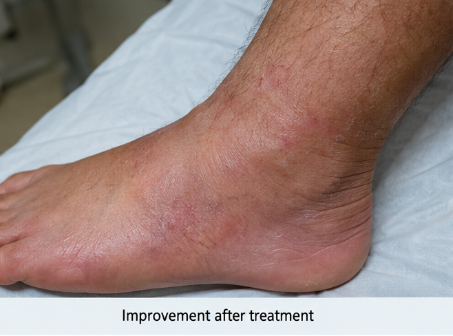
He was advised regarding appropriate wound hygiene, skin care, and prompt treatment of future breaks in the skin to minimize the risk of recurrent cellulitis.
Discussion
Cellulitis is a common bacterial infection of the skin and subcutaneous tissues. The lower extremities are particularly susceptible, and the condition typically manifests with erythema, warmth, swelling, and tenderness.
Disruption of the normal skin barrier is an important initiating factor. Even minor trauma may provide an entry point for bacteria. Other predisposing conditions include lymphedema, chronic edema, venous insufficiency, obesity, fungal infections of the feet, and previous cellulitis.
Streptococci and Staphylococcus aureus are among the principal organisms associated with cellulitis. Microbiological confirmation is frequently unavailable in uncomplicated cases because there may be no accessible purulent material for culture. Consequently, cellulitis remains predominantly a clinical diagnosis.
An important part of assessment is differentiating cellulitis from other causes of a red, swollen leg. Deep-vein thrombosis, venous eczema, lymphedema, lipodermatosclerosis, erysipelas, and superficial thrombophlebitis may resemble cellulitis.
The distinction between non-purulent cellulitis and an abscess is also important. Abscesses generally require drainage, whereas uncomplicated non-purulent cellulitis is primarily managed with systemic antimicrobial therapy.
Assessment of disease severity is essential when determining the appropriate treatment setting. Patients who are clinically stable and lack significant systemic manifestations may often be managed on an outpatient basis. Hospitalization and intravenous therapy may be required in patients with systemic infection, hemodynamic instability, significant comorbidities, failure of initial treatment, or concern for deeper infection.
Necrotizing soft-tissue infection must be considered in rapidly progressive or unusually painful presentations. Severe pain disproportionate to examination findings, rapidly spreading inflammation, systemic toxicity, bullae, skin necrosis, or crepitus should prompt urgent evaluation and surgical assessment.
Clinical resolution of cellulitis may be gradual. Local inflammation can persist during the initial treatment period even when antimicrobial therapy is effective. Limb elevation and management of associated edema and underlying predisposing factors are therefore important components of treatment.
In the present case, a minor abrasion was considered the probable portal of bacterial entry. The combination of unilateral erythema, warmth, edema, and tenderness without an abscess or features of necrotizing infection supported the diagnosis of uncomplicated non-purulent cellulitis. Prompt treatment and supportive measures resulted in progressive resolution without significant complications.
Conclusion
Cellulitis is an acute bacterial infection of the dermis and subcutaneous tissues that commonly affects the lower extremities. Typical manifestations include erythema, warmth, swelling, tenderness, and pain, with systemic symptoms occurring in some patients.
Diagnosis is predominantly clinical. Careful examination should identify possible portals of entry, predisposing conditions, and findings suggestive of abscess formation or deeper infection.
Important differential diagnoses include deep-vein thrombosis, venous stasis dermatitis, erysipelas, abscess, and necrotizing soft-tissue infection.
Prompt antimicrobial therapy, limb elevation, appropriate wound care, symptomatic management, and close clinical monitoring generally result in favorable outcomes in uncomplicated disease. In this patient, early recognition and appropriate treatment were associated with progressive resolution of inflammation and an uncomplicated recovery.
References
-
Sullivan T, de Barra E. Diagnosis and management of cellulitis. Clin Med (Lond). 2018;18(2):160–163. https://pmc.ncbi.nlm.nih.gov/articles/PMC6303460/
-
Santer M. Management of cellulitis: current practice and research questions. Br J Gen Pract. 2018;68(677):595–596. https://pmc.ncbi.nlm.nih.gov/articles/PMC6255235/
-
Kwak YG, Choi SH, Kim T, et al. Clinical guidelines for the antibiotic treatment for community-acquired skin and soft tissue infection. Infect Chemother. 2017;49(4):301–325. https://pmc.ncbi.nlm.nih.gov/articles/PMC5754343/
-
Sartelli M, Guirao X, Hardcastle TC, et al. 2018 WSES/SIS-E consensus conference: recommendations for the management of skin and soft-tissue infections. World J Emerg Surg. 2018;13:58. https://pmc.ncbi.nlm.nih.gov/articles/PMC6295010/
-
Sartelli M, Guirao X, Hardcastle TC, et al. WSES/GAIS/WSIS/SIS-E/AAST global clinical pathways for patients with skin and soft tissue infections. World J Emerg Surg. 2022;17:3. https://pmc.ncbi.nlm.nih.gov/articles/PMC8761341/
Read more such content on @ Hidoc Dr | Medical Learning App for Doctors
Recommended News For You
Recommended Articles For You
Featured News
Featured Articles
Featured Events
Featured KOL Videos
1.
Regular physical activity before cancer diagnosis may lower progression and death risks
2.
Medication Self-Injection Embraced by Breast Cancer Patients
3.
Financial hardship for cancer survivors due to high-cost immunotherapies, especially for blood cancer patients
4.
FDA Advisors Split on Regimens for Breast, Prostate Cancers
5.
ctDNA-Guided Adjuvant Therapy Cuts Bladder Cancer Costs
1.
RDW-CV vs. MCV: Knowing the Difference Can Help You Better Understand Your Health
2.
Clinical Pathways in Hematology in Clinical Decision-Making
3.
Immune Surveillance Screening for Early Cancer Risk Detection
4.
Unlocking the Mysteries of Lymphoblastic Lymphoma: A Journey Into the Unknown
5.
AI-Driven Digital Pathology in Oncology: From Histology Images to Predictive Treatment Biomarkers
1.
International Conference on Innovations in Critical Care for Oncology and Cardiology
2.
International Conference on Oncology, Cardiology and Critical Care Policy
3.
International Conference on Oncology, Cancer Prevention and Public Health
4.
International Conference on Cancer Nursing and Rehabilitation Strategies
5.
International Conference on Best Practices in Oncology, Cardiology and Critical Care
1.
How Advances in Cancer Research Are Improving Treatment Options
2.
Navigating the Complexities of Ph Negative ALL - Part II
3.
Signs and Symptoms of Leukemia
4.
Updates on Standard V/S High Risk Myeloma Treatment- The Next Part
5.
Virtual Case Study on Elephantiasis of Lower Limb- An Initiative by Hidoc Dr.

Address :
Hidoc Dr. Inc. | Delaware C Corp | 1309 Coffeen Ave. Suite 1200, Sheridan WY, 82801
Phone :
+1-415-463-3094
Email :
anishagadia@hidoc.co
© Copyright 2026 Hidoc Dr. Inc.
Terms & Conditions - LLP | Inc. | Privacy Policy - LLP | Inc. | Account Deactivation

To get started please enter your email ID
Welcome to Hidoc Dr.
Join to enhance your clinical skills and gain specialized in-depth medical knowledge