Oral Candidiasis in an Immunocompromised Adult: A Case Report
OthersPage Navigation
Abstract
Oral candidiasis is one of the most common opportunistic fungal infections affecting the oral cavity and is predominantly caused by Candida albicans. Although it may occur in healthy individuals, the condition is more frequently encountered in immunocompromised patients, those receiving prolonged antibiotic or corticosteroid therapy, individuals with poorly controlled diabetes mellitus, and denture wearers. Clinical manifestations range from asymptomatic white plaques to painful erythematous lesions that interfere with eating and speaking. Early diagnosis and prompt antifungal therapy are essential to prevent disease progression and recurrence. We report the case of a 56-year-old man with poorly controlled type 2 diabetes mellitus who presented with painful white plaques over the tongue and buccal mucosa associated with burning sensation and dysphagia. Clinical examination and microbiological investigations confirmed pseudomembranous oral candidiasis. The patient was successfully treated with topical antifungal therapy, optimization of glycemic control, and maintenance of oral hygiene, resulting in complete clinical resolution. This case highlights the importance of identifying predisposing factors and providing comprehensive management to prevent recurrence.
Introduction
Oral candidiasis is a superficial fungal infection of the oral mucosa caused primarily by Candida albicans, a commensal organism that becomes pathogenic under favorable conditions. Alterations in host immunity, disruption of the normal oral microbiota, xerostomia, systemic illnesses, and prolonged use of broad-spectrum antibiotics or corticosteroids predispose individuals to infection. The disease commonly presents as pseudomembranous, erythematous, hyperplastic, or angular cheilitis variants.
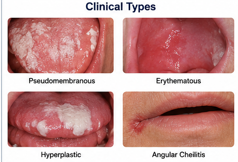
Patients often complain of burning sensation, altered taste, soreness, dysphagia, or difficulty eating. Diagnosis is generally based on characteristic clinical findings supported by potassium hydroxide (KOH) preparation, fungal culture, or cytological examination when necessary. Management includes elimination of predisposing factors, topical or systemic antifungal therapy depending on disease severity, and reinforcement of oral hygiene measures.
Case Report
A 56-year-old man presented to the dental outpatient department with complaints of painful white patches over the tongue and inner cheeks for ten days. He reported a persistent burning sensation while eating spicy food, unpleasant taste in the mouth, and mild difficulty swallowing. The symptoms had progressively worsened despite using antiseptic mouthwash.
The patient had a ten-year history of poorly controlled type 2 diabetes mellitus and had recently completed a two-week course of broad-spectrum antibiotics for a lower respiratory tract infection. He denied tobacco or alcohol use and had no previous history of similar oral lesions.
General examination revealed stable vital signs. Intraoral examination demonstrated multiple creamy white, curd-like plaques involving the dorsal surface of the tongue, bilateral buccal mucosa, and soft palate. The plaques could be gently scraped off, leaving an erythematous and mildly bleeding surface beneath. Mild angular fissuring was also noted at both corners of the mouth. No cervical lymphadenopathy was present.
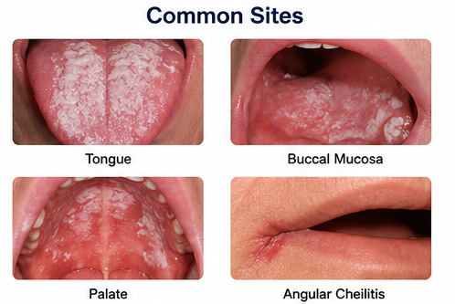
Laboratory investigations showed elevated fasting blood glucose of 218 mg/dL and glycated hemoglobin (HbA1c) of 9.2%, indicating poor glycemic control. Complete blood count revealed mild neutrophilic leukocytosis, while renal and liver function tests were within normal limits. Oral swab microscopy using 10% KOH preparation demonstrated budding yeast cells with pseudohyphae.
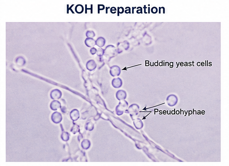
Fungal culture on Sabouraud dextrose agar yielded growth consistent with Candida albicans, confirming the diagnosis of pseudomembranous oral candidiasis.
Management and Outcome
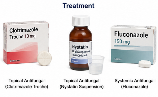
The patient was prescribed topical clotrimazole troches (10 mg) to be dissolved in the mouth five times daily for fourteen days.
He was also advised to use chlorhexidine mouth rinse, maintain meticulous oral hygiene, clean the tongue regularly, and avoid smoking, alcohol, and excessively spicy foods during treatment.
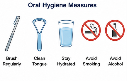
The patient was referred to an endocrinologist for optimization of diabetic management, and dietary counseling was provided to improve glycemic control. Adequate hydration was encouraged to minimize oral dryness, and the importance of completing the full course of antifungal therapy was emphasized.
At the two-week follow-up visit, the burning sensation had resolved completely, and the white plaques had disappeared. Repeat oral examination showed healthy oral mucosa without residual lesions. At three months, there was no recurrence, and improved glycemic control was documented with a reduction in HbA1c levels.
Follow-up
Two Weeks
- Complete disappearance of oral white plaques.
- Resolution of burning sensation and dysphagia.
- Improved oral comfort and appetite.
One Month
- Healthy oral mucosa without residual erythema.
- No evidence of fungal recurrence.
- Improved oral hygiene practices.
Three Months
The patient remained asymptomatic with no recurrence of oral candidiasis. Glycemic control had improved significantly, and regular dental follow-up was advised to monitor for future episodes.
Discussion
Oral candidiasis develops when local or systemic factors disrupt the balance between the host immune response and the normal oral microbial flora, allowing excessive proliferation of Candida species. Poorly controlled diabetes mellitus, prolonged antibiotic therapy, corticosteroid use, xerostomia, immunosuppression, and denture use are among the most important predisposing factors. Hyperglycemia promotes fungal growth by increasing salivary glucose concentration and impairing neutrophil function, thereby facilitating mucosal colonization.
Pseudomembranous candidiasis, commonly known as oral thrush, is the most frequent clinical presentation. It is characterized by removable white plaques that reveal erythematous mucosa on scraping. Although diagnosis is usually clinical, microbiological confirmation may be helpful in recurrent, atypical, or treatment-resistant cases.
Topical antifungal agents such as clotrimazole and nystatin remain first-line therapy for uncomplicated disease, whereas systemic fluconazole is reserved for extensive, recurrent, or refractory infections. Successful management requires not only antifungal therapy but also correction of underlying risk factors such as uncontrolled diabetes, poor oral hygiene, and unnecessary antibiotic exposure. Patient education regarding oral hygiene and regular follow-up plays a crucial role in preventing recurrence.
Prognosis
The prognosis of oral candidiasis is excellent when predisposing factors are addressed and appropriate antifungal therapy is initiated promptly. Most patients experience complete clinical resolution within one to two weeks. However, recurrent infections may occur in individuals with persistent immunosuppression, uncontrolled diabetes, or continued use of predisposing medications. Regular follow-up and management of underlying systemic conditions significantly improve long-term outcomes.
Conclusion
Oral candidiasis is a common opportunistic fungal infection that should be suspected in patients presenting with removable white oral plaques, burning sensation, and associated systemic risk factors. Early diagnosis through careful clinical examination and microbiological confirmation when indicated enables prompt initiation of antifungal therapy. Comprehensive management, including elimination of predisposing factors, optimization of systemic health, and reinforcement of oral hygiene practices, is essential for achieving complete recovery and minimizing recurrence.
References
- Akpan A, Morgan R. Oral candidiasis. Postgraduate Medical Journal. 2002;78(922):455–459. https://pmc.ncbi.nlm.nih.gov/articles/PMC1742467/
- Taylor M, Brizuela M, Raja A. Oral Candidiasis. StatPearls Publishing; Updated July 4, 2023. https://www.ncbi.nlm.nih.gov/books/NBK545282/
- Hellstein JW, Marek CL. Candidiasis: Red and White Manifestations in the Oral Cavity. Head and Neck Pathology. 2019;13(1):25–32. https://pmc.ncbi.nlm.nih.gov/articles/PMC6405794/
- Millsop JW, Fazel N. Oral candidiasis. Clinics in Dermatology. 2016;34(4):487–49 https://pubmed.ncbi.nlm.nih.gov/27343964/
- Pappas PG, Kauffman CA, Andes DR, et al. Clinical Practice Guideline for the Management of Candidiasis: 2016 Update by the Infectious Diseases Society of America. Clinical Infectious Diseases. 2016;62(4):e1–e50. https://academic.oup.com/cid/article/62/4/e1/2462830
Read more such content on @ Hidoc Dr | Medical Learning App for Doctors
Recommended News For You
Recommended Articles For You
Featured News
Featured Articles
Featured Events
Featured KOL Videos
1.
Year in Review: Non-Small Cell Lung Cancer
2.
Study suggests around 40% of postmenopausal hormone positive breast cancers are linked to excess body fat
3.
The need for more Latinx participants in Alzheimer's trials is urgent.
4.
Why palliative care goes hand in hand with treatment for people with cancer: Q&A
5.
MRD-Guided Azacitidine May Delay Relapse in AML, MDS
1.
Exploring the Benefits of Teclistamab for Treating Advanced Cancer
2.
The Danger of Methemoglobinemia and How to Prevent It
3.
Deciphering FFR: A Comprehensive Guide to Understanding Its Meaning
4.
Red Blood Cell Microparticles: Tiny Warriors Against Bleeding in the Brain
5.
Artificial Intelligence in Oncology: Current Trends, Challenges and Future Outlook
1.
Asian Symposium on Advancement in Hematology and Oncology
2.
Asian Symposium on Advancement in Hematology and Oncology
3.
Asian Symposium on Advancement in Hematology and Oncology
4.
International Cancer Conference
5.
Asian Symposium on Advancement in Hematology and Oncology
1.
Daratumumab, Lenalidomide, and Dexamethasone (DRd) Versus Lenalidomide and Dexamethasone (Rd) in MRD Negativity
2.
Lorlatinib in the Management of 1st line ALK+ mNSCLC (CROWN TRIAL Update)
3.
Thromboprophylaxis In Medical Settings
4.
Post Progression Approaches After First-line Third-Generaion ALK Inhibitors
5.
Molecular Contrast: EGFR Axon 19 vs. Exon 21 Mutations - Part VII

Address :
Hidoc Dr. Inc. | Delaware C Corp | 1309 Coffeen Ave. Suite 1200, Sheridan WY, 82801
Phone :
+1-415-463-3094
Email :
anishagadia@hidoc.co
© Copyright 2026 Hidoc Dr. Inc.
Terms & Conditions - LLP | Inc. | Privacy Policy - LLP | Inc. | Account Deactivation

To get started please enter your email ID
Welcome to Hidoc Dr.
Join to enhance your clinical skills and gain specialized in-depth medical knowledge