Left Ventricular Hypertrophy Associated With Long-Standing Hypertension
OthersPage Navigation
Introduction
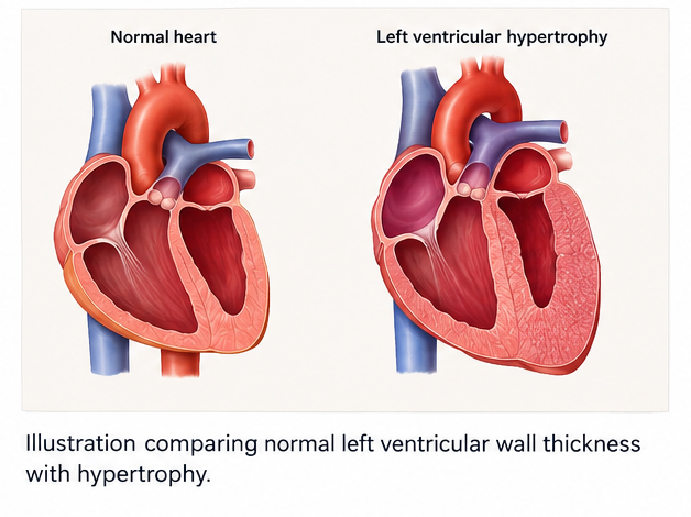
Left ventricular hypertrophy (LVH) is a structural cardiac abnormality characterized by an increase in left ventricular myocardial mass. It commonly develops in response to chronic pressure or volume overload, with long-standing hypertension being one of the most important acquired causes.
Persistent elevation of systemic blood pressure increases left ventricular afterload. In response, the myocardium undergoes adaptive remodeling involving cardiomyocyte hypertrophy, changes in ventricular geometry, and, over time, myocardial fibrosis. LVH may initially preserve systolic function but can progressively impair ventricular relaxation and contribute to diastolic dysfunction.
Patients with LVH may remain asymptomatic or may present with exertional dyspnea, reduced exercise tolerance, chest discomfort, palpitations, or symptoms of heart failure. LVH is clinically important because it is associated with increased cardiovascular morbidity and mortality and may coexist with atrial fibrillation, heart failure, coronary artery disease, stroke, and other cardiovascular complications.
Electrocardiography may provide evidence suggestive of LVH, while transthoracic echocardiography is commonly used to assess left ventricular mass, wall thickness, chamber dimensions, geometry, and systolic and diastolic function. Cardiac magnetic resonance imaging provides more detailed assessment of ventricular structure and myocardial tissue characteristics when required.
Management focuses primarily on identification and treatment of the underlying cause, particularly adequate blood pressure control. Lifestyle modification and antihypertensive therapy can reduce left ventricular mass and may lead to regression of LVH.
Case Report
A 58-year-old man presented to the outpatient cardiology clinic with a six-month history of gradually progressive exertional breathlessness and reduced exercise tolerance. He reported becoming short of breath while climbing two flights of stairs, although he remained comfortable at rest.
He also described occasional episodes of a sensation of forceful heartbeat, particularly after physical activity. There was no history of syncope, orthopnea, paroxysmal nocturnal dyspnea, pedal edema, or recent chest pain.
The patient had been diagnosed with hypertension approximately 12 years earlier but acknowledged inconsistent adherence to antihypertensive medication. He had no previous history of myocardial infarction, valvular heart disease, congenital heart disease, or known heart failure.
He had type 2 diabetes mellitus for six years and was receiving oral medication. He was overweight and had a sedentary lifestyle. He was a former smoker with a 10-pack-year history and had stopped smoking five years earlier.
On examination, the patient was alert and comfortable at rest. His blood pressure was 168/96 mmHg, pulse rate was 82 beats/minute, respiratory rate was 18 breaths/minute, and oxygen saturation was 97% on room air.
Cardiovascular examination revealed a sustained apical impulse that was mildly displaced laterally. A soft fourth heart sound was audible at the apex. There was no significant cardiac murmur, elevated jugular venous pressure, or peripheral edema.
The initial clinical impression was poorly controlled chronic hypertension with possible hypertensive cardiac remodeling.
Investigations
Laboratory investigations showed a hemoglobin level of 14.1 g/dL, leukocyte count of 7,800 cells/mm³, and platelet count of 274,000 cells/mm³.
Serum sodium was 139 mmol/L, potassium was 4.2 mmol/L, bicarbonate was 24 mmol/L, blood urea nitrogen was 18 mg/dL, and serum creatinine was 1.0 mg/dL.
Fasting plasma glucose was 126 mg/dL, and glycated hemoglobin was 7.1%. The lipid profile showed total cholesterol of 196 mg/dL, low-density lipoprotein cholesterol of 121 mg/dL, high-density lipoprotein cholesterol of 42 mg/dL, and triglycerides of 164 mg/dL.
Thyroid function tests were within normal limits. High-sensitivity cardiac troponin-I was not elevated.
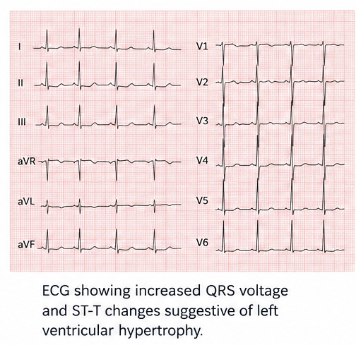
A 12-lead electrocardiogram demonstrated sinus rhythm with increased QRS voltage and secondary ST-T abnormalities in the lateral leads, findings suggestive of left ventricular hypertrophy with repolarization changes.
Twenty-four-hour ambulatory blood pressure monitoring demonstrated persistent elevation of blood pressure, with an average daytime blood pressure of 154/91 mmHg and nighttime blood pressure of 142/82 mmHg.
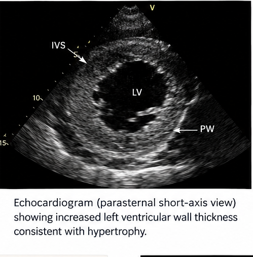
Transthoracic echocardiography demonstrated increased interventricular septal thickness of 13 mm and posterior wall thickness of 12 mm. The left ventricular end-diastolic diameter was 49 mm. Left ventricular ejection fraction was 62%.
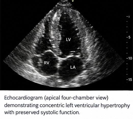
The calculated left ventricular mass index was 128 g/m², consistent with increased left ventricular mass. Relative wall thickness was increased, indicating a concentric pattern of left ventricular hypertrophy.
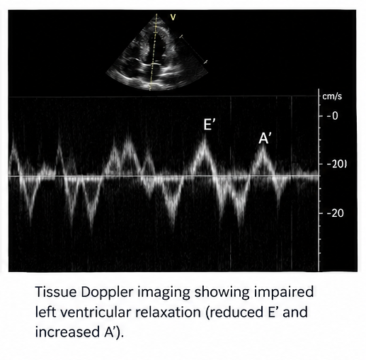
The left atrium was mildly enlarged. Doppler and tissue Doppler assessment demonstrated impaired left ventricular relaxation with evidence of grade I diastolic dysfunction. There was no significant valvular stenosis or regurgitation.
No regional wall-motion abnormality was identified.
Based on the clinical, electrocardiographic, and echocardiographic findings, the patient was diagnosed with hypertension-associated concentric left ventricular hypertrophy with preserved left ventricular systolic function.
Differential Diagnosis
Hypertrophic Cardiomyopathy
Hypertrophic cardiomyopathy may produce increased left ventricular wall thickness and can clinically resemble hypertensive LVH. However, the patient's history of long-standing poorly controlled hypertension, concentric ventricular geometry, absence of a characteristic asymmetric hypertrophy pattern, and lack of a significant left ventricular outflow tract obstruction favored hypertensive remodeling.
Aortic Stenosis
Aortic stenosis can produce pressure overload and secondary concentric LVH. However, echocardiography demonstrated no significant aortic valve stenosis or elevated transvalvular gradient, making this diagnosis unlikely.
Infiltrative Cardiomyopathy
Conditions such as cardiac amyloidosis may produce increased ventricular wall thickness and diastolic dysfunction. However, the patient's clinical profile, absence of systemic features suggestive of infiltrative disease, preserved ventricular function, and typical hypertensive remodeling pattern did not support an infiltrative cardiomyopathy.
Ischemic Heart Disease
Coronary artery disease may cause exertional dyspnea and structural cardiac abnormalities. However, there was no history of typical angina, cardiac biomarkers were normal, and echocardiography showed no regional wall-motion abnormality.
Athlete's Heart
Physiological cardiac remodeling associated with intensive athletic training may result in increased left ventricular mass. The patient had no history of sustained endurance or competitive athletic activity, and the presence of poorly controlled hypertension and concentric hypertrophy favored pathological remodeling.
Management and Outcome
The patient was advised regarding the importance of sustained blood pressure control and adherence to antihypertensive therapy.
Antihypertensive treatment was initiated with an angiotensin-converting enzyme inhibitor, and additional therapy with a calcium channel blocker was subsequently introduced because blood pressure remained above the desired treatment range.
The patient was counseled regarding dietary sodium reduction, weight management, regular moderate physical activity, smoking avoidance, and adherence to treatment for diabetes and other cardiovascular risk factors.
Home blood pressure monitoring was recommended, and medication adherence was reviewed at each follow-up visit.
Because the patient had no evidence of decompensated heart failure, significant valvular disease, myocardial ischemia, or clinically important arrhythmia, hospitalization was not required.
At the first four-week follow-up, the patient's blood pressure had decreased to 142/84 mmHg. His exertional breathlessness had improved, although he continued to experience occasional mild symptoms during strenuous activity.
The antihypertensive regimen was continued and subsequently adjusted to achieve sustained blood pressure control.
At three months, his office blood pressure was 128/78 mmHg. He reported substantially improved exercise tolerance and no further episodes of significant palpitations.
A repeat echocardiographic assessment after continued blood pressure control demonstrated a reduction in left ventricular mass compared with the baseline examination, although mild concentric remodeling persisted. Left ventricular ejection fraction remained preserved.
The patient was advised to continue long-term blood pressure monitoring and cardiovascular risk-factor modification.
Discussion
Left ventricular hypertrophy is an important manifestation of cardiac remodeling associated with chronic pressure overload. Hypertension is a major cause, although LVH can also occur with aortic valve disease, hypertrophic cardiomyopathy, chronic kidney disease, and other conditions associated with increased ventricular workload.
The development of hypertensive LVH involves both mechanical and neurohormonal mechanisms. Persistent elevation of systemic vascular resistance increases left ventricular afterload and stimulates cardiomyocyte hypertrophy. Neurohormonal activation, particularly involving the renin-angiotensin-aldosterone system, together with myocardial fibrosis, contributes to progressive structural remodeling.
The clinical presentation can be variable. Many patients with LVH remain asymptomatic for prolonged periods. Others develop exertional dyspnea or reduced exercise tolerance as ventricular stiffness increases and diastolic filling becomes impaired. Atrial enlargement and increased myocardial electrical heterogeneity may also contribute to the development of atrial fibrillation and other rhythm disturbances.
Electrocardiography can provide an initial indication of LVH through increased QRS voltage and associated repolarization abnormalities. However, ECG-based criteria do not correlate perfectly with anatomical LV mass. Echocardiography provides a more direct assessment of ventricular wall thickness, chamber dimensions, LV mass, geometry, systolic function, and diastolic function. Cardiac magnetic resonance imaging can provide further structural and tissue characterization when the diagnosis remains uncertain.
LVH may present with different geometric patterns. Concentric LVH is characterized by increased LV mass together with increased relative wall thickness, whereas eccentric LVH is characterized by increased LV mass with relatively less increase in wall thickness. Concentric remodeling represents increased relative wall thickness without an increase in LV mass.
The distinction between hypertensive LVH and other causes of increased wall thickness is clinically important. Hypertrophic cardiomyopathy, infiltrative disorders, storage diseases, and valvular disease may require different diagnostic investigations and management strategies.
The primary therapeutic approach in hypertensive LVH is effective management of the underlying blood pressure elevation. Lifestyle measures include dietary modification, sodium restriction, appropriate physical activity, weight management, avoidance of tobacco exposure, and adherence to prescribed treatment.
Antihypertensive therapy can reduce left ventricular mass. Evidence indicates that successful blood pressure reduction is associated with regression of LVH, while randomized-trial meta-analysis has demonstrated lower rates of LVH and greater regression with more intensive blood pressure lowering compared with standard treatment, although treatment targets must be individualized.
The regression of LVH is clinically relevant because persistent or newly developed LVH has been associated with cardiovascular events. A meta-analysis of hypertensive patients found that regression of echocardiographic LVH was associated with a lower risk of subsequent cardiovascular events compared with persistent or newly developed LVH.
More recent observational data have similarly shown that regression of LVH during antihypertensive therapy is associated with fewer cardiovascular events, including heart failure hospitalization and cardiovascular mortality. However, these associations should not be interpreted as proof that LVH regression itself independently causes the reduction in risk.
The 2024 European Society of Cardiology hypertension guideline emphasizes comprehensive cardiovascular risk assessment and appropriate blood pressure management in patients with hypertension.
In the present case, prolonged inadequately controlled hypertension was associated with concentric LVH and early diastolic dysfunction despite preserved systolic function. Improvement in blood pressure control was accompanied by improvement in symptoms and subsequent reduction in left ventricular mass.
This case highlights the importance of identifying hypertensive cardiac remodeling before the development of overt heart failure. Regular assessment of blood pressure, cardiovascular risk factors, symptoms, and, when clinically indicated, cardiac structure and function can help guide long-term management.
Conclusion
Left ventricular hypertrophy is an important manifestation of chronic cardiovascular remodeling, most commonly associated with sustained hypertension.
In this patient, long-standing poorly controlled hypertension was associated with increased left ventricular mass, concentric geometry, and mild diastolic dysfunction despite preserved systolic function.
Electrocardiography provided evidence suggestive of LVH, while echocardiography confirmed increased left ventricular mass and characterized the ventricular geometry and functional abnormalities.
Effective blood pressure control, adherence to antihypertensive therapy, lifestyle modification, and management of associated cardiovascular risk factors are central to treatment.
Regression of LVH during treatment is associated with improved cardiovascular risk profiles and represents an important marker of response to long-term management.
Early recognition of hypertensive cardiac remodeling may help prevent progression to heart failure, atrial fibrillation, and other cardiovascular complications.
References
- Messerli FH, Rimoldi SF, Bangalore S. The transition from hypertension to heart failure: role of left ventricular hypertrophy. J Am Coll Cardiol. 2017;69(10):1317–1325. https://pubmed.ncbi.nlm.nih.gov/28279253/
- Williams B, Mancia G, Spiering W, et al. 2024 ESC Guidelines for the management of elevated blood pressure and hypertension. Eur Heart J. 2024. https://www.escardio.org/Guidelines/Clinical-Practice-Guidelines/2024-ESC-Guidelines-for-the-management-of-elevated-blood-pressure-and-hypertension
- Cuspidi C, Sala C, Negri F, Mancia G, Morganti A. Prevalence of left-ventricular hypertrophy in hypertension: an updated review of echocardiographic studies. J Hum Hypertens. 2012;26:343–349. https://pubmed.ncbi.nlm.nih.gov/22089122/
- Pierdomenico SD, Cuccurullo F. Risk reduction after regression of echocardiographic left ventricular hypertrophy in hypertension: a meta-analysis. Am J Hypertens. 2010;23(8):876–881. https://pubmed.ncbi.nlm.nih.gov/20414193/
- Kim HM, Hwang IC, Choi HM, Yoon YE, Cho GY. Prognostic implication of left ventricular hypertrophy regression after antihypertensive therapy in patients with hypertension. Front Cardiovasc Med. 2022;9:1082008. https://pmc.ncbi.nlm.nih.gov/articles/PMC9807809/
- Hu J, Zhou Y. Effect of intensive blood pressure lowering on left ventricular hypertrophy in patients with hypertension: a meta-analysis of randomized trials. Blood Press. 2023;32(1):2242501. https://pubmed.ncbi.nlm.nih.gov/37652401/
- O'Rourke MF, Safar ME. Relationship between aortic stiffening and microvascular disease in brain and kidney: cause and logic of therapy. Hypertension. 2005;46:200–204. https://pubmed.ncbi.nlm.nih.gov/15939800/
- Mutai H, et al. Hypertension and left ventricular geometry: diagnosis, prognosis, and management. 2026. https://pubmed.ncbi.nlm.nih.gov/42376431/
Read more such content on @ Hidoc Dr | Medical Learning App for Doctors
Recommended News For You
Recommended Articles For You
Featured News
Featured Articles
Featured Events
Featured KOL Videos
1.
Early cancer detection during pregnancy and the postpartum period may be aided by breast milk examination.
2.
Global warming could be driving up women's cancer risk, find researchers
3.
Use of social media during pregnancy may increase depression.
4.
Pickleball program boosts health and wellness for cancer survivors, study finds
5.
Researchers can now forecast how prostate cancer bone metastases will react to radium-223 treatment.
1.
The Mysterious Pain of Scrotal Hematoma: A Guide to Diagnosis and Treatment
2.
Base Editing for Rare Blood Diseases: Mechanisms, Clinical Applications, and Future Directions
3.
Immune Escape Landscapes in Solid Tumors
4.
Case Study: The Role of Machine Learning in the Detection of Skin Cancer
5.
How EPCORITAMAB is Revolutionizing the Treatment of Cancer
1.
International Conference on Innovations in Critical Care for Oncology and Cardiology
2.
International Conference on Oncology, Cardiology and Critical Care Policy
3.
International Conference on Oncology, Cancer Prevention and Public Health
4.
International Conference on Cancer Nursing and Rehabilitation Strategies
5.
International Conference on Best Practices in Oncology, Cardiology and Critical Care
1.
From Relapse to Remission : Chasing the Invisible and Redefining Long-Term Survival in Adult R/R B-Cell ALL
2.
Evolving Space of First-Line Treatment for Urothelial Carcinoma- Case Discussion
3.
How Nutrition Can Support Patients During Cancer Care
4.
An Eagles View - Evidence-based Discussion on Iron Deficiency Anemia- Panel Discussion III
5.
Recent Data Analysis for First-Line Treatment of ALK+ NSCLC

Address :
Hidoc Dr. Inc. | Delaware C Corp | 1309 Coffeen Ave. Suite 1200, Sheridan WY, 82801
Phone :
+1-415-463-3094
Email :
anishagadia@hidoc.co
© Copyright 2026 Hidoc Dr. Inc.
Terms & Conditions - LLP | Inc. | Privacy Policy - LLP | Inc. | Account Deactivation

To get started please enter your email ID
Welcome to Hidoc Dr.
Join to enhance your clinical skills and gain specialized in-depth medical knowledge