Diphtheria Presenting With Progressive Sore Throat, Cervical Swelling, and a Gray-White Pharyngeal Pseudomembrane: A Case Report
OthersPage Navigation
Introduction
Diphtheria is an acute, potentially life-threatening bacterial infection caused primarily by toxigenic strains of Corynebacterium diphtheriae. Although widespread childhood immunization has substantially reduced its incidence, diphtheria continues to occur, particularly in populations with incomplete vaccination coverage.
Respiratory diphtheria typically affects the tonsils, pharynx, larynx, or nasal passages. The characteristic clinical feature is the formation of a thick, gray-white pseudomembrane over the affected mucosal surface. Attempts to remove the membrane may result in bleeding because of its firm adherence to the underlying tissue.
The severity of diphtheria is largely related to production of diphtheria toxin. After local production, the toxin may enter the circulation and cause systemic complications, particularly myocarditis and neurological dysfunction. Extensive cervical lymphadenopathy and soft-tissue edema can produce the characteristic “bull-neck” appearance seen in severe disease.
Early clinical recognition is critical because treatment with diphtheria antitoxin should not be delayed while awaiting microbiological confirmation when respiratory diphtheria is strongly suspected. Antibiotic therapy, respiratory isolation, airway monitoring, and evaluation of close contacts are additional important components of management.
This report describes a patient presenting with progressive sore throat, fever, cervical swelling, and a firmly adherent pharyngeal pseudomembrane who was subsequently diagnosed with respiratory diphtheria.
Case Report
A 12-year-old boy presented to the emergency department with a four-day history of sore throat, low-grade fever, progressive difficulty swallowing, and increasing swelling of the neck.

The illness initially began with generalized weakness, reduced appetite, and throat discomfort. During the following two days, the patient developed increasing pain while swallowing and noticed progressive swelling around the upper neck.
His parents reported that his voice had become slightly muffled and that he had developed increasing difficulty swallowing solid food. There was no history of significant cough, vomiting, diarrhea, skin rash, or recent trauma.
On the day of presentation, the patient appeared tired and unwell. His parents also reported that his childhood vaccination schedule had been incomplete, although precise vaccination records were unavailable.
On examination, the patient was conscious and oriented but appeared moderately ill. His temperature was 38.1°C, pulse rate was 108 beats/min, respiratory rate was 22 breaths/min, and blood pressure was 104/68 mmHg. Oxygen saturation was 97% on room air.
Examination of the neck demonstrated bilateral cervical lymphadenopathy with surrounding soft-tissue swelling. The swelling produced a visibly broadened appearance of the neck.
Oropharyngeal examination revealed erythematous and swollen tonsillar and pharyngeal mucosa. A thick gray-white membrane was present over both tonsils and extended onto the posterior pharyngeal wall.
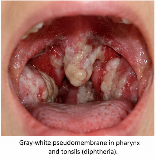
The membrane was firmly adherent to the underlying mucosa. Gentle manipulation produced slight bleeding, raising strong clinical suspicion of a diphtheritic pseudomembrane.
There was no stridor at presentation, although the extent of pharyngeal involvement raised concern regarding possible progression to upper-airway obstruction.
Cardiovascular examination demonstrated tachycardia without an obvious murmur. Respiratory examination revealed bilateral air entry without significant wheezing or crepitations.
The combination of an incomplete immunization history, cervical swelling, systemic illness, and a firmly adherent gray-white pharyngeal pseudomembrane was strongly suggestive of respiratory diphtheria.
Investigations
Because diphtheria was clinically suspected, appropriate throat and nasopharyngeal specimens were obtained for microbiological examination before antibiotic administration whenever feasible.
Swabs were carefully collected from the pharyngeal lesion, including the margin and area beneath the pseudomembrane, without attempting forceful removal.
Laboratory investigations demonstrated mild leukocytosis with neutrophilic predominance. Renal and hepatic parameters were within acceptable limits at presentation.
A throat specimen demonstrated organisms morphologically compatible with Corynebacterium species.

Subsequent culture identified Corynebacterium diphtheriae.

Testing for toxigenicity was performed because identification of C. diphtheriae alone does not establish toxin production. Detection of the diphtheria toxin gene together with appropriate microbiological assessment supported the diagnosis of toxigenic C. diphtheriae infection.
An electrocardiogram was obtained at baseline because myocarditis is an important systemic complication of diphtheria. Initial ECG did not demonstrate a significant conduction abnormality.
Cardiac monitoring was continued during hospitalization, and cardiac biomarkers were assessed because myocardial involvement may develop after the initial respiratory illness.
Based on the characteristic clinical presentation and microbiological findings, a diagnosis of respiratory diphtheria caused by toxigenic Corynebacterium diphtheriae was established.
Differential Diagnosis
Streptococcal Pharyngitis
Group A streptococcal pharyngitis commonly presents with fever, sore throat, tonsillar inflammation, and cervical lymphadenopathy. Tonsillar exudates may be present. However, a thick, firmly adherent gray pseudomembrane extending beyond the tonsils is much more suggestive of diphtheria.
Infectious Mononucleosis
Infectious mononucleosis may cause fever, severe pharyngitis, tonsillar exudates, and cervical lymphadenopathy. Generalized lymphadenopathy, marked fatigue, hepatosplenomegaly, and characteristic hematological findings may help distinguish it from diphtheria.
Peritonsillar Abscess
A peritonsillar abscess may present with fever, severe unilateral throat pain, dysphagia, muffled voice, and difficulty opening the mouth. Examination generally demonstrates unilateral peritonsillar swelling with displacement of the uvula rather than a diffuse adherent pseudomembrane.
Epiglottitis
Epiglottitis can produce fever, severe throat discomfort, dysphagia, drooling, and potentially rapid airway compromise. The characteristic adherent pharyngeal membrane and cervical lymphadenopathy observed in this patient favored diphtheria.
Vincent Angina
Necrotizing infections of the oropharynx can produce ulcerative lesions and membrane-like material. However, the clinical context, characteristic pseudomembrane, vaccination history, and microbiological identification of toxigenic C. diphtheriae supported diphtheria.
Management and Outcome
Because respiratory diphtheria was strongly suspected clinically, the patient was immediately placed under appropriate respiratory isolation precautions.
Diphtheria antitoxin was administered promptly after appropriate assessment because circulating toxin should be neutralized as early as possible. Treatment was initiated without waiting for final microbiological confirmation.

Antibacterial therapy effective against C. diphtheriae was also started to eradicate the organism, limit transmission, and prevent continued toxin production.
The patient's airway was closely monitored because progressive pseudomembrane formation and cervical soft-tissue edema can result in potentially life-threatening airway obstruction.
No attempt was made to forcibly remove the pseudomembrane because manipulation can cause bleeding and may worsen local tissue injury or precipitate airway complications.
Supportive treatment included adequate hydration, nutritional support, fever management, and careful monitoring of respiratory and cardiovascular status.
Serial electrocardiographic monitoring was performed because diphtheria toxin can cause myocarditis, conduction abnormalities, and arrhythmias. The patient remained hemodynamically stable without evidence of clinically significant myocardial involvement during hospitalization.
Neurological status was also monitored for features such as palatal weakness, cranial neuropathy, limb weakness, or other manifestations of toxin-mediated neuropathy.
Over the following days, fever subsided and the patient's throat discomfort gradually improved. Cervical swelling decreased progressively, and the pseudomembrane began to resolve.
The patient did not develop airway obstruction, significant cardiac complications, or neurological deficits.
Microbiological clearance was subsequently assessed according to recommended public-health protocols. Close household contacts were identified for clinical assessment, appropriate antimicrobial prophylaxis, vaccination review, and microbiological testing when indicated.
The patient's immunization status was reassessed, and completion of age-appropriate diphtheria-containing vaccination was advised because recovery from diphtheria does not reliably provide protective immunity.
Discussion
Diphtheria remains an important infectious disease because respiratory infection can progress rapidly and toxin-mediated complications may occur even when the initial local illness appears relatively limited.
Toxigenic Corynebacterium diphtheriae colonizes the respiratory mucosa and produces diphtheria toxin. Local tissue injury results in inflammation, necrosis, and formation of a pseudomembrane composed of fibrin, inflammatory cells, bacteria, and necrotic epithelial tissue.
The resulting membrane is typically gray or white and is characteristically firmly adherent to the underlying mucosa. Bleeding after attempted removal is an important clinical clue.
The tonsils and pharynx are frequently involved in respiratory diphtheria. In more extensive disease, the pseudomembrane may spread into the nasopharynx or larynx, increasing the risk of airway obstruction.
Cervical lymphadenopathy and surrounding soft-tissue edema can produce marked neck swelling. Severe swelling may result in the classic “bull-neck” appearance and is generally associated with substantial local disease.
Systemic toxicity results primarily from absorption of diphtheria toxin. The toxin inhibits cellular protein synthesis and may damage multiple organs.
Myocarditis is among the most serious complications. Patients may develop tachycardia, conduction disturbances, arrhythmias, heart failure, or cardiovascular collapse. Because myocardial injury can develop after the onset of the respiratory illness, continued cardiac surveillance is important.
Neurological complications may occur later and include palatal paralysis, cranial nerve dysfunction, peripheral neuropathy, and motor weakness. Recovery from neurological involvement may require weeks or months.
Diagnosis of respiratory diphtheria is primarily clinical during the initial management period. The presence of a characteristic adherent pseudomembrane, particularly in a patient with absent or incomplete immunization, should prompt immediate consideration of diphtheria.
Microbiological testing is important for confirmation and public-health surveillance. Specimens should be collected from the affected pharyngeal or nasal areas, including beneath or around the pseudomembrane when this can be performed safely.
Identification of C. diphtheriae should be followed by assessment for toxigenicity because disease severity is closely associated with toxin production.
Treatment requires both antitoxin and antimicrobial therapy. Diphtheria antitoxin neutralizes circulating unbound toxin but cannot reverse toxin that has already entered cells. Consequently, early administration is important and should not be postponed when clinical suspicion is high.
Antibiotics eliminate the organism and reduce transmission but do not substitute for antitoxin in toxin-mediated respiratory disease.
Infection-control and public-health measures are particularly important because respiratory diphtheria can spread through close contact and respiratory secretions. Patients require appropriate isolation until they are considered noninfectious according to applicable public-health guidance.
Close contacts should be identified promptly. Management may include clinical surveillance, microbiological testing, antimicrobial prophylaxis, and administration of diphtheria-containing vaccine when immunization is incomplete or not up to date.
Vaccination remains the most effective strategy for prevention. Protection can decline over time, making completion of primary vaccination and recommended booster doses important.
The present case demonstrates the classical progression of respiratory diphtheria, with sore throat followed by an adherent pharyngeal pseudomembrane and progressive cervical swelling. Recognition of these findings allowed early antitoxin therapy, antibiotic treatment, isolation, and monitoring for potentially serious systemic complications.
Conclusion
Diphtheria should be considered in patients presenting with progressive pharyngitis associated with a thick, firmly adherent gray-white pseudomembrane, particularly when vaccination is incomplete or uncertain.
Early recognition is critical because diphtheria toxin can cause potentially fatal cardiac, neurological, and respiratory complications. Treatment should be initiated on the basis of strong clinical suspicion rather than being delayed until microbiological confirmation is available.
Diphtheria antitoxin, appropriate antibiotics, respiratory isolation, airway surveillance, and monitoring for toxin-mediated complications form the major components of management.
Evaluation and prophylactic management of close contacts are also essential for limiting transmission.
In this patient, recognition of the characteristic pseudomembrane and cervical swelling led to prompt treatment and monitoring, followed by clinical recovery without major cardiac, neurological, or airway complications.
References
- Sharma NC, Efstratiou A, Mokrousov I, Mutreja A, Das B, Ramamurthy T. Diphtheria. Nat Rev Dis Primers. 2019;5(1):81. https://pubmed.ncbi.nlm.nih.gov/31727996/
- Centers for Disease Control and Prevention. Clinical Guidance for Diphtheria. https://www.cdc.gov/diphtheria/hcp/clinical-guidance/index.html
- World Health Organization. Diphtheria. https://www.who.int/health-topics/diphtheria
- Truelove SA, Keegan LT, Moss WJ, et al. Clinical and epidemiological aspects of diphtheria: a systematic review and pooled analysis. Clin Infect Dis. 2020;71(1):89–97. https://pmc.ncbi.nlm.nih.gov/articles/PMC7312233/
- Acosta AM, Moro PL, Hariri S, Tiwari TSP. Diphtheria. In: Epidemiology and Prevention of Vaccine-Preventable Diseases. Centers for Disease Control and Prevention. https://www.cdc.gov/pinkbook/hcp/table-of-contents/chapter-7-diphtheria.html
Read more such content on @ Hidoc Dr | Medical Learning App for Doctors
Recommended News For You
Recommended Articles For You
Featured News
Featured Articles
Featured Events
Featured KOL Videos
1.
Early cancer detection during pregnancy and the postpartum period may be aided by breast milk examination.
2.
Global warming could be driving up women's cancer risk, find researchers
3.
Use of social media during pregnancy may increase depression.
4.
Pickleball program boosts health and wellness for cancer survivors, study finds
5.
Researchers can now forecast how prostate cancer bone metastases will react to radium-223 treatment.
1.
The Mysterious Pain of Scrotal Hematoma: A Guide to Diagnosis and Treatment
2.
Base Editing for Rare Blood Diseases: Mechanisms, Clinical Applications, and Future Directions
3.
Immune Escape Landscapes in Solid Tumors
4.
Case Study: The Role of Machine Learning in the Detection of Skin Cancer
5.
How EPCORITAMAB is Revolutionizing the Treatment of Cancer
1.
International Conference on Innovations in Critical Care for Oncology and Cardiology
2.
International Conference on Oncology, Cardiology and Critical Care Policy
3.
International Conference on Oncology, Cancer Prevention and Public Health
4.
International Conference on Cancer Nursing and Rehabilitation Strategies
5.
International Conference on Best Practices in Oncology, Cardiology and Critical Care
1.
From Relapse to Remission : Chasing the Invisible and Redefining Long-Term Survival in Adult R/R B-Cell ALL
2.
Evolving Space of First-Line Treatment for Urothelial Carcinoma- Case Discussion
3.
How Nutrition Can Support Patients During Cancer Care
4.
An Eagles View - Evidence-based Discussion on Iron Deficiency Anemia- Panel Discussion III
5.
Recent Data Analysis for First-Line Treatment of ALK+ NSCLC

Address :
Hidoc Dr. Inc. | Delaware C Corp | 1309 Coffeen Ave. Suite 1200, Sheridan WY, 82801
Phone :
+1-415-463-3094
Email :
anishagadia@hidoc.co
© Copyright 2026 Hidoc Dr. Inc.
Terms & Conditions - LLP | Inc. | Privacy Policy - LLP | Inc. | Account Deactivation

To get started please enter your email ID
Welcome to Hidoc Dr.
Join to enhance your clinical skills and gain specialized in-depth medical knowledge