Benign Paroxysmal Positional Vertigo in a Middle-Aged Woman
OthersPage Navigation
Introduction
Benign paroxysmal positional vertigo (BPPV) is one of the most common causes of peripheral vertigo. It is characterised by brief, recurrent episodes of a spinning sensation triggered by changes in the position of the head relative to gravity. Common triggering movements include turning over in bed, lying down, getting up from bed, bending forward, and looking upward.
BPPV occurs when calcium carbonate crystals, known as otoconia, become displaced from the utricle and enter one of the semicircular canals. Movement of these particles during changes in head position produces abnormal stimulation of the vestibular system, resulting in vertigo and positional nystagmus. The posterior semicircular canal is affected most frequently.
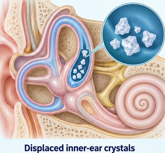
Although BPPV is not generally life-threatening, it can significantly affect mobility, sleep, daily activities, and quality of life. It may also increase the risk of falls, particularly among older adults. Diagnosis is based primarily on the clinical history and positional tests. Appropriate repositioning manoeuvres can provide rapid symptom relief without the need for extensive investigations or prolonged medication.
Case Report
A 58-year-old woman presented to the outpatient department with recurrent episodes of dizziness for five days. She described the dizziness as a sudden spinning sensation that occurred whenever she turned onto her right side while lying in bed. Similar episodes occurred when she got out of bed in the morning, bent forward to pick up an object, or looked upward.
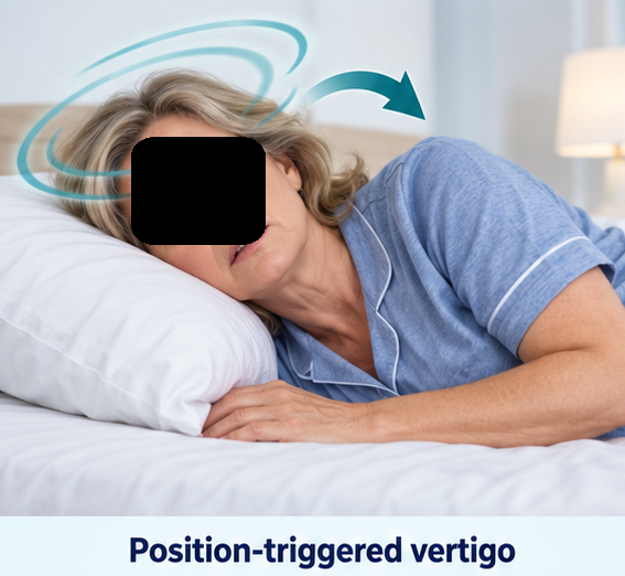
Each episode began suddenly and lasted approximately 20–30 seconds. The symptoms resolved spontaneously when she kept her head still. She experienced mild nausea during some episodes but did not report vomiting. The attacks were more frequent during the morning and had caused significant anxiety. She had started sleeping in a semi-upright position because she feared provoking another episode.
There was no history of continuous dizziness, loss of consciousness, falls, seizures, headache, photophobia, double vision, difficulty speaking, difficulty swallowing, facial weakness, limb weakness, numbness, or impaired coordination. She denied hearing loss, tinnitus, ear pain, ear discharge, or a sensation of fullness in either ear.
The patient had no recent history of fever, upper respiratory tract infection, head trauma, ear surgery, or prolonged bed rest. She had hypertension that was adequately controlled with amlodipine. There was no history of diabetes mellitus, migraine, stroke, cervical spine disease, or a previous vestibular disorder.
On examination, the patient was conscious, oriented, and haemodynamically stable. Her pulse rate was 78 beats per minute, blood pressure was 132/78 mmHg while supine, respiratory rate was 16 breaths per minute, and oxygen saturation was 98% on room air. Her blood pressure remained stable after standing, making orthostatic hypotension unlikely.
Otoscopy showed normal external auditory canals and intact tympanic membranes. A bedside assessment demonstrated no obvious hearing impairment. There was no spontaneous nystagmus while the patient was sitting upright.
Neurological examination revealed normal higher mental functions and cranial nerve findings. Muscle strength, tone, sensation, coordination, and deep tendon reflexes were normal. The finger–nose and heel–shin tests were performed without difficulty. Her gait was cautious but stable, and the Romberg test was negative. No truncal ataxia or focal neurological deficit was observed.
The brief episodes triggered by specific head movements, combined with the absence of auditory and neurological abnormalities, raised a strong clinical suspicion of BPPV.
Investigations
The Dix–Hallpike test was performed to evaluate the posterior semicircular canals. Testing on the left side did not produce vertigo or nystagmus. When the patient’s head was turned 45 degrees to the right and she was rapidly lowered into a supine head-hanging position, she developed intense vertigo after a latency of approximately three seconds.

Examination of her eyes revealed transient upbeating torsional nystagmus, with the upper poles of the eyes rotating toward the right ear. The nystagmus gradually increased and then subsided within approximately 20 seconds. The symptoms and nystagmus became less intense when the test was repeated.
The supine roll test did not produce horizontal nystagmus on either side. These findings were consistent with canalithiasis involving the right posterior semicircular canal.
Routine blood investigations and neuroimaging were not performed because the patient demonstrated typical positional symptoms and characteristic nystagmus without focal neurological abnormalities or other warning signs.
Differential Diagnosis
Vestibular Neuritis
Vestibular neuritis generally causes sudden and continuous vertigo lasting several hours or days. It may be accompanied by nausea, vomiting, gait instability, and spontaneous unidirectional nystagmus. In this patient, the vertigo was brief, occurred only after positional changes, and was absent while she remained still.
Meniere Disease
Meniere disease is associated with recurrent attacks of vertigo that usually last from several minutes to hours. These attacks are commonly accompanied by fluctuating hearing loss, tinnitus, or aural fullness. The absence of auditory symptoms made Meniere disease unlikely.
Vestibular Migraine
Vestibular migraine can cause recurrent vertigo and sensitivity to movement. However, the patient had no previous history of migraine and did not experience headache, photophobia, phonophobia, or visual aura. The characteristic nystagmus observed during the Dix–Hallpike test supported BPPV.
Orthostatic Hypotension
Orthostatic hypotension may cause dizziness or light-headedness after standing. However, it does not usually cause a spinning sensation while turning in bed or produce positional nystagmus. The patient’s blood pressure did not fall significantly after standing.
Central Positional Vertigo
Lesions affecting the cerebellum or brainstem may produce positional vertigo and nystagmus. A central cause was considered unlikely because the nystagmus had a short latency, was transient and fatigable, and followed the expected pattern of posterior-canal BPPV. The patient also had no neurological deficits, severe headache, persistent imbalance, or other central warning signs.
Management and Outcome
A right-sided Epley canalith repositioning manoeuvre was performed during the same visit. The patient was guided through a sequence of head and body positions intended to move the displaced otoconia from the posterior semicircular canal back into the vestibule.
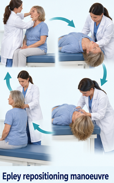
She experienced brief vertigo during the initial positions, but the symptoms gradually settled as the manoeuvre was completed. After resting for several minutes, the right Dix–Hallpike test was repeated. It no longer produced vertigo or observable nystagmus.
The patient reported substantial improvement and was able to stand and walk without difficulty. Vestibular suppressant medication was not prescribed because her symptoms had responded to the repositioning manoeuvre. She was informed that such medications do not correct the underlying mechanical cause of BPPV and may produce sedation.
She was counselled regarding the possibility of recurrence and advised to move carefully until she felt confident. Measures to reduce fall hazards at home were discussed. She was also advised to seek urgent medical attention if she developed continuous dizziness, severe headache, double vision, difficulty speaking, limb weakness, sensory loss, hearing loss, loss of consciousness, or severe gait instability.
At the one-week follow-up visit, the patient reported complete resolution of the spinning episodes. She was sleeping normally and had resumed her usual daily activities. Repeat Dix–Hallpike testing on the right side remained negative. At six weeks, she continued to remain asymptomatic and had experienced no falls or neurological symptoms.
Discussion
This case demonstrates the characteristic clinical presentation of posterior-canal BPPV. The patient experienced brief, recurrent episodes of vertigo triggered by turning in bed and other changes in head position. The presence of transient torsional upbeating nystagmus during the right Dix–Hallpike test established the affected canal and side.
In posterior-canal BPPV, displaced otoconia move within the semicircular canal after changes in head position. This movement produces abnormal endolymphatic flow and inappropriate stimulation of the vestibular system. The resulting symptoms are typically delayed by a few seconds, last less than one minute, and decrease with repeated testing.
A detailed history is important for distinguishing BPPV from other causes of dizziness. Continuous vertigo, focal neurological abnormalities, severe gait instability, atypical nystagmus, new hearing loss, or severe headache should prompt evaluation for alternative peripheral or central disorders.
Canalith repositioning is the preferred treatment for posterior-canal BPPV. The Epley manoeuvre is simple, inexpensive, and often produces rapid relief. It directly addresses the underlying mechanical abnormality and may be repeated when symptoms persist or recur.
Routine use of vestibular suppressant medication is generally discouraged because it does not reposition displaced otoconia. Routine imaging is also unnecessary when the history and positional findings are typical and no warning signs are present. Persistent or atypical symptoms require reassessment and possible neurological or vestibular investigation.

Conclusion
BPPV should be considered in patients with brief, recurrent vertigo triggered by changes in head position. A characteristic response during the Dix–Hallpike test can establish the diagnosis of posterior-canal BPPV.
In this patient, right posterior-canal BPPV was successfully treated with a single Epley manoeuvre. Early recognition and appropriate repositioning can provide rapid symptom relief while preventing unnecessary investigations and prolonged medication use.
References
- Bhattacharyya N, Gubbels SP, Schwartz SR, et al. Clinical practice guideline: Benign paroxysmal positional vertigo (update). Otolaryngology–Head and Neck Surgery. 2017;156(3 Suppl):S1–S47. https://pubmed.ncbi.nlm.nih.gov/28248609/
- von Brevern M, Bertholon P, Brandt T, et al. Benign paroxysmal positional vertigo: Diagnostic criteria. Journal of Vestibular Research. 2015;25(3–4):105–117. https://pubmed.ncbi.nlm.nih.gov/26756126/
- You P, Instrum R, Parnes L. Benign paroxysmal positional vertigo. Laryngoscope Investigative Otolaryngology. 2019;4(1):116–123. https://pmc.ncbi.nlm.nih.gov/articles/PMC6383320/
- Kim HJ, Park J, Kim JS. Update on benign paroxysmal positional vertigo. Journal of Neurology. 2021;268(5):1995–2000. https://pmc.ncbi.nlm.nih.gov/articles/PMC7684151/
- Parnes LS, Agrawal SK, Atlas J. Diagnosis and management of benign paroxysmal positional vertigo. Canadian Medical Association Journal. 2003;169(7):681–693. https://www.cmaj.ca/content/169/7/681
Read more such content on @ Hidoc Dr | Medical Learning App for Doctors
Recommended News For You
Recommended Articles For You
Featured News
Featured Articles
Featured Events
Featured KOL Videos
1.
New Nanoparticles Can Destroy Undruggable Cancer Proteins
2.
Brain MRI Surveillance Alone Helps Preserve Cognition in Small Cell Lung Cancer
3.
NEET SS Counseling 2023: MCC provides information on DNB SS Medical Oncology seats available at ESIC Medical College and Hospital Faridabad.
4.
Belzutifan Plus Pembro Approved for Adjuvant RCC
5.
Using MRD Status to Deescalate Multiple Myeloma Therapy
1.
Diagnosis and Treatment of Follicular Thyroid Cancer: A Comprehensive Guide
2.
Obesity as a major risk factor for cancer
3.
Unraveling the Genetic Mystery of Hereditary Spherocytosis
4.
Advanced Pathways in Oncology for Better Care
5.
Essential Updates in Hematology in Daily Practice
1.
International Conference on Oncology, Cancer Prevention and Public Health
2.
International Conference on Cancer Nursing and Rehabilitation Strategies
3.
International Conference on Best Practices in Oncology, Cardiology and Critical Care
4.
International Conference on Innovations in Critical Care for Oncology and Cardiology
5.
International Symposium on Oncology, Cardiology and Critical Care Innovations
1.
Guideline Recommendations of Lorlatinib as First-Line Treatment for ALK+ NSCLC
2.
Breaking Ground: ALK-Positive Lung Cancer Front-Line Management - Part I
3.
Understanding Anemia and Its Common Causes
4.
Targeting Oncologic Drivers with Dacomitinib: Further Discussion on Lung Cancer Treatment
5.
Early Cancer Detection Saves Lives

Address :
Hidoc Dr. Inc. | Delaware C Corp | 1309 Coffeen Ave. Suite 1200, Sheridan WY, 82801
Phone :
+1-415-463-3094
Email :
anishagadia@hidoc.co
© Copyright 2026 Hidoc Dr. Inc.
Terms & Conditions - LLP | Inc. | Privacy Policy - LLP | Inc. | Account Deactivation

To get started please enter your email ID
Welcome to Hidoc Dr.
Join to enhance your clinical skills and gain specialized in-depth medical knowledge