Malignant Melanoma Presenting as a Rapidly Enlarging Pigmented Skin Lesion: A Case Report
OthersPage Navigation
Abstract
Melanoma is an aggressive malignant neoplasm arising from melanocytes, the pigment-producing cells of the skin. Although it accounts for a smaller proportion of skin cancers, melanoma is responsible for a significant majority of skin cancer-related deaths because of its potential for early invasion and metastatic spread. Risk factors include ultraviolet radiation exposure, fair skin, multiple or atypical nevi, family history, and genetic susceptibility. Early recognition of suspicious pigmented lesions and prompt histopathological evaluation are essential for improving outcomes. We report the case of a 52-year-old woman who presented with a rapidly enlarging and changing pigmented lesion on the upper back. Dermoscopic examination revealed asymmetry, irregular borders, multiple colors, and an atypical pigment network. Excisional biopsy confirmed invasive malignant melanoma. The patient underwent wide local excision with appropriate surgical margins and sentinel lymph node biopsy, which showed no evidence of regional nodal metastasis. The patient remains disease-free on follow-up. This case highlights the importance of recognizing evolving pigmented lesions and early surgical management of localized melanoma.
Introduction
Melanoma is a malignant tumor originating from melanocytes and most commonly develops in the skin, although it may also arise in mucosal surfaces, the eye, and other sites. Cutaneous melanoma is associated with ultraviolet radiation exposure and may develop de novo or within a pre-existing melanocytic nevus.
Clinically, melanoma may present as a new pigmented lesion or as a pre-existing mole demonstrating changes in size, shape, color, or surface characteristics. The ABCDE criteria—Asymmetry, Border irregularity, Color variation, Diameter greater than 6 mm, and Evolution—are commonly used to identify suspicious lesions. The "ugly duckling" sign, in which one lesion appears different from the patient's other nevi, may also raise clinical suspicion.
Early-stage melanoma is primarily managed surgically, while advanced disease may require systemic therapies, including immune checkpoint inhibitors and targeted therapies for tumors harboring specific molecular alterations. Early diagnosis and accurate staging remain critical for determining prognosis and guiding treatment.
Case Report
A 52-year-old woman presented to the dermatology clinic with a pigmented lesion on the upper back that had progressively increased in size over approximately eight months. She reported recent changes in the lesion's color and shape, along with occasional itching. There was no history of bleeding, ulceration, or significant pain.
The patient had no previous history of melanoma or other malignancy. She reported intermittent sun exposure but no history of chronic immunosuppression. There was no significant family history of melanoma or other skin cancers.
On physical examination, a solitary, irregularly pigmented lesion measuring approximately 1.7 cm in diameter was identified over the upper back. The lesion demonstrated asymmetry, irregular borders, and areas of dark brown, black, and reddish pigmentation. No clinically palpable regional lymphadenopathy was noted.
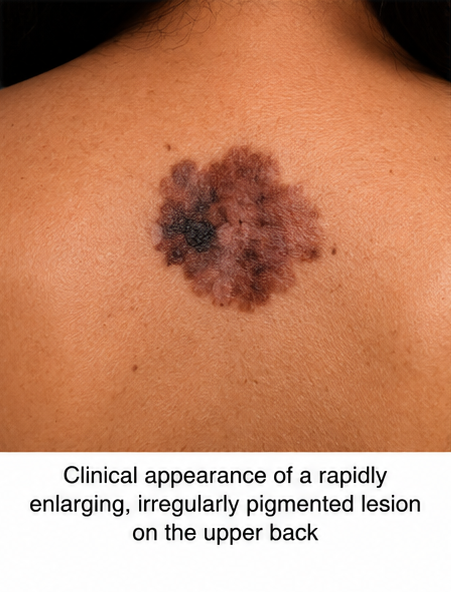
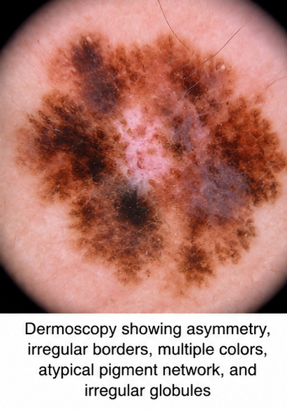
Dermoscopy revealed an asymmetric lesion with an atypical pigment network, irregular globules, multiple pigmentation patterns, and areas of regression. Based on the clinical and dermoscopic findings, melanoma was suspected.
An excisional biopsy of the lesion was performed. Histopathological examination demonstrated atypical melanocytes arranged in nests and single cells with invasion into the dermis. The tumor showed marked cytological atypia and mitotic activity, confirming the diagnosis of invasive cutaneous malignant melanoma.
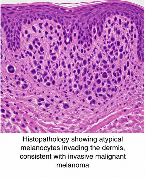
The Breslow tumor thickness was 1.2 mm, with no ulceration identified. The surgical margins of the initial biopsy were not considered adequate for definitive local control. Sentinel lymph node evaluation was therefore planned as part of the staging workup.
Management and Outcome
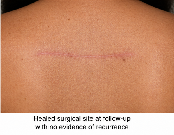
The patient underwent wide local excision of the primary melanoma with appropriate margins based on tumor thickness. Sentinel lymph node biopsy was performed concurrently.
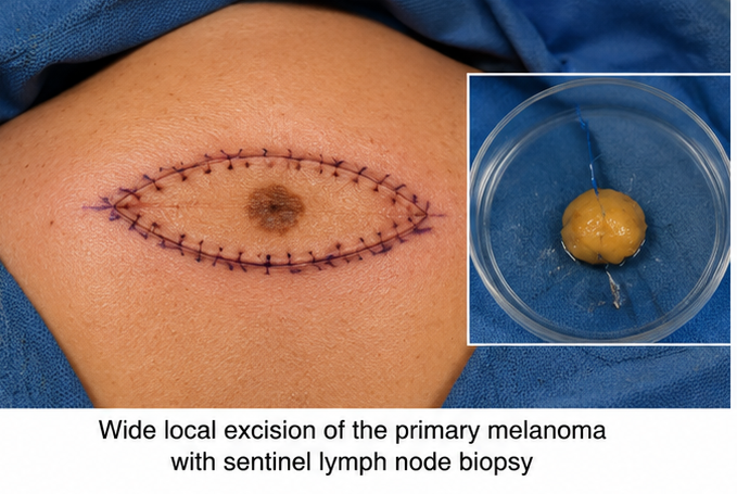
Histopathological examination of the excision specimen demonstrated complete removal of the primary tumor with clear surgical margins. The sentinel lymph node was negative for metastatic melanoma.
Systemic staging investigations, including appropriate imaging, showed no evidence of distant metastatic disease. Based on the clinical, histopathological, and radiological findings, the patient was diagnosed with localized invasive cutaneous melanoma without regional or distant metastasis.
Given the absence of nodal involvement and distant disease, no immediate systemic adjuvant therapy was initiated. The patient was advised regarding regular dermatological surveillance, sun-protection measures, and self-examination of the skin and regional lymph node areas.
Follow-up
One Month
- Surgical wound healed without complications.
- No residual or recurrent pigmentation was identified.
- Regional lymph node examination was unremarkable.
- The patient was counseled regarding sun protection.
Three Months
- No evidence of local recurrence.
- No clinically detectable regional lymphadenopathy.
- Dermatological examination showed no suspicious new lesions.
- The patient remained clinically well.
Six Months
- No evidence of melanoma recurrence.
- No regional or distant metastatic disease identified.
- Continued periodic dermatological surveillance was advised.
- The patient demonstrated good adherence to sun-protection measures.
Discussion
Melanoma is an aggressive skin malignancy with a significant potential for local invasion and metastatic spread. The incidence of melanoma has increased in many populations, with ultraviolet radiation exposure representing an important modifiable risk factor.
The clinical diagnosis of melanoma relies on careful examination of suspicious pigmented lesions. Changes in asymmetry, border, color, diameter, or evolution should prompt further evaluation. Dermoscopy improves diagnostic accuracy by allowing visualization of subsurface structures that may not be apparent on routine examination.
Histopathological examination remains the definitive method for diagnosing melanoma. Important prognostic factors include Breslow thickness, ulceration, mitotic activity, and regional lymph node involvement. Breslow thickness is particularly important because increasing tumor thickness is associated with a higher risk of metastatic disease.
Surgical excision remains the cornerstone of treatment for localized cutaneous melanoma. Wide local excision with appropriate margins provides definitive local control. Sentinel lymph node biopsy is used for staging in selected patients and helps identify occult regional metastasis that may not be clinically apparent.
Patients with regional or metastatic melanoma may require systemic treatment. Modern therapeutic approaches include immune checkpoint inhibitors targeting pathways such as PD-1 and CTLA-4, as well as molecularly targeted therapies for tumors with actionable mutations, including BRAF alterations.
The present case emphasizes the importance of evaluating an evolving pigmented lesion. The patient's lesion demonstrated multiple clinical warning signs, including asymmetry, irregular borders, color variation, and progressive evolution. Early biopsy and appropriate surgical management allowed diagnosis at a localized stage, when the potential for curative treatment was high.
Prognosis
The prognosis of melanoma depends primarily on disease stage at diagnosis and pathological features such as tumor thickness, ulceration, and lymph node involvement. Patients diagnosed with localized melanoma generally have substantially better outcomes than those presenting with regional or distant metastatic disease.
In the present case, the relatively thin primary tumor, absence of ulceration, negative sentinel lymph node, and lack of distant metastasis were favorable prognostic features. Following complete surgical excision, the patient requires long-term clinical surveillance because melanoma survivors remain at risk for local recurrence, metastatic disease, and development of new primary melanomas.
Conclusion
Melanoma should be considered in patients presenting with new or changing pigmented skin lesions, particularly those demonstrating asymmetry, irregular borders, color variation, increasing size, or evolution over time. Clinical examination combined with dermoscopy can help identify lesions requiring biopsy, while histopathological evaluation is essential for definitive diagnosis and staging.
Early detection remains one of the most important factors influencing melanoma outcomes. Wide local excision is the primary treatment for localized disease, while sentinel lymph node biopsy provides valuable prognostic and staging information in appropriately selected patients. Advanced melanoma may require immunotherapy or targeted systemic treatment based on disease stage and molecular characteristics.
Regular skin examination, patient education, avoidance of excessive ultraviolet radiation, and prompt evaluation of suspicious or changing lesions are essential components of melanoma prevention and early detection. Long-term follow-up is important to identify recurrence and detect new primary melanomas at an early stage.
References
- Swetter SM, Tsao H, Bichakjian CK, et al. Guidelines of care for the management of primary cutaneous melanoma. Journal of the American Academy of Dermatology. 2019;80(1):208–250. https://pubmed.ncbi.nlm.nih.gov/30392755/
- Michielin O, van Akkooi ACJ, Ascierto PA, Dummer R, Keilholz U. Cutaneous melanoma: ESMO Clinical Practice Guidelines for diagnosis, treatment and follow-up. Annals of Oncology. 2019;30(12):1884–1901. https://pubmed.ncbi.nlm.nih.gov/31566661/
- Gershenwald JE, Scolyer RA, Hess KR, et al. Melanoma staging: Evidence-based changes in the American Joint Committee on Cancer eighth edition cancer staging manual. CA: A Cancer Journal for Clinicians. 2017. https://pubmed.ncbi.nlm.nih.gov/28094846/
- Coit DG, Thompson JA, Albertini MR, et al. Cutaneous Melanoma, Version 2.2019, NCCN Clinical Practice Guidelines in Oncology. Journal of the National Comprehensive Cancer Network. 2019;17(4):367–402. https://pubmed.ncbi.nlm.nih.gov/30959471/
- Schadendorf D, van Akkooi ACJ, Berking C, et al. Melanoma. The Lancet. 2015;385(9980):971–983. https://pubmed.ncbi.nlm.nih.gov/25454313/
Read more such content on @ Hidoc Dr | Medical Learning App for Doctors
Recommended News For You
Recommended Articles For You
Featured News
Featured Articles
Featured Events
Featured KOL Videos
1.
Year in Review: Non-Small Cell Lung Cancer
2.
Study suggests around 40% of postmenopausal hormone positive breast cancers are linked to excess body fat
3.
The need for more Latinx participants in Alzheimer's trials is urgent.
4.
Why palliative care goes hand in hand with treatment for people with cancer: Q&A
5.
MRD-Guided Azacitidine May Delay Relapse in AML, MDS
1.
Exploring the Benefits of Teclistamab for Treating Advanced Cancer
2.
The Danger of Methemoglobinemia and How to Prevent It
3.
Deciphering FFR: A Comprehensive Guide to Understanding Its Meaning
4.
Red Blood Cell Microparticles: Tiny Warriors Against Bleeding in the Brain
5.
Artificial Intelligence in Oncology: Current Trends, Challenges and Future Outlook
1.
Asian Symposium on Advancement in Hematology and Oncology
2.
Asian Symposium on Advancement in Hematology and Oncology
3.
Asian Symposium on Advancement in Hematology and Oncology
4.
International Cancer Conference
5.
Asian Symposium on Advancement in Hematology and Oncology
1.
Daratumumab, Lenalidomide, and Dexamethasone (DRd) Versus Lenalidomide and Dexamethasone (Rd) in MRD Negativity
2.
Lorlatinib in the Management of 1st line ALK+ mNSCLC (CROWN TRIAL Update)
3.
Thromboprophylaxis In Medical Settings
4.
Post Progression Approaches After First-line Third-Generaion ALK Inhibitors
5.
Molecular Contrast: EGFR Axon 19 vs. Exon 21 Mutations - Part VII

Address :
Hidoc Dr. Inc. | Delaware C Corp | 1309 Coffeen Ave. Suite 1200, Sheridan WY, 82801
Phone :
+1-415-463-3094
Email :
anishagadia@hidoc.co
© Copyright 2026 Hidoc Dr. Inc.
Terms & Conditions - LLP | Inc. | Privacy Policy - LLP | Inc. | Account Deactivation

To get started please enter your email ID
Welcome to Hidoc Dr.
Join to enhance your clinical skills and gain specialized in-depth medical knowledge