Retrocalcaneal Bursitis: Chronic Heel Pain Associated with Inflammation of the Retrocalcaneal Bursa – A Case Report
SurgeryPage Navigation
Abstract
Retrocalcaneal bursitis is an inflammatory disorder involving the retrocalcaneal bursa located between the Achilles tendon and the calcaneus. It commonly results from repetitive mechanical stress, overuse injuries, biomechanical abnormalities, or inflammatory conditions. Patients typically present with posterior heel pain, swelling, tenderness, and impaired mobility. Early diagnosis is essential to prevent chronic pain and functional limitations.
We present the case of a 45-year-old woman with progressive posterior heel pain and swelling for six months. Clinical examination demonstrated tenderness and swelling in the retrocalcaneal region. Ultrasonography and magnetic resonance imaging (MRI) confirmed inflammation and fluid accumulation within the retrocalcaneal bursa with mild Achilles tendon involvement. Conservative treatment including nonsteroidal anti-inflammatory drugs (NSAIDs), physiotherapy, footwear modification, and activity adjustment resulted in significant clinical improvement and restoration of function.
This case highlights the importance of accurate diagnosis and multidisciplinary management of retrocalcaneal bursitis to achieve favorable long-term outcomes.
Introduction
Retrocalcaneal bursitis is a common cause of posterior heel pain caused by inflammation of the bursa situated between the Achilles tendon and the posterior calcaneus. The bursa serves as a cushioning structure that minimizes friction during ankle movement.
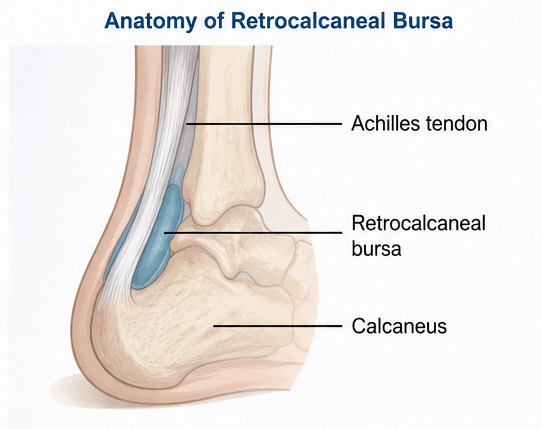
The condition may develop due to repetitive microtrauma, excessive physical activity, tight calf musculature, improper footwear, obesity, or structural abnormalities such as Haglund deformity. It may also occur in association with inflammatory arthropathies including rheumatoid arthritis and spondyloarthritis.
Common risk factors include:
-
Repetitive running and jumping activities
-
Tight Achilles tendon
-
Poorly fitting footwear
-
Haglund deformity
-
Obesity
-
Inflammatory arthritis
Patients usually present with posterior heel pain, swelling, tenderness, and difficulty with walking or sports activities. Although diagnosis is often clinical, imaging studies help confirm the diagnosis and identify associated tendon abnormalities.
Case Report
Patient History
A 45-year-old female presented to the orthopedic clinic with:
-
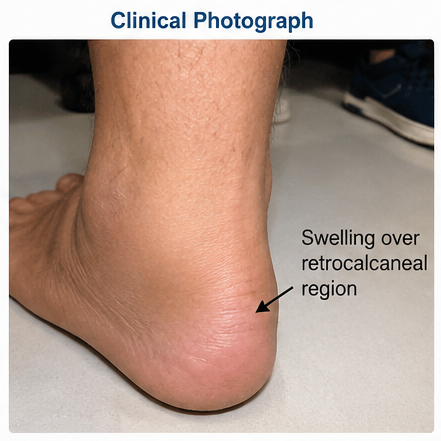
Progressive pain at the back of the right heel for six months
-
Swelling near the Achilles tendon insertion
-
Difficulty walking long distances
-
Pain while climbing stairs
-
Morning stiffness lasting approximately 20 minutes
Symptoms gradually worsened despite rest and over-the-counter analgesics. The patient noted increased discomfort while wearing closed-back footwear and during prolonged standing.
Past medical history included:
-
Obesity (BMI 31 kg/m²)
-
Mild hypertension
There was no history of trauma, inflammatory arthritis, gout, infection, or previous ankle surgery.
Clinical Examination
The patient walked with a mild antalgic gait.
Vital signs were stable:
-
Blood pressure: 136/84 mmHg
-
Pulse rate: 78 beats/minute
-
Temperature: Afebrile
Local examination revealed:
-
Mild swelling over the posterior heel
-
Tenderness anterior to the Achilles tendon insertion
-
Local warmth
-
Pain on compression of the retrocalcaneal space
Ankle dorsiflexion was mildly restricted and plantar flexion produced discomfort. Toe-rise testing reproduced symptoms.
Clinical Evaluation
Differential Diagnosis
The following conditions were considered:
-
Retrocalcaneal bursitis
-
Achilles tendinopathy
-
Haglund syndrome
-
Calcaneal stress fracture
-
Plantar fasciitis
-
Septic bursitis
-
Inflammatory arthropathy
The location of pain and swelling strongly suggested retrocalcaneal bursitis.
Investigations
Laboratory Evaluation
Laboratory studies showed:
-
Hemoglobin: 13.2 g/dL
-
White blood cell count: 7,400/mm³
-
ESR: 18 mm/hr
-
CRP: 4.2 mg/L
Autoimmune screening including rheumatoid factor, anti-CCP antibodies, and antinuclear antibodies was negative.
Plain Radiography
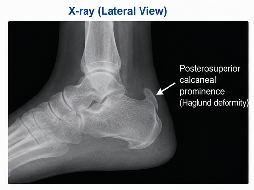
Lateral ankle radiographs demonstrated:
-
Mild posterosuperior calcaneal prominence
-
No fracture
-
Preserved joint spaces
These findings suggested a possible Haglund deformity contributing to mechanical irritation.
Ultrasonography
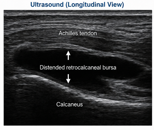
Musculoskeletal ultrasound revealed:
-
Distended retrocalcaneal bursa
-
Fluid accumulation within the bursa
-
Synovial thickening
-
Increased Doppler vascularity
-
Mild Achilles tendon insertional inflammation
These findings were consistent with active bursitis.
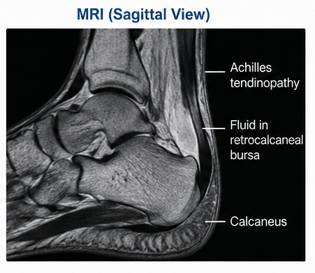
Magnetic Resonance Imaging
MRI demonstrated:
-
Fluid-filled retrocalcaneal bursa
-
Thickened bursal wall
-
Peribursal edema
-
Mild insertional Achilles tendinopathy
-
Reactive soft tissue inflammation
No tendon rupture, osteomyelitis, or neoplastic lesion was identified.
Diagnosis
Based on clinical and imaging findings, the patient was diagnosed with: Chronic Retrocalcaneal Bursitis Associated with Mechanical Irritation and Mild Achilles Tendinopathy
Management and Outcome
Initial Treatment
Conservative treatment was initiated, including:
-
Oral NSAIDs
-
Activity modification
-
Ice application
-
Heel lifts
-
Footwear modification
-
Physiotherapy
-
Achilles tendon stretching exercises
-
Weight reduction counseling
The patient was advised to avoid prolonged walking, running, and high-impact activities.
Physiotherapy Program
The rehabilitation program focused on:
-
Gastrocnemius stretching
-
Soleus stretching
-
Achilles tendon flexibility exercises
-
Calf strengthening
-
Proprioceptive training
Clinical Course
After six weeks of treatment:
-
Pain decreased substantially
-
Swelling improved
-
Walking tolerance increased
-
Ankle mobility improved
No complications occurred during treatment.
Follow-Up
At 6 Weeks
The patient reported:
-
Approximately 50% reduction in pain
-
Improved gait
-
Reduced tenderness
At 3 Months
Further improvement was observed:
-
Near-complete resolution of swelling
-
Restoration of functional mobility
-
Pain score reduction from 8/10 to 2/10
At 12 Months
Long-term follow-up demonstrated:
-
Complete return to daily activities
-
No recurrence of severe symptoms
-
Sustained functional improvement
-
Continued adherence to stretching exercises
Discussion
Retrocalcaneal bursitis develops when repetitive friction and mechanical loading trigger inflammation within the bursa between the Achilles tendon and calcaneus. The resulting inflammatory response leads to fluid accumulation, synovial thickening, edema, and pain.
Biomechanical abnormalities such as a tight Achilles tendon or Haglund deformity increase pressure within the retrocalcaneal space and predispose patients to chronic inflammation.
Clinical Features
Common symptoms include:
-
Posterior heel pain
-
Swelling near the Achilles tendon insertion
-
Tenderness on palpation
-
Difficulty walking
-
Pain during exercise
-
Morning stiffness
Differential Diagnosis
Achilles Tendinopathy: Usually presents with tendon thickening and tenderness directly over the tendon.
Haglund Syndrome: Characterized by a posterosuperior calcaneal prominence causing mechanical irritation.
Plantar Fasciitis: Produces pain on the plantar aspect of the heel rather than posteriorly.
Calcaneal Stress Fracture: Associated with persistent pain and radiographic evidence of bone injury.
Septic Bursitis: Often accompanied by fever, erythema, elevated inflammatory markers, and infection.
Treatment Options
Most patients respond to conservative management, including:
-
NSAIDs
-
Rest and activity modification
-
Ice therapy
-
Orthotic support
-
Heel lifts
-
Physiotherapy
-
Weight reduction
Persistent cases may require ultrasound-guided corticosteroid injections, although caution is necessary because of the potential risk of Achilles tendon rupture.
Surgical intervention, including bursectomy or correction of Haglund deformity, is reserved for refractory cases.
Complications
Potential complications include:
-
Chronic heel pain
-
Recurrent bursitis
-
Reduced mobility
-
Achilles tendinopathy
-
Tendon degeneration
-
Functional impairment
Early treatment significantly reduces these risks.
Prognosis
The prognosis of retrocalcaneal bursitis is generally excellent when diagnosed early and treated appropriately. Outcomes depend on symptom duration, underlying biomechanical abnormalities, treatment compliance, and associated Achilles tendon pathology. Most patients experience substantial symptom relief and return to normal activities with conservative management.
Conclusion
Retrocalcaneal bursitis is an important cause of posterior heel pain that can significantly impair mobility and quality of life. This case illustrates the role of mechanical stress and calcaneal prominence in the development of bursal inflammation. Clinical assessment combined with ultrasonography and MRI facilitates accurate diagnosis and exclusion of other causes of heel pain. Early conservative treatment, including anti-inflammatory therapy, physiotherapy, footwear modification, and activity adjustment, can achieve excellent clinical outcomes and prevent long-term disability.
References
-
Stephens MM. Haglund's deformity and retrocalcaneal bursitis. Orthopedic Clinics of North America. 1994;25(1):41-46. https://pubmed.ncbi.nlm.nih.gov/8291403/
-
Benjamin M, McGonagle D. The anatomical basis for disease localization in spondyloarthropathy. Journal of Anatomy. 2001;199(5):503-526. https://pubmed.ncbi.nlm.nih.gov/11760883/
-
Maffulli N, Oliva F, Del Buono A. Achilles tendon disorders and related pathologies. Clinics in Sports Medicine. 2015;34(4):607-624. https://pubmed.ncbi.nlm.nih.gov/26321459/
-
American Academy of Orthopaedic Surgeons (AAOS). Heel Pain and Achilles Tendon Disorders. https://orthoinfo.aaos.org
-
Miller TT. Imaging of heel pain. Radiologic Clinics of North America. 2008;46(6):1001-1016. https://pubmed.ncbi.nlm.nih.gov/18922388/
-
The British Orthopaedic Foot & Ankle Society. Retrocalcaneal Bursitis Information. https://www.bofas.org.uk
Read more such content on @ Hidoc Dr | Medical Learning App for Doctors
Recommended News For You
Recommended Articles For You
Featured News
Featured Articles
Featured Events
Featured KOL Videos
1.
The Boom in ADHD Coaching Has Few Rules
2.
Cancer patients with HIV have demonstrated safety with immune checkpoint inhibitors.
3.
The FDA approves Enhertu for HER2-positive cancers, regardless of tumor type.
4.
Medicaid expansion tied to smaller declines in early-stage cancer detection
5.
Specialist Referrals Are Critical for Functional High-Risk Multiple Myeloma
1.
Mastering Prostate Cancer Care: CME, Certification, Digital Tools, and Evidence-Based Strategies
2.
Pembrolizumab Plus Lenvatinib in EBV-Associated Advanced Intrahepatic Cholangiocarcinoma: Case Study
3.
Targeting the Tumor Microenvironment: Next-Generation Strategies for Immuno-Oncology Success
4.
Unpacking the Importance of White Blood Cells (WBCs) in the Body
5.
Essential Updates in Hematology in Daily Practice

Address :
Hidoc Dr. Inc. | Delaware C Corp | 1309 Coffeen Ave. Suite 1200, Sheridan WY, 82801
Phone :
+1-415-463-3094
Email :
anishagadia@hidoc.co
© Copyright 2026 Hidoc Dr. Inc.
Terms & Conditions - LLP | Inc. | Privacy Policy - LLP | Inc. | Account Deactivation

To get started please enter your email ID
Welcome to Hidoc Dr.
Join to enhance your clinical skills and gain specialized in-depth medical knowledge