Nephrotic Syndrome Presenting with Generalized Edema and Massive Proteinuria: A Case Report
OthersPage Navigation
Abstract
Nephrotic syndrome is a clinical disorder characterized by massive proteinuria, hypoalbuminemia, generalized edema, hyperlipidemia, and lipiduria resulting from increased glomerular permeability. It represents a common manifestation of various primary and secondary glomerular diseases and can affect both children and adults. Early recognition and timely management are essential to prevent complications such as acute kidney injury, thromboembolism, severe infections, and progression to chronic kidney disease. We report the case of a 34-year-old man who presented with progressive facial puffiness, bilateral lower-limb swelling, frothy urine, and weight gain over two weeks. Clinical examination demonstrated generalized pitting edema with normal blood pressure. Laboratory investigations revealed nephrotic-range proteinuria, severe hypoalbuminemia, hypercholesterolemia, and preserved renal function. Renal biopsy established the diagnosis of minimal change disease. The patient was managed with corticosteroids, angiotensin-converting enzyme inhibitors, diuretics, dietary sodium restriction, and supportive therapy, resulting in complete clinical remission. This case emphasizes the importance of prompt diagnosis, renal biopsy when indicated, and individualized treatment to preserve renal function and reduce long-term complications.
Introduction
Nephrotic syndrome is a glomerular disorder characterized by urinary protein excretion exceeding 3.5 g/day, hypoalbuminemia, generalized edema, hyperlipidemia, and lipiduria. The syndrome results from damage to the glomerular filtration barrier, leading to excessive urinary protein loss and reduced plasma oncotic pressure. Primary causes include minimal change disease, focal segmental glomerulosclerosis, and membranous nephropathy, whereas secondary causes include diabetes mellitus, systemic lupus erythematosus, infections, malignancies, and certain medications.
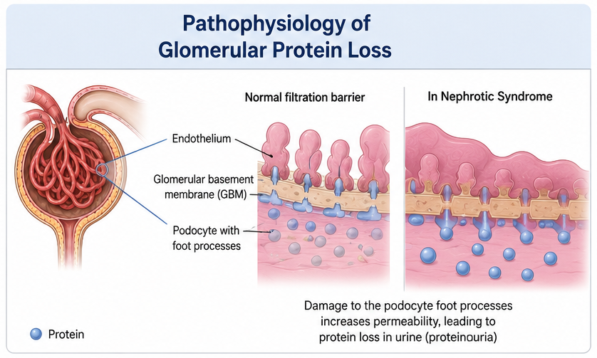
Patients commonly present with progressive swelling of the face and lower limbs, frothy urine, fatigue, weight gain, and reduced exercise tolerance. Because nephrotic syndrome predisposes patients to thromboembolic events, severe infections, dyslipidemia, and progressive renal dysfunction, early diagnosis and comprehensive management are essential. Diagnosis relies on clinical findings supported by laboratory evaluation, urine protein quantification, renal imaging, and kidney biopsy when appropriate. Treatment depends on the underlying etiology and includes immunosuppressive therapy for selected primary glomerular diseases, together with supportive measures aimed at controlling edema, proteinuria, hypertension, and cardiovascular risk factors.
Case Report
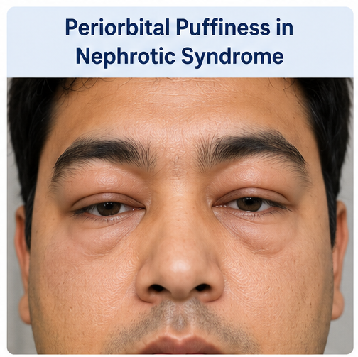
A 34-year-old man presented to the nephrology outpatient department with progressive swelling around the eyes and both lower limbs for approximately two weeks.
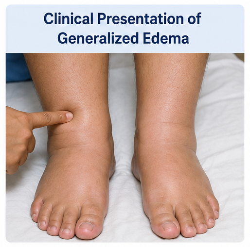
He also complained of frothy urine, generalized weakness, reduced appetite, and an unintentional weight gain of nearly five kilograms during the preceding fortnight.

There was no history of fever, hematuria, dysuria, breathlessness, joint pain, skin rash, recent sore throat, diabetes mellitus, hypertension, or previous renal disease.
On physical examination, the patient was afebrile with stable vital signs. Blood pressure was 126/78 mmHg, pulse rate was 82 beats per minute, and oxygen saturation was 99% on room air. Periorbital puffiness, bilateral pitting pedal edema extending to the knees, and mild abdominal distension consistent with ascites were noted. Cardiovascular, respiratory, and neurological examinations were otherwise unremarkable.
Laboratory investigations demonstrated nephrotic-range proteinuria of 7.2 g/24 hours, serum albumin of 2.1 g/dL, total serum protein of 4.8 g/dL, serum cholesterol of 328 mg/dL, triglycerides of 286 mg/dL, and preserved renal function with serum creatinine of 0.9 mg/dL. Urinalysis revealed 4+ proteinuria with oval fat bodies and lipid casts but no significant hematuria. Complete blood count, liver function tests, blood glucose, complement levels, and thyroid profile were within normal limits. Serological investigations for hepatitis B, hepatitis C, HIV, antinuclear antibody, and anti-double stranded DNA antibodies were negative.
Renal ultrasonography demonstrated normal-sized kidneys with preserved corticomedullary differentiation and no evidence of obstruction. Owing to the absence of secondary causes, a percutaneous renal biopsy was performed.
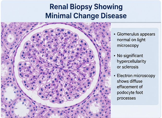
Light microscopy showed minimal glomerular abnormalities, while electron microscopy demonstrated diffuse podocyte foot process effacement without immune complex deposition, confirming the diagnosis of minimal change disease presenting as nephrotic syndrome.
Management and Outcome
The patient was admitted for initiation of therapy and close monitoring. Oral prednisolone was started at 1 mg/kg/day as first-line treatment for minimal change disease. Furosemide was administered to reduce edema, while ramipril was initiated to decrease intraglomerular pressure and reduce proteinuria. Atorvastatin was prescribed for severe hyperlipidemia, and prophylactic proton pump inhibitor therapy was provided during corticosteroid treatment.
Dietary counseling emphasized sodium restriction, adequate protein intake, avoidance of excessive fluid consumption, and regular weight monitoring. The patient was advised regarding medication adherence, infection prevention, and early reporting of symptoms suggestive of thromboembolic complications.
Within three weeks, pedal edema and facial swelling reduced markedly, urinary protein excretion declined substantially, and serum albumin gradually improved. At eight weeks, urinary protein became negative on dipstick testing, serum albumin normalized to 3.9 g/dL, lipid profile improved significantly, and complete clinical remission was achieved without treatment-related adverse effects.
Follow-up
One Month
- Significant reduction in generalized edema.
- Marked decrease in urinary protein excretion.
- Improved appetite and energy levels.
- Gradual normalization of serum albumin.
Three Months
- Complete remission of proteinuria.
- Resolution of pedal edema and periorbital swelling.
- Improved lipid profile.
- Normal renal function maintained.
Six Months
The patient remained asymptomatic with normal renal function and no recurrence of edema or proteinuria. Corticosteroids were gradually tapered, and regular nephrology follow-up was continued for early detection of relapse.
Discussion
Nephrotic syndrome develops following disruption of the glomerular filtration barrier, resulting in excessive urinary protein loss and subsequent hypoalbuminemia. Reduced plasma oncotic pressure promotes movement of fluid into the interstitial space, leading to generalized edema. Simultaneously, hepatic synthesis of lipoproteins increases, producing hyperlipidemia that is characteristic of the syndrome.
Minimal change disease is the leading cause of nephrotic syndrome in children but also accounts for a significant proportion of adult cases. Electron microscopy demonstrating diffuse podocyte foot process effacement remains the hallmark pathological feature. Adults often require renal biopsy to exclude other glomerular disorders before initiating immunosuppressive therapy.
Management consists of disease-specific treatment together with supportive care. Corticosteroids remain the cornerstone of therapy for minimal change disease and achieve remission in most patients. Angiotensin-converting enzyme inhibitors or angiotensin receptor blockers reduce proteinuria and provide long-term renal protection. Diuretics effectively control edema, whereas statins are beneficial for persistent dyslipidemia. Patients should also receive counseling regarding dietary sodium restriction, vaccination where appropriate, and prompt treatment of infections because urinary loss of immunoglobulins increases susceptibility to bacterial infections.
Untreated nephrotic syndrome may lead to serious complications including acute kidney injury, venous thromboembolism, spontaneous bacterial peritonitis, severe dyslipidemia, accelerated cardiovascular disease, and progressive chronic kidney disease. Regular follow-up with assessment of proteinuria, serum albumin, renal function, and blood pressure is essential for detecting relapse and preventing long-term morbidity.
Prognosis
The prognosis depends on the underlying etiology, treatment response, and development of complications. Minimal change disease generally has an excellent prognosis, with most patients achieving complete remission following corticosteroid therapy. However, relapses may occur and require prolonged follow-up. Early diagnosis, adherence to treatment, and regular nephrological monitoring significantly improve long-term renal outcomes and quality of life.
Conclusion
Nephrotic syndrome is a potentially reversible glomerular disorder when recognized and treated promptly. Progressive edema, frothy urine, and heavy proteinuria should immediately raise clinical suspicion. Comprehensive evaluation including laboratory investigations and renal biopsy, when indicated, enables accurate diagnosis and targeted therapy. Early corticosteroid treatment in minimal change disease, together with supportive measures such as diuretics, renin-angiotensin system blockade, lipid-lowering therapy, dietary modification, and close follow-up, can achieve sustained remission, preserve renal function, and prevent life-threatening complications.
References
- Kodner, C. (2016). Diagnosis and management of nephrotic syndrome in adults. American Family Physician, 93(6), 479–485. https://www.aafp.org/pubs/afp/issues/2016/0315/p479.html
- Kidney Disease: Improving Global Outcomes (KDIGO) Glomerular Diseases Work Group. (2021). KDIGO 2021 clinical practice guideline for the management of glomerular diseases. Kidney International, 100(4S), S1–S276. https://kdigo.org/guidelines/glomerular-diseases/
- Rovin, B. H., Adler, S. G., Barratt, J., et al. (2021). Executive summary of the KDIGO 2021 guideline for the management of glomerular diseases. Kidney International, 100(4), 753–779. https://www.kidney-international.org/article/S0085-2538(21)00832-8/fulltext
- Hull, R. P., & Goldsmith, D. J. A. (2008). Nephrotic syndrome in adults. BMJ, 336(7654), 1185–1189. https://www.bmj.com/content/336/7654/1185
- Glassock, R. J. (2007). Prophylactic anticoagulation in nephrotic syndrome: A clinical conundrum. Journal of the American Society of Nephrology, 18(8), 2221–2225. https://journals.lww.com/jasn/fulltext/2007/08000/prophylactic_anticoagulation_in_nephrotic.3.aspx
Read more such content on @ Hidoc Dr | Medical Learning App for Doctors
Recommended News For You
Recommended Articles For You
Featured News
Featured Articles
Featured Events
Featured KOL Videos
1.
Year in Review: Non-Small Cell Lung Cancer
2.
Study suggests around 40% of postmenopausal hormone positive breast cancers are linked to excess body fat
3.
The need for more Latinx participants in Alzheimer's trials is urgent.
4.
Why palliative care goes hand in hand with treatment for people with cancer: Q&A
5.
MRD-Guided Azacitidine May Delay Relapse in AML, MDS
1.
Exploring the Benefits of Teclistamab for Treating Advanced Cancer
2.
The Danger of Methemoglobinemia and How to Prevent It
3.
Deciphering FFR: A Comprehensive Guide to Understanding Its Meaning
4.
Red Blood Cell Microparticles: Tiny Warriors Against Bleeding in the Brain
5.
Artificial Intelligence in Oncology: Current Trends, Challenges and Future Outlook
1.
Asian Symposium on Advancement in Hematology and Oncology
2.
Asian Symposium on Advancement in Hematology and Oncology
3.
Asian Symposium on Advancement in Hematology and Oncology
4.
International Cancer Conference
5.
Asian Symposium on Advancement in Hematology and Oncology
1.
Daratumumab, Lenalidomide, and Dexamethasone (DRd) Versus Lenalidomide and Dexamethasone (Rd) in MRD Negativity
2.
Lorlatinib in the Management of 1st line ALK+ mNSCLC (CROWN TRIAL Update)
3.
Thromboprophylaxis In Medical Settings
4.
Post Progression Approaches After First-line Third-Generaion ALK Inhibitors
5.
Molecular Contrast: EGFR Axon 19 vs. Exon 21 Mutations - Part VII

Address :
Hidoc Dr. Inc. | Delaware C Corp | 1309 Coffeen Ave. Suite 1200, Sheridan WY, 82801
Phone :
+1-415-463-3094
Email :
anishagadia@hidoc.co
© Copyright 2026 Hidoc Dr. Inc.
Terms & Conditions - LLP | Inc. | Privacy Policy - LLP | Inc. | Account Deactivation

To get started please enter your email ID
Welcome to Hidoc Dr.
Join to enhance your clinical skills and gain specialized in-depth medical knowledge