Lobar Pneumonia Requiring Hospitalization And Intravenous Antibiotic Therapy
OthersPage Navigation
Introduction
Lobar pneumonia is an acute infection of the lung characterized by consolidation involving a substantial portion of a pulmonary lobe. It is most commonly caused by bacterial pathogens, although viral and atypical organisms may produce similar clinical and radiological presentations. Streptococcus pneumoniae remains an important cause of community-acquired pneumonia.

Clinical manifestations commonly include fever, productive cough, pleuritic chest pain, dyspnea, fatigue, and constitutional symptoms. Examination may reveal tachypnea, hypoxemia, bronchial breath sounds, crackles, increased vocal resonance, and localized dullness to percussion.
Diagnosis is based on the clinical presentation together with imaging demonstrating a new pulmonary infiltrate or consolidation. Chest radiography remains an important initial investigation, while computed tomography may be useful when the diagnosis is uncertain or complications such as pleural effusion, abscess, or necrosis are suspected.
Severity assessment is essential because pneumonia may progress to respiratory failure, sepsis, septic shock, or multiorgan dysfunction. Treatment includes appropriate antimicrobial therapy, oxygen and supportive care when required, and monitoring for clinical deterioration.
Case Report
A 62-year-old man presented to the emergency department with a four-day history of high-grade fever, productive cough, right-sided pleuritic chest pain, and progressively worsening shortness of breath. The cough was associated with yellowish sputum. He also reported generalized weakness, reduced appetite, and chills.
His symptoms had initially been managed at home with paracetamol and oral fluids, but the fever persisted and his breathlessness increased over the preceding 24 hours.
The patient had a history of hypertension and type 2 diabetes mellitus controlled with oral medication. He had no previous history of chronic respiratory disease, tuberculosis, bronchiectasis, malignancy, or recent hospitalization. He was a former smoker with a 15-pack-year history and had stopped smoking eight years earlier.
On examination, the patient appeared acutely ill and mildly dehydrated. His temperature was 39.2°C, pulse rate was 112 beats/minute, blood pressure was 102/66 mmHg, respiratory rate was 30 breaths/minute, and oxygen saturation was 88% on room air.
Respiratory examination revealed reduced air entry over the right lower chest with bronchial breath sounds, coarse crackles, and increased vocal resonance over the right lower lung field. There was no clinical evidence of pneumothorax or significant pleural effusion.
The initial clinical impression was community-acquired pneumonia with right lower-lobe consolidation and hypoxemic respiratory compromise.
Investigations
Laboratory investigations showed a hemoglobin level of 12.8 g/dL, leukocyte count of 17,600 cells/mm³ with marked neutrophilia, and platelet count of 286,000 cells/mm³.
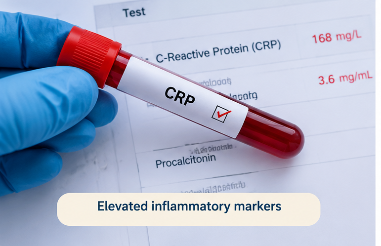
Serum sodium was 133 mmol/L, potassium was 3.8 mmol/L, bicarbonate was 21 mmol/L, blood urea nitrogen was 28 mg/dL, and serum creatinine was 1.2 mg/dL. C-reactive protein was markedly elevated at 168 mg/L, while procalcitonin was 3.6 ng/mL.
Arterial blood gas analysis showed a pH of 7.44, partial pressure of oxygen of 58 mmHg, and partial pressure of carbon dioxide of 34 mmHg, consistent with hypoxemia and mild respiratory alkalosis.
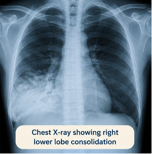
Chest radiography demonstrated dense homogeneous air-space consolidation involving the right lower lobe with visible air bronchograms. There was no significant pleural effusion or pneumothorax.
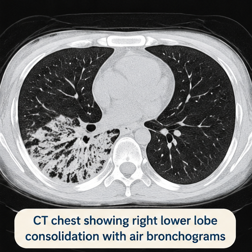
Because of the severity of symptoms and concern regarding possible complications, contrast-enhanced computed tomography of the chest was performed. It demonstrated extensive right lower-lobe consolidation with air bronchograms and surrounding inflammatory ground-glass changes. No lung abscess, cavitation, pulmonary embolism, or significant pleural collection was identified.
Blood cultures were obtained before antibiotic administration. Sputum was sent for Gram staining and bacterial culture. Respiratory viral testing for influenza and SARS-CoV-2 was negative.
The findings supported a diagnosis of severe community-acquired right lower-lobe lobar pneumonia.
Differential Diagnosis
Pulmonary Tuberculosis
Tuberculosis may produce cough, fever, constitutional symptoms, and pulmonary infiltrates. However, the acute four-day presentation, high-grade fever, neutrophilic leukocytosis, lobar consolidation, and absence of cavitation or chronic constitutional symptoms made acute bacterial pneumonia more likely.
Pulmonary Embolism
Pulmonary embolism may present with dyspnea and pleuritic chest pain. However, the prominent fever, purulent sputum, marked inflammatory response, and lobar consolidation with air bronchograms favored pneumonia. CT pulmonary angiography showed no pulmonary embolism.
Acute Bronchitis
Acute bronchitis may cause cough and sputum production but generally does not produce focal lobar consolidation or significant hypoxemia. The radiological findings therefore argued against uncomplicated bronchitis.
Cardiogenic Pulmonary Edema
Pulmonary edema can cause dyspnea and bilateral pulmonary opacities. However, the unilateral lobar consolidation, high fever, purulent sputum, and inflammatory laboratory findings were inconsistent with isolated cardiogenic pulmonary edema.
Lung Abscess
A lung abscess may complicate bacterial pneumonia and produce fever and productive cough. CT imaging showed consolidation without cavitation or a fluid-filled cavity, making established lung abscess unlikely.
Management and Outcome
The patient was admitted to the hospital because of hypoxemia, tachypnea, systemic inflammatory response, and underlying diabetes mellitus.
Supplemental oxygen was administered through a nasal cannula, maintaining oxygen saturation above 92%. Intravenous balanced crystalloid fluids were given cautiously because of the risk of pulmonary fluid accumulation. Antipyretics and appropriate analgesia were administered for fever and pleuritic chest pain.
Empirical intravenous antibacterial therapy was initiated promptly with ceftriaxone and azithromycin. The antibiotic regimen was subsequently reviewed according to the patient's clinical response and microbiological findings.
Blood culture grew Streptococcus pneumoniae, which was susceptible to the administered antimicrobial therapy. Sputum culture also supported pneumococcal infection.
The patient's respiratory status and inflammatory markers were monitored closely. There was no indication for mechanical ventilation, vasopressor therapy, or intensive care admission.

During the first 48 hours, the fever gradually subsided and the respiratory rate decreased. Oxygen requirements progressively declined. The serum lactate remained within normal limits, and renal function was preserved.
By the third hospital day, oxygen saturation was 94% on minimal supplemental oxygen. His cough persisted but sputum production had decreased substantially. Chest examination showed improving air entry with residual crackles over the right lower zone.
Intravenous antibiotics were continued initially and subsequently converted to oral therapy after clinical stabilization. Oral intake was gradually increased, and the patient was encouraged to mobilize and perform appropriate pulmonary hygiene.
The patient was discharged after seven days of hospitalization in stable condition with instructions to complete the prescribed antimicrobial course and attend follow-up.
At the four-week follow-up, he reported resolution of fever, dyspnea, and pleuritic chest pain. His cough had markedly improved. A follow-up chest radiograph demonstrated substantial resolution of the right lower-lobe consolidation.
Discussion
Lobar pneumonia represents a pattern of pulmonary infection in which inflammatory exudation fills the alveolar spaces, producing consolidation of a defined anatomical region. The clinical presentation may vary according to the causative organism, host factors, disease severity, and presence of complications.
In this patient, the combination of acute high-grade fever, productive cough, pleuritic chest pain, hypoxemia, neutrophilic leukocytosis, elevated inflammatory markers, and focal bronchial respiratory signs strongly supported pneumonia. The chest radiograph demonstrated characteristic right lower-lobe air-space consolidation with air bronchograms.
Chest radiography remains an important diagnostic investigation in suspected community-acquired pneumonia. Lung ultrasound can also support diagnosis, particularly in patients who are critically ill or when rapid bedside assessment is required. CT provides greater anatomical detail and can identify complications or alternative diagnoses when the initial assessment is inconclusive.
Severity assessment is important when determining the appropriate site of care. Clinical parameters including respiratory rate, blood pressure, oxygen saturation, mental status, age, and comorbidities contribute to risk assessment. Scores such as CURB-65 or CRB65 can complement, but should not replace, clinical judgment.
Microbiological investigations are not required routinely in every patient with mild community-acquired pneumonia. They become more relevant in moderate- or high-severity disease, suspected sepsis, treatment failure, or when identification of a pathogen may allow antimicrobial narrowing.
Prompt antimicrobial treatment is central to management of bacterial pneumonia. Empirical treatment should consider disease severity, relevant comorbidities, recent antibiotic exposure, local antimicrobial resistance patterns, and the possibility of atypical pathogens. Once microbiological results become available, therapy should be reassessed and narrowed when appropriate.
Supportive management is equally important. Hypoxemia should be corrected with supplemental oxygen, while fluid administration should be individualized according to hemodynamic status and risk of fluid overload. Patients should be monitored for respiratory deterioration, sepsis, pleural complications, and treatment failure.
Potential complications include parapneumonic effusion, empyema, lung abscess, necrotizing pneumonia, respiratory failure, bacteremia, septic shock, and multiorgan dysfunction.
Radiological resolution may lag behind clinical improvement. Therefore, persistent radiographic opacity shortly after treatment does not necessarily indicate ongoing infection when the patient is clinically improving. Follow-up imaging should be guided by the clinical course and relevant risk factors.
In this patient, early recognition, timely antimicrobial therapy, oxygen supplementation, microbiological confirmation, and close monitoring resulted in progressive clinical and radiological recovery without respiratory failure or other major complications.
Conclusion
Lobar pneumonia can produce significant systemic illness and hypoxemic respiratory compromise, particularly in older adults and patients with underlying comorbidities.
In this patient, acute fever, productive cough, pleuritic chest pain, focal respiratory findings, inflammatory laboratory abnormalities, and right lower-lobe consolidation established the diagnosis of community-acquired lobar pneumonia.
Early severity assessment, prompt antimicrobial treatment, appropriate microbiological evaluation, oxygen therapy, and close monitoring were central to management.
Recognition of clinical deterioration and complications remains essential because community-acquired pneumonia may progress rapidly to respiratory failure, sepsis, or other serious complications.
References
- Metlay JP, Waterer GW, Long AC, et al. Diagnosis and Treatment of Adults with Community-acquired Pneumonia. An Official Clinical Practice Guideline of the American Thoracic Society and Infectious Diseases Society of America. Am J Respir Crit Care Med. 2019;200(7):e45–e67. https://pubmed.ncbi.nlm.nih.gov/31573350/
- National Institute for Health and Care Excellence. Pneumonia: diagnosis and management. NICE Guideline NG250. Published September 2025. https://www.nice.org.uk/guidance/NG250
- Washington L, O'Sullivan-Murphy B, Christensen JD, McAdams HP. Radiographic Imaging of Community-Acquired Pneumonia: A Case-Based Review. Infect Dis Clin North Am. 2024;38(1):19–33. https://pubmed.ncbi.nlm.nih.gov/38280764/
- Metlay JP, Waterer GW, Long AC, et al. Diagnosis and Treatment of Adults with Community-acquired Pneumonia. Am J Respir Crit Care Med. 2019;200(7):e45–e67. https://pmc.ncbi.nlm.nih.gov/articles/PMC6812437/
- National Institute for Health and Care Excellence. Pneumonia: diagnosis and management — Recommendations. NICE Guideline NG250. https://www.nice.org.uk/guidance/NG250/chapter/recommendations
Read more such content on @ Hidoc Dr | Medical Learning App for Doctors
Recommended News For You
Recommended Articles For You
Featured News
Featured Articles
Featured Events
Featured KOL Videos
1.
Regular physical activity before cancer diagnosis may lower progression and death risks
2.
Medication Self-Injection Embraced by Breast Cancer Patients
3.
Financial hardship for cancer survivors due to high-cost immunotherapies, especially for blood cancer patients
4.
FDA Advisors Split on Regimens for Breast, Prostate Cancers
5.
ctDNA-Guided Adjuvant Therapy Cuts Bladder Cancer Costs
1.
RDW-CV vs. MCV: Knowing the Difference Can Help You Better Understand Your Health
2.
Clinical Pathways in Hematology in Clinical Decision-Making
3.
Immune Surveillance Screening for Early Cancer Risk Detection
4.
Unlocking the Mysteries of Lymphoblastic Lymphoma: A Journey Into the Unknown
5.
AI-Driven Digital Pathology in Oncology: From Histology Images to Predictive Treatment Biomarkers
1.
International Conference on Innovations in Critical Care for Oncology and Cardiology
2.
International Conference on Oncology, Cardiology and Critical Care Policy
3.
International Conference on Oncology, Cancer Prevention and Public Health
4.
International Conference on Cancer Nursing and Rehabilitation Strategies
5.
International Conference on Best Practices in Oncology, Cardiology and Critical Care
1.
How Advances in Cancer Research Are Improving Treatment Options
2.
Navigating the Complexities of Ph Negative ALL - Part II
3.
Signs and Symptoms of Leukemia
4.
Updates on Standard V/S High Risk Myeloma Treatment- The Next Part
5.
Virtual Case Study on Elephantiasis of Lower Limb- An Initiative by Hidoc Dr.

Address :
Hidoc Dr. Inc. | Delaware C Corp | 1309 Coffeen Ave. Suite 1200, Sheridan WY, 82801
Phone :
+1-415-463-3094
Email :
anishagadia@hidoc.co
© Copyright 2026 Hidoc Dr. Inc.
Terms & Conditions - LLP | Inc. | Privacy Policy - LLP | Inc. | Account Deactivation

To get started please enter your email ID
Welcome to Hidoc Dr.
Join to enhance your clinical skills and gain specialized in-depth medical knowledge