Electric Burn Presenting With Deep Partial-Thickness Thermal Injury to the Upper Limb: A Case Report
OthersPage Navigation
Abstract
Electrical burns are complex injuries that can result in extensive tissue damage disproportionate to the apparent surface injury. High-voltage and low-voltage electrical exposures may produce cutaneous burns, deep muscle necrosis, neurovascular injury, cardiac arrhythmias, and compartment syndrome. Prompt evaluation is essential because internal tissue destruction may not be immediately evident. We report the case of a 35-year-old male electrician who presented after accidental contact with a live electrical wire while performing maintenance work. He sustained burns to the right hand and forearm, associated with severe pain and swelling. Examination revealed entry and exit wounds with deep partial-thickness burns involving the right upper limb. Cardiac monitoring and laboratory investigations showed no evidence of arrhythmia or rhabdomyolysis. The patient underwent fluid resuscitation, wound care, analgesia, tetanus prophylaxis, and surgical debridement followed by split-thickness skin grafting. The postoperative recovery was uneventful, with satisfactory wound healing and restoration of limb function through rehabilitation. This case highlights the importance of comprehensive evaluation of electrical burn injuries, early identification of occult tissue damage, and multidisciplinary management to optimize functional outcomes.
Introduction
Electrical burns are unique injuries caused by the passage of electrical current through the body, producing both thermal and non-thermal tissue damage. Although electrical burns account for a relatively small proportion of burn injuries, they are associated with significant morbidity because the extent of internal tissue destruction often exceeds the visible skin injury.
The severity of electrical injury depends on multiple factors, including voltage, type of current, duration of contact, tissue resistance, and current pathway through the body. Injuries may range from superficial skin burns to deep muscle necrosis, vascular thrombosis, nerve injury, fractures secondary to violent muscle contractions, compartment syndrome, and life-threatening cardiac arrhythmias.
Patients commonly present with localized burns at the entry and exit sites, severe pain, swelling, and varying degrees of neurological deficits. Cardiac evaluation is particularly important in patients with high-voltage injuries or suspected transthoracic current passage. Laboratory investigations are performed to assess muscle injury, renal function, and rhabdomyolysis.
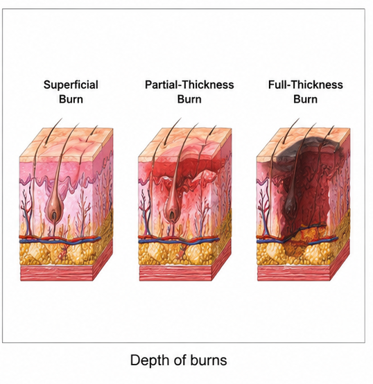
Management requires prompt stabilization, fluid resuscitation when indicated, pain control, wound care, infection prevention, surgical debridement of nonviable tissue, and reconstruction when necessary. Early rehabilitation plays a vital role in preserving limb function and preventing long-term disability.
We report a case of electrical burn involving the upper limb, highlighting the clinical presentation, evaluation, surgical management, and functional recovery.
Case Report
A 35-year-old male electrician presented to the emergency department after sustaining an accidental electrical injury while repairing industrial wiring. The patient reported direct contact between his right hand and an exposed live electrical cable, followed by immediate severe pain and involuntary muscle contraction before being separated from the power source.
He complained of pain, swelling, and blackened areas over the right hand and forearm. There was no history of loss of consciousness, chest pain, palpitations, seizures, or respiratory distress.
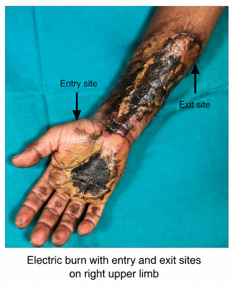
The patient had no significant past medical history and was not taking any regular medications. He had received routine childhood immunizations but could not recall his most recent tetanus vaccination.
On examination, the patient was alert and hemodynamically stable. Burn wounds were present over the right palm and distal forearm, with a smaller wound over the elbow region suggestive of an exit site. The affected areas demonstrated deep partial-thickness burns with surrounding edema and tenderness. Capillary refill and distal pulses were preserved, while sensory and motor function remained intact.
Electrocardiography showed normal sinus rhythm without evidence of arrhythmia. Laboratory investigations, including complete blood count, serum electrolytes, renal function tests, creatine kinase, and urinalysis, were within normal limits. There was no evidence of myoglobinuria or acute kidney injury.
Based on the history of electrical exposure and clinical examination, the patient was diagnosed with an electrical burn involving deep partial-thickness injury of the right upper limb without systemic complications.
Management and Outcome
Initial management included stabilization, intravenous fluids, adequate analgesia, wound irrigation, sterile dressings, tetanus prophylaxis, and continuous cardiac monitoring during the observation period.
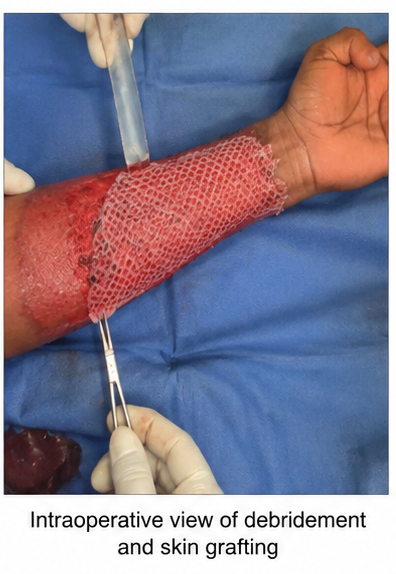
Serial examinations demonstrated progressive demarcation of nonviable tissue. The patient subsequently underwent surgical debridement of devitalized tissue followed by split-thickness skin grafting over the affected areas.
Postoperatively, wound care was continued with regular dressing changes, infection surveillance, and physiotherapy aimed at preserving hand mobility and preventing contracture formation.
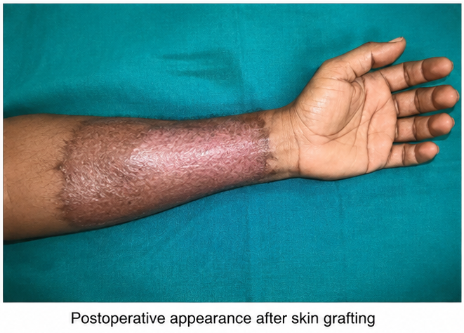
The skin graft healed satisfactorily without significant infection or graft failure. Functional recovery improved progressively with supervised rehabilitation, and the patient regained satisfactory hand movement and grip strength.
Follow-up
One Week
- Pain and swelling had decreased significantly.
- Skin graft remained viable with satisfactory wound healing.
- No evidence of wound infection was observed.
- Gentle physiotherapy was initiated.
One Month
- Complete graft healing was achieved.
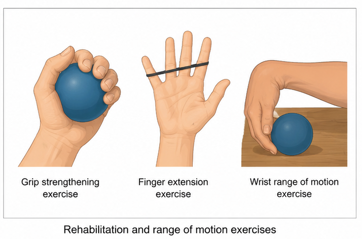
- Range of motion of the wrist and fingers improved.
- Grip strength progressively increased.
- The patient resumed most routine daily activities.
Six Months
- Stable wound healing with mature scar formation.
- No recurrent wound breakdown was observed.
- Functional use of the affected hand remained satisfactory.
- Continued scar management and strengthening exercises were advised.
Discussion
Electrical burns differ substantially from conventional thermal burns because the electrical current travels through tissues of varying resistance, producing deep internal injury despite relatively limited skin involvement. Skin, muscle, nerves, and blood vessels may all be affected depending on the current pathway.
The present case demonstrates a typical occupational electrical injury involving the upper limb. The presence of characteristic entry and exit wounds raised suspicion for deeper tissue injury, warranting careful clinical assessment despite the relatively limited external burn surface.
Initial evaluation of electrical burn patients should prioritize airway, breathing, circulation, and neurological assessment. Cardiac monitoring is recommended for patients with high-voltage exposure, loss of consciousness, documented arrhythmias, or suspected transthoracic current passage. Laboratory evaluation is useful for detecting rhabdomyolysis, electrolyte abnormalities, and renal dysfunction.
Serial clinical examination remains essential because tissue necrosis may continue to evolve during the first several days after injury. Early recognition of compartment syndrome is particularly important, as delayed diagnosis may result in permanent functional impairment.
Management focuses on adequate resuscitation, meticulous wound care, prevention of infection, timely surgical debridement, and reconstruction when indicated. Split-thickness skin grafting remains an effective reconstructive option following removal of nonviable tissue.
Rehabilitation constitutes an integral component of treatment. Early physiotherapy, splinting when appropriate, scar management, and range-of-motion exercises help minimize contracture formation and maximize long-term functional recovery.
This case also highlights the importance of workplace electrical safety measures, including appropriate insulation, personal protective equipment, equipment maintenance, and adherence to occupational safety protocols to reduce the risk of preventable electrical injuries.
Prognosis
The prognosis following electrical burn injury depends on the voltage involved, duration of exposure, extent of deep tissue injury, and presence of associated systemic complications. Patients without significant cardiac injury, compartment syndrome, or extensive muscle necrosis generally achieve favorable outcomes with timely multidisciplinary management.
Long-term follow-up remains important to monitor scar maturation, identify functional limitations, and detect late neurological or musculoskeletal complications that may require additional intervention.
Conclusion
Electrical burns are potentially severe injuries that may produce extensive internal tissue damage despite relatively limited cutaneous findings. Careful clinical evaluation, cardiac assessment when indicated, serial wound examination, and early surgical intervention are essential components of management.
This case illustrates the successful treatment of a deep partial-thickness electrical burn through timely debridement, skin grafting, comprehensive wound care, and structured rehabilitation. Early recognition and multidisciplinary management remain critical for minimizing complications, preserving limb function, and improving long-term outcomes.
References
- Lee RC. Injury by electrical forces: pathophysiology, manifestations, and therapy. Curr Probl Surg. 1997;34(9):677-764. https://pubmed.ncbi.nlm.nih.gov/9298483/
- Purdue GF, Hunt JL. Electrocardiographic monitoring after electrical injury. J Trauma. 1986;26(2):166-167. https://pubmed.ncbi.nlm.nih.gov/3944967/
- Arnoldo BD, Purdue GF, Kowalske K, Helm PA, Burris A, Hunt JL. Electrical injuries: a 20-year review. J Burn Care Res. 2006;27(4):479-484. https://pubmed.ncbi.nlm.nih.gov/16819353/
- American Burn Association. Practice Guidelines for Burn Care. https://ameriburn.org/
- Koumbourlis AC. Electrical injuries. Crit Care Med. 2002;30(11 Suppl):S424-S430. https://pubmed.ncbi.nlm.nih.gov/12528787/
- Cooper MA. Emergent care of lightning and electrical injuries. Semin Neurol. 1995;15(3):268-278. https://pubmed.ncbi.nlm.nih.gov/8564535/
- Waldmann V, Narayanan K, Combes N, Jost D, Jouven X, Marijon E. Electrical cardiac injuries: current concepts and management. Eur Heart J. 2018;39(16):1459-1465. https://pubmed.ncbi.nlm.nih.gov/29293817/
Read more such content on @ Hidoc Dr | Medical Learning App for Doctors
Recommended News For You
Recommended Articles For You
Featured News
Featured Articles
Featured Events
Featured KOL Videos
1.
Study suggests exercise could reduce breast cancer recurrence
2.
Dense breasts: What it means and what women can do about screening
3.
Exercise may help breast cancer survivors manage weight
4.
For patients with prostate cancer, long-term follow-up helps identify treatment side effects.
5.
New CAR T-Cell Therapy Approved for Leukemia
1.
Evolutionary Bottlenecks in Tumor Progression
2.
PARP Inhibitors in Testicular Cancer: Tackling Cisplatin Resistance with Precision
3.
Understanding Vaso Occlusive Crisis: Symptoms, Causes, and Management Strategies
4.
Studying Lactic Acid in Pediatric Tumor Microenvironments: Experimental Approaches Explored
5.
Uncovering the Mysteries of Breast Cancer: The Journey to a Cure
1.
Asian Symposium on Advancement in Hematology and Oncology (ASAHO)
2.
International Cancer Conference
3.
Asian Symposium on Advancement in Hematology and Oncology (ASAHO)
4.
Asian Symposium on Advancement in Hematology and Oncology
5.
Asian Symposium on Advancement in Hematology and Oncology
1.
Preventing Blood Clots: The Importance of Venous Thromboembolism Management
2.
Navigating the Complexities of Ph Negative ALL - Part III
3.
An Eagles View - Evidence-based Discussion on Iron Deficiency Anemia- Panel Discussion IV
4.
A Comprehensive Guide to First Line Management of ALK Positive Lung Cancer - Part VIII
5.
Breaking Ground: ALK-Positive Lung Cancer Front-Line Management - Part II

Address :
Hidoc Dr. Inc. | Delaware C Corp | 1309 Coffeen Ave. Suite 1200, Sheridan WY, 82801
Phone :
+1-415-463-3094
Email :
anishagadia@hidoc.co
© Copyright 2026 Hidoc Dr. Inc.
Terms & Conditions - LLP | Inc. | Privacy Policy - LLP | Inc. | Account Deactivation

To get started please enter your email ID
Welcome to Hidoc Dr.
Join to enhance your clinical skills and gain specialized in-depth medical knowledge