Fracture of the Right Middle Finger Distal Phalanx: A Case Report of Traumatic Injury, Diagnostic Evaluation, and Management
OthersPage Navigation
Abstract
Distal phalanx fractures are common injuries of the hand and frequently occur following direct trauma, crush injuries, or impact to the fingertip. Clinical manifestations commonly include pain, swelling, tenderness, and limitation of movement. Associated nail-bed or soft-tissue injury may also occur depending on the mechanism and severity of trauma. Plain radiography is generally sufficient to establish the diagnosis and assess fracture configuration, displacement, and joint involvement.
We report a case of a young adult who presented with pain and swelling involving the right middle finger following trauma. Clinical examination demonstrated localized tenderness and swelling over the distal phalanx with painful finger movements. Radiographs of the right hand demonstrated a fracture involving the distal phalanx of the middle finger without significant displacement or intra-articular extension. There was no evidence of neurovascular compromise.
The patient was managed conservatively with immobilization of the distal interphalangeal joint, analgesia, activity modification, and subsequent mobilization. Follow-up demonstrated progressive improvement in pain and swelling with satisfactory fracture healing and restoration of finger function.
This case highlights the importance of appropriate clinical examination, radiographic assessment, immobilization, and follow-up in patients presenting with distal phalanx fractures.
Introduction
Distal phalanx fractures are among the most frequently encountered fractures of the hand. The distal phalanx is particularly vulnerable to injury because of its exposed position and its role in fingertip function. Injuries commonly occur following direct impact, falls, sports-related trauma, workplace accidents, or crush mechanisms.
The clinical presentation depends on the mechanism and fracture pattern. Patients typically present with localized pain, swelling, tenderness, and difficulty moving the affected finger. Subungual hematoma, nail-bed injury, or an open wound may accompany more severe injuries.
Radiographic evaluation is important for confirming the fracture and determining its location, displacement, comminution, and relationship to the distal interphalangeal joint. Most uncomplicated, stable distal phalanx fractures can be treated conservatively with appropriate immobilization.
Careful assessment remains necessary because inadequate treatment may result in persistent pain, stiffness, deformity, reduced grip function, or impaired fingertip sensation.
Case Report
A young adult presented with pain and swelling involving the right middle finger following trauma. The patient reported direct injury to the fingertip, followed by immediate pain and gradually increasing swelling.
There was no history of previous fracture or surgery involving the affected finger. No associated injury involving the wrist or other fingers was reported.
Clinical examination demonstrated swelling and localized tenderness over the distal aspect of the right middle finger. Tenderness was maximal over the distal phalanx. Movement at the distal interphalangeal joint was painful and mildly restricted because of discomfort.
The overlying skin was intact, with no active bleeding or significant soft-tissue wound. There was no obvious rotational or angular deformity.
Distal neurovascular examination was normal. Capillary refill was preserved, and there was no significant sensory deficit.
Radiographic examination of the right middle finger was performed to characterize the underlying osseous injury.
Investigations
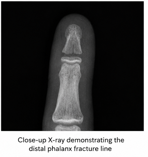
Plain radiographs of the right middle finger demonstrated a fracture involving the distal phalanx.
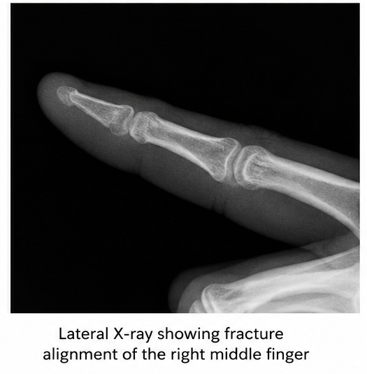
The fracture showed no significant displacement or angulation. Alignment of the distal phalanx was maintained, without significant involvement of the distal interphalangeal joint.
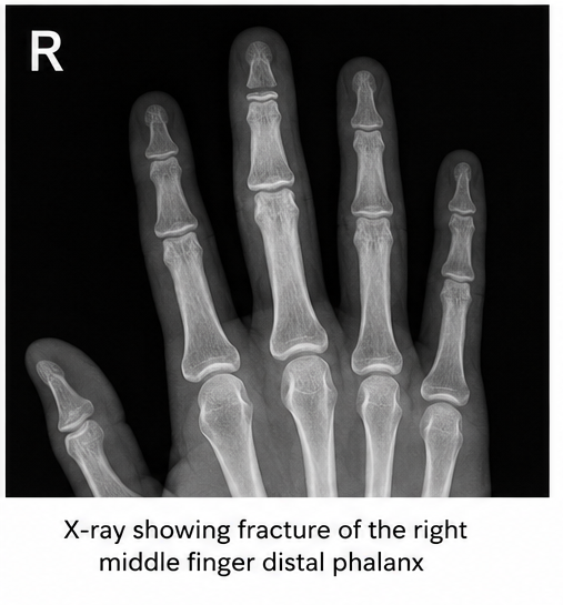
There was associated soft-tissue swelling surrounding the distal aspect of the finger.
No additional fracture or dislocation was identified.
Clinical examination was also performed to assess for associated nail-bed injury, tendon dysfunction, and neurovascular compromise.
The combined clinical and radiographic findings supported the diagnosis of a fracture of the right middle finger distal phalanx.
Diagnosis
The diagnosis was based on:
- History of trauma to the right middle finger
- Localized pain and swelling
- Tenderness over the distal phalanx
- Painful and restricted distal interphalangeal joint movement
- Radiographic evidence of distal phalanx fracture
- Preserved fracture alignment
- Absence of significant neurovascular compromise
The findings were consistent with an uncomplicated fracture of the distal phalanx of the right middle finger.
Management and Outcome
The patient was managed conservatively because the fracture was stable and did not demonstrate significant displacement.
The distal interphalangeal joint was immobilized with an appropriate finger splint to protect the fracture and maintain alignment during healing. The proximal interphalangeal joint was kept mobile where possible to minimize unnecessary stiffness.

Analgesic medication was prescribed for pain control. The patient was advised to elevate the affected hand during the early phase of injury to help reduce swelling.
Activities involving direct pressure, repetitive use, or additional trauma to the affected finger were temporarily restricted.
The patient was advised regarding appropriate splint care and instructed to seek medical attention if increasing pain, swelling, numbness, discoloration, or other concerning symptoms developed.
At subsequent follow-up, pain and swelling progressively decreased. Repeat clinical assessment demonstrated satisfactory alignment and improvement in finger movement.
Gradual mobilization was initiated following adequate clinical healing to restore distal interphalangeal joint movement and overall finger function.
Follow-up
One Week
- Reduction in pain and swelling
- Maintained finger alignment
- No neurovascular abnormality
- Splint maintained
- No evidence of secondary displacement
Three Weeks
- Significant improvement in tenderness
- Reduced soft-tissue swelling
- Stable clinical alignment
- Progressive fracture healing
- Continued protection of the affected finger
Six Weeks
- Minimal residual tenderness
- Satisfactory fracture healing
- Improved distal interphalangeal joint movement
- Gradual return to routine activities
- No significant deformity
Three Months
- Stable fracture union
- Functional range of finger movement
- No significant residual pain
- Preserved fingertip sensation
- Return to normal daily activities

Discussion
Distal phalanx fractures represent a common form of hand trauma and frequently result from direct impact or crush mechanisms. Because the distal phalanx supports the fingertip and nail complex, associated soft-tissue and nail-bed injuries should always be considered.
Clinical assessment should include evaluation of the skin, nail plate, nail bed, distal interphalangeal joint, flexor and extensor tendon function, capillary refill, and sensation. Identification of an open fracture or significant nail-bed injury may alter management.
Plain radiographs are the primary imaging modality for evaluating suspected distal phalanx fractures. Anteroposterior and lateral views help determine the fracture location, displacement, angulation, comminution, and possible articular involvement.
Treatment depends on fracture morphology and associated injuries. Stable, minimally displaced fractures can generally be managed conservatively with immobilization and appropriate follow-up. Immobilization primarily protects the distal phalanx and distal interphalangeal joint while allowing unaffected joints to remain mobile.
Displaced, unstable, significantly intra-articular, or open fractures may require specialist evaluation and, in selected cases, operative stabilization.
Potential complications include persistent tenderness, stiffness, malunion, nail deformity, chronic pain, sensory disturbance, infection in open injuries, and reduced finger function.
Early recognition, appropriate immobilization, and timely mobilization after adequate healing can help minimize these complications and facilitate restoration of hand function.
Prognosis
The prognosis of uncomplicated distal phalanx fractures is generally favorable when fracture alignment is maintained and appropriate treatment is provided.
Healing depends on the fracture pattern, degree of displacement, soft-tissue involvement, and adherence to immobilization and rehabilitation.
Stable fractures without significant joint, tendon, nail-bed, or neurovascular involvement usually demonstrate satisfactory healing with conservative management.
Persistent stiffness or tenderness may occur during recovery, particularly following prolonged immobilization. Appropriate rehabilitation and gradual return to activity can help restore functional movement.
Conclusion
Fracture of the distal phalanx of the right middle finger is a common traumatic hand injury that may present with localized pain, swelling, tenderness, and restricted movement.
This case highlights the importance of careful clinical examination and radiographic evaluation to determine fracture characteristics and identify associated soft-tissue, nail-bed, tendon, or neurovascular injuries.
Stable and minimally displaced distal phalanx fractures can generally be managed successfully with appropriate immobilization, analgesia, activity modification, and follow-up. Gradual mobilization following fracture healing is important for restoring finger movement and function.
Early diagnosis and appropriate management can provide satisfactory fracture healing while minimizing the risk of persistent pain, stiffness, deformity, and functional impairment.
References
- Nellans KW, Chung KC. Hand fractures and their management. Plast Reconstr Surg. 2013;132(2):275e–285e. https://pubmed.ncbi.nlm.nih.gov/23897347/
- Oetgen ME, Dodds SD. Non-operative treatment of common finger injuries. Curr Rev Musculoskelet Med. 2008;1(2):97–102. https://pubmed.ncbi.nlm.nih.gov/19468880/
- Meals C, Meals R. Hand fractures: a review of current treatment strategies. J Hand Surg Am. 2013;38(5):1021–1031. https://pubmed.ncbi.nlm.nih.gov/23618466/
- AO Foundation. Distal phalanx fractures – principles of treatment. https://surgeryreference.aofoundation.org/orthopedic-trauma/adult-trauma/hand-distal-phalanges
Read more such content on @ Hidoc Dr | Medical Learning App for Doctors
Recommended News For You
Recommended Articles For You
Featured News
Featured Articles
Featured Events
Featured KOL Videos
1.
Extended Maintenance with Imatinib Enhances Disease-Free Survival in High-Risk GIST.
2.
Prostate cancer therapies: AI-based image analysis detects early organ damage
3.
Applications are being accepted for TN Health's post-doctoral fellowship program in gynecologic oncology and minimal access surgery; complete details are available here.
4.
OA Tied to Arthritis AE Risk in ICI-Treated Patients
5.
Dr. Prerana S. Nesargi's message for Childhood Cancer Awareness Month 2023 is to increase understanding of pediatric oncology.
1.
The Statistical Paradigm of Leukemia: From Evolving Epidemiology and Molecular Stratification to Precision Therapies and Future Horizons in Leukemia
2.
Hematopoietic Recovery After Childhood Therapy: Mechanisms, Clinical Implications, and Emerging Strategies
3.
Oncology Partnerships: Driving Innovation Through Strategic Collaborations and Pharma Engagement USA
4.
Understanding Verrucous Carcinoma: Symptoms, Diagnosis, and Treatment Options
5.
Genomic Control of Erythropoietic Stem Cell Renewal
1.
Asian Symposium on Advancement in Hematology and Oncology (ASAHO)
2.
International Cancer Conference
3.
Asian Symposium on Advancement in Hematology and Oncology (ASAHO)
4.
Asian Symposium on Advancement in Hematology and Oncology
5.
Asian Symposium on Advancement in Hematology and Oncology
1.
Management of 1st line ALK+ mNSCLC (CROWN TRIAL Update) - Part IV
2.
Summary of The Impact of CDK4/6 Inhibition in HR+/HER2- Metastatic Breast Cancer: Insights from PALOMA-2
3.
Efficient Management of First line ALK-rearranged NSCLC - Part VI
4.
Treatment Paradigm for Patients with R/R Adult B-cell ALL- Expert Discussions
5.
A Continuation to Deep Dive Into EGFR Mutation Positive Non-Small Cell Lung Cancer

Address :
Hidoc Dr. Inc. | Delaware C Corp | 1309 Coffeen Ave. Suite 1200, Sheridan WY, 82801
Phone :
+1-415-463-3094
Email :
anishagadia@hidoc.co
© Copyright 2026 Hidoc Dr. Inc.
Terms & Conditions - LLP | Inc. | Privacy Policy - LLP | Inc. | Account Deactivation

To get started please enter your email ID
Welcome to Hidoc Dr.
Join to enhance your clinical skills and gain specialized in-depth medical knowledge