Anemia of Chronic Disease Associated with Active Rheumatoid Arthritis
OthersPage Navigation
Introduction
Anemia of chronic disease, also called anemia of inflammation, develops in association with persistent inflammatory conditions, including autoimmune disorders, chronic infections, and malignancies. It usually presents as mild-to-moderate normocytic, normochromic anemia, although microcytosis may develop with prolonged disease.
Unlike uncomplicated iron-deficiency anemia, this condition involves restricted availability of iron despite preserved or increased iron stores. Inflammation also suppresses red blood cell production and shortens erythrocyte survival. Patients may experience fatigue, pallor, exertional breathlessness, and reduced functional capacity.
Rheumatoid arthritis provides an important clinical setting for anemia of chronic disease. However, anemia in these patients may also reflect nutritional deficiency, gastrointestinal blood loss, renal dysfunction, or treatment-related toxicity. Identifying the predominant mechanism is essential for appropriate management.
Case Report
A 54-year-old woman presented to the internal medicine outpatient department with progressive fatigue, generalized weakness, and breathlessness on exertion for three months.
Initially, she noticed difficulty completing household activities and required frequent rest after walking. Over the following weeks, she developed occasional palpitations while climbing stairs. There was no breathlessness at rest, chest pain, syncope, or orthopnea.
She had been diagnosed with seropositive rheumatoid arthritis six years earlier. During the preceding four months, she experienced increasing pain and swelling of the wrists, metacarpophalangeal joints, and proximal interphalangeal joints. Morning stiffness lasted approximately 90 minutes.
Her prescribed treatment included weekly methotrexate with folic acid supplementation. However, she reported inconsistent adherence during the previous six months. She occasionally used an over-the-counter nonsteroidal anti-inflammatory drug for joint pain.
She denied hematemesis, melena, rectal bleeding, hematuria, recurrent epistaxis, or recent surgery. She had been postmenopausal for three years and reported no vaginal bleeding. There was no history of fever, night sweats, significant weight loss, chronic diarrhea, or restrictive dietary practices.
She had no known chronic kidney disease, liver disease, thyroid disorder, or previous hematological illness.
General examination revealed conjunctival and palmar pallor. Her pulse was 94 beats/minute, blood pressure was 118/74 mmHg, and oxygen saturation was 98% on room air. There was no jaundice, lymphadenopathy, pedal edema, or hepatosplenomegaly.
Musculoskeletal examination demonstrated symmetrical tenderness and swelling of both wrists and several small joints of the hands. Grip strength was reduced because of pain. Cardiovascular and respiratory examinations revealed no additional abnormalities.
Investigations
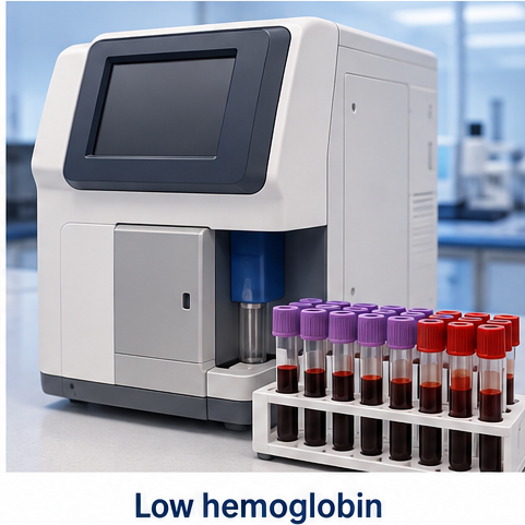
A complete blood count demonstrated moderate anemia with a normal mean corpuscular volume. The peripheral blood smear showed predominantly normocytic, normochromic erythrocytes without schistocytes, spherocytes, or abnormal circulating cells.Serum vitamin B12 and folate concentrations were within laboratory reference ranges. Renal function, liver function, and thyroid-stimulating hormone were normal.

Serum lactate dehydrogenase, indirect bilirubin, and haptoglobin did not suggest hemolysis. Urinalysis showed no hematuria.
The soluble transferrin receptor concentration was within the assay-specific reference range, providing additional support against a substantial absolute iron deficiency component. This result was interpreted alongside the clinical findings and other iron indices.
The combination of active rheumatoid arthritis, elevated inflammatory markers,
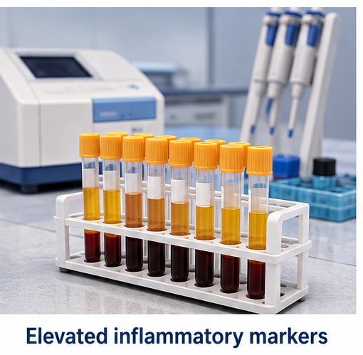
low serum iron and transferrin saturation, reduced iron-binding capacity, and elevated ferritin supported a working diagnosis of anemia of chronic disease.
Bone marrow examination was deferred because the blood smear and remaining cell lines did not suggest a primary marrow disorder.
Differential Diagnosis
Iron-deficiency anemia
Gastrointestinal blood loss was considered because of intermittent nonsteroidal anti-inflammatory drug use. However, reduced iron-binding capacity, elevated ferritin, and a normal soluble transferrin receptor favored inflammation as the predominant mechanism.
Elevated ferritin alone cannot exclude coexisting iron deficiency because ferritin increases during inflammation. Further assessment for blood loss would be necessary if anemia persisted, worsened, or developed additional features of iron depletion.
Combined iron deficiency and anemia of inflammation
A mixed anemia remained a consideration. Low transferrin saturation can occur in both conditions, making it insufficient to distinguish them independently. Interpretation requires the overall iron profile and inflammatory context.
Anemia associated with chronic kidney disease
This was unlikely because renal function was normal and there was no established renal disease.
Vitamin B12 or folate deficiency
Normal vitamin concentrations and the absence of macrocytosis made these diagnoses less likely.
Drug-related marrow suppression
Methotrexate toxicity was considered. However, the absence of leukopenia, thrombocytopenia, mucositis, or renal dysfunction made clinically significant generalized marrow suppression less likely in this presentation.
Hemolytic anemia
The low reticulocyte response, unremarkable hemolysis markers, and absence of characteristic smear abnormalities argued against hemolysis.
Management and Outcome
The patient was counselled that the anemia was most consistent with persistent inflammation associated with inadequately controlled rheumatoid arthritis.
A rheumatology review was arranged to reassess disease activity, medication adherence, and treatment tolerability. Her disease-modifying treatment was optimized after reviewing the required safety investigations. The weekly methotrexate schedule and prescribed folic acid supplementation were reinforced.
Unsupervised nonsteroidal anti-inflammatory drug use was discouraged, and a supervised pain-management plan was established.
Iron supplementation was not initiated solely on the basis of the low serum iron concentration because absolute iron deficiency had not been demonstrated. Nutritional counselling supported a balanced diet, while follow-up testing was planned to reassess iron status if recovery was inadequate.
Neither transfusion nor an erythropoiesis-stimulating agent was used in this illustrative case. The patient was clinically stable, and management initially focused on controlling the underlying inflammatory disease.
At four weeks, joint swelling and morning stiffness had decreased. Fatigue was less pronounced, hemoglobin had increased to 10.2 g/dL, and C-reactive protein had fallen to 19 mg/L.
At twelve weeks, hemoglobin was 11.8 g/dL and C-reactive protein was 6 mg/L. Exercise tolerance and daily functioning had improved.
This parallel improvement supported the working diagnosis, although treatment response alone would not establish the cause of anemia.
Discussion
Hepcidin plays a central role in inflammation-associated iron restriction. Inflammatory signaling increases hepatic hepcidin production, which reduces ferroportin-mediated iron export from intestinal cells and macrophages.
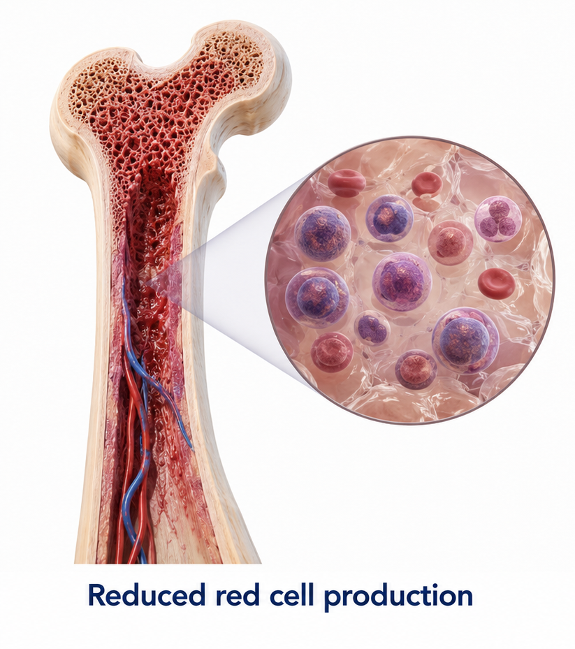
Consequently, less iron reaches the circulation and becomes available for erythropoiesis. Inflammatory cytokines also impair erythroid activity and the response to erythropoietin.
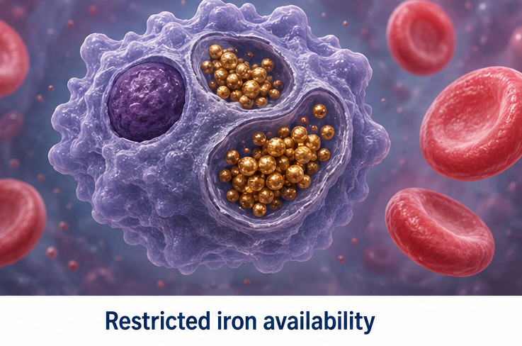
The resulting laboratory pattern can appear contradictory: circulating iron is low while ferritin is normal or elevated. This reflects restricted iron availability and the acute-phase behavior of ferritin. A low reticulocyte production index indicates that marrow output is insufficient for the degree of anemia.
In rheumatoid arthritis, anemia should prompt assessment of both disease activity and alternative causes. Research examining iron status and hepcidin in rheumatoid arthritis has demonstrated that anemia of chronic disease and iron deficiency may occur separately or together.
Control of inflammation is central to treatment. A comparative study of tocilizumab and tumor necrosis factor inhibitors reported improvements in anemia and reductions in hepcidin alongside improvement in rheumatoid arthritis activity. These findings support the relationship between inflammatory control and iron availability; they do not mean that every patient with anemia requires biologic therapy.
Additional anemia-directed treatment depends on the underlying disorder, symptoms, severity, and evidence of concurrent deficiency. Persistent anemia despite improved inflammatory control warrants renewed investigation rather than automatic attribution to chronic disease.
Conclusion
Anemia of chronic disease should be considered in patients with persistent inflammation and an inadequate reticulocyte response. In this illustrative case, active rheumatoid arthritis and the characteristic iron profile supported the diagnosis.
Assessment must still address coexisting iron deficiency, blood loss, nutritional deficiencies, renal dysfunction, and medication effects. Controlling the underlying inflammatory disorder, together with appropriate hematological follow-up, can improve hemoglobin levels and functional capacity.
References
- Nemeth E, Ganz T. Anemia of inflammation. Hematology/Oncology Clinics of North America. 2014;28(4):671–68 https://pmc.ncbi.nlm.nih.gov/articles/PMC4115203/
- Khalaf W, Al-Rubaie HA, Shihab S. Studying anemia of chronic disease and iron deficiency in patients with rheumatoid arthritis by iron status and circulating hepcidin. Hematology Reports. 2019;11(1):7708. https://pmc.ncbi.nlm.nih.gov/articles/PMC6434328/
- Dignass A, Farrag K, Stein J. Limitations of serum ferritin in diagnosing iron deficiency in inflammatory conditions. International Journal of Chronic Diseases. 2018;2018:9394060. https://pmc.ncbi.nlm.nih.gov/articles/PMC5878890/
- Weiss G, Ganz T, Goodnough LT. Anemia of inflammation. Blood. 2019;133(1):40–50. https://pmc.ncbi.nlm.nih.gov/articles/PMC6536698/
- Song SNJ, Iwahashi M, Tomosugi N, et al. Comparative evaluation of the effects of treatment with tocilizumab and TNF-α inhibitors on serum hepcidin, anemia response and disease activity in rheumatoid arthritis patients. Arthritis Research & Therapy. 2013;15:R141. https://pmc.ncbi.nlm.nih.gov/articles/PMC3978580/
Read more such content on @ Hidoc Dr | Medical Learning App for Doctors
Recommended News For You
Recommended Articles For You
Featured News
Featured Articles
Featured Events
Featured KOL Videos
1.
Prostate cancer death risk is higher in late teens and early 20s weight gain.
2.
Bayer is transforming pharmaceutical research into women's health
3.
Biomarker panel offers hope for early pancreatic cancer detection
4.
A small rise in cancer was observed following the 1986 Chernobyl accident.
5.
Added Plinabulin Boosts OS as Later-Line Treatment for EGFR Wild-Type NSCLC
1.
Beyond the Brain Fog: The Complex Neurological Challenges and Therapeutic Advances
2.
Cancer Trials 2025: Vaccines, Metabolic Targets, and the Microbiome in Oncology
3.
Immune Microenvironment Biomarkers for Early Tumor Evolution
4.
Transformative Methods in Oncology and Quality Improvement
5.
Unveiling the Mystery of Epstein-Barr Virus: What We Know So Far

Address :
Hidoc Dr. Inc. | Delaware C Corp | 1309 Coffeen Ave. Suite 1200, Sheridan WY, 82801
Phone :
+1-415-463-3094
Email :
anishagadia@hidoc.co
© Copyright 2026 Hidoc Dr. Inc.
Terms & Conditions - LLP | Inc. | Privacy Policy - LLP | Inc. | Account Deactivation

To get started please enter your email ID
Welcome to Hidoc Dr.
Join to enhance your clinical skills and gain specialized in-depth medical knowledge