Acute Cholangitis Secondary to Choledocholithiasis Presenting With Fever, Jaundice and Right Upper Quadrant Pain: A Case Report
OthersPage Navigation
Introduction
Acute cholangitis is an acute bacterial infection of the biliary tract that usually develops in the presence of biliary obstruction. Obstruction increases intrabiliary pressure, promotes bacterial proliferation, and may facilitate systemic dissemination of infection. Without timely treatment, the condition can progress to sepsis, organ dysfunction, and septic shock.
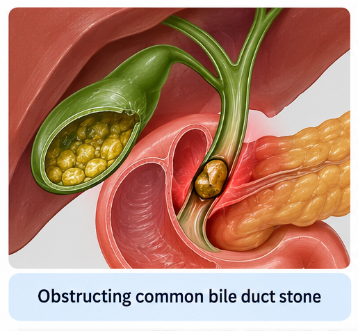
Choledocholithiasis is one of the most common causes of acute cholangitis. Other causes include benign or malignant biliary strictures, obstructed biliary stents, pancreaticobiliary tumors, and complications following biliary interventions.
The classical clinical presentation consists of fever, right upper quadrant abdominal pain, and jaundice, collectively known as Charcot's triad. However, not all patients demonstrate all three manifestations. Severe disease may additionally present with hypotension and altered mental status, forming Reynolds' pentad.
Diagnosis is based on a combination of clinical features, evidence of systemic inflammation and cholestasis on laboratory investigations, and imaging demonstrating biliary dilatation or an underlying cause of obstruction.
Management requires rapid stabilization, appropriate antimicrobial therapy, and relief of biliary obstruction when indicated. Endoscopic retrograde cholangiopancreatography (ERCP) is commonly used to achieve biliary drainage and treat obstructing common bile duct stones.
This report describes a patient presenting with fever, jaundice, and right upper quadrant abdominal pain who was diagnosed with acute cholangitis secondary to an obstructing common bile duct stone.
Case Report
A 58-year-old man presented to the emergency department with fever, chills, right upper quadrant abdominal pain, and yellowish discoloration of the eyes for two days.
The abdominal pain had developed suddenly and was predominantly localized to the right upper quadrant and epigastric region. It was continuous, moderate to severe in intensity, and was associated with nausea and two episodes of non-bilious vomiting.
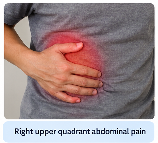
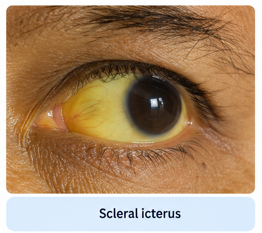
Several hours after the onset of abdominal pain, the patient developed high-grade fever associated with chills and rigors. His family subsequently noticed yellowish discoloration of his sclera and dark-colored urine.
He reported reduced appetite and generalized weakness but denied hematemesis, melena, diarrhea, or significant abdominal distension.
The patient had experienced intermittent episodes of postprandial right upper quadrant discomfort during the preceding several months but had not previously undergone evaluation for gallstone disease.
There was no previous history of jaundice, pancreatitis, cholangitis, biliary surgery, or ERCP.
On examination, the patient appeared acutely ill and febrile. His temperature was 39.1°C, pulse rate was 108 beats/min, blood pressure was 108/68 mmHg, respiratory rate was 20 breaths/min, and oxygen saturation was 97% on room air.
Scleral icterus was evident.
Abdominal examination revealed tenderness in the right upper quadrant and epigastric region without guarding or rigidity. No palpable abdominal mass was identified. Bowel sounds were present.
The combination of fever, right upper quadrant pain, and jaundice was consistent with Charcot's triad and raised strong clinical suspicion of acute cholangitis.
Investigations
Laboratory investigations demonstrated leukocytosis with neutrophilic predominance and elevated inflammatory markers, supporting an acute systemic inflammatory response.
Liver function tests demonstrated a predominantly cholestatic pattern, with elevated serum bilirubin, alkaline phosphatase, and gamma-glutamyl transferase. Serum aminotransferases were also moderately elevated.
Renal function and serum electrolytes were assessed to evaluate the patient's general condition and identify possible organ dysfunction.
Blood cultures were obtained before initiation of antimicrobial therapy.
Abdominal ultrasonography demonstrated cholelithiasis with dilatation of the common bile duct and mild intrahepatic biliary dilatation. These findings raised suspicion of distal common bile duct obstruction.
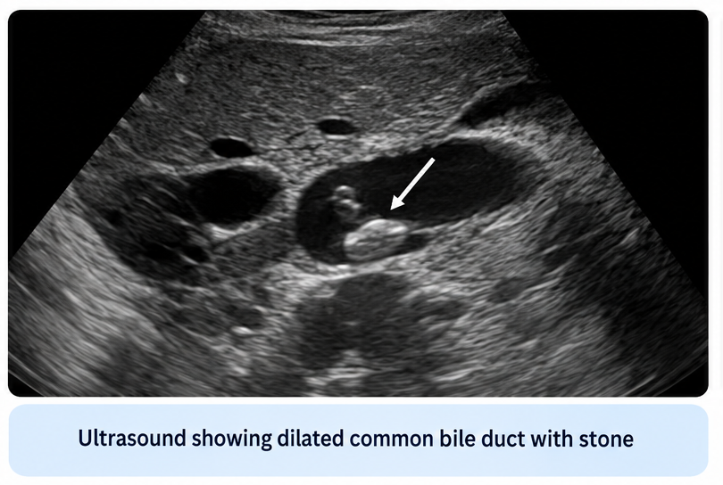
Magnetic resonance cholangiopancreatography (MRCP) subsequently demonstrated a dilated common bile duct with an obstructing calculus in its distal portion, consistent with choledocholithiasis.
The presence of systemic inflammation, biochemical evidence of cholestasis, and imaging evidence of biliary obstruction supported the diagnosis of acute cholangitis secondary to choledocholithiasis.
Differential Diagnosis
Acute Cholecystitis
Acute cholecystitis commonly presents with fever and right upper quadrant abdominal pain and may therefore resemble acute cholangitis. However, significant jaundice and common bile duct dilatation are less characteristic of uncomplicated acute cholecystitis. Imaging in this patient demonstrated obstruction of the common bile duct.
Gallstone Pancreatitis
Gallstone pancreatitis may cause severe epigastric or upper abdominal pain associated with vomiting and abnormal liver function tests. However, the combination of fever, jaundice, inflammatory changes, and documented biliary obstruction favored acute cholangitis.
Acute Viral Hepatitis
Acute viral hepatitis can produce jaundice, constitutional symptoms, and elevated liver enzymes. However, marked aminotransferase elevation is generally more prominent, whereas imaging evidence of common bile duct obstruction and biliary dilatation favors an obstructive biliary process.
Malignant Biliary Obstruction
Pancreatic, ampullary, or cholangiocarcinoma can produce obstructive jaundice and may be complicated by cholangitis. However, imaging in this patient demonstrated an obstructing common bile duct stone without evidence of an underlying malignant lesion.
Liver Abscess
A pyogenic liver abscess can present with fever, right upper quadrant pain, leukocytosis, and abnormal liver function tests. Imaging, however, did not demonstrate a focal hepatic collection and instead identified biliary obstruction due to choledocholithiasis.
Management and Outcome
The patient was admitted and immediately started on supportive management.
Intravenous fluids were administered to maintain adequate circulating volume and tissue perfusion. The patient was kept under close monitoring for blood pressure, heart rate, urine output, temperature, and signs of developing organ dysfunction.
Empirical intravenous broad-spectrum antibiotic therapy targeting common biliary pathogens was initiated after blood cultures were obtained.
Analgesics, antipyretics, antiemetics, and correction of fluid and electrolyte abnormalities were provided as required.
Because imaging demonstrated an obstructing common bile duct calculus in the setting of acute cholangitis, biliary decompression was planned.
ERCP demonstrated an obstructing stone within the distal common bile duct with infected bile draining following biliary access. Endoscopic sphincterotomy was performed, followed by extraction of the common bile duct stone and biliary drainage.
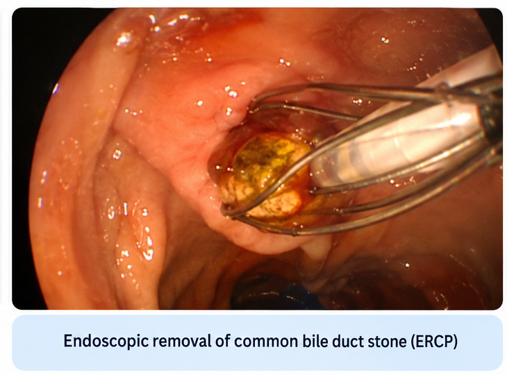
Following biliary decompression, the patient's clinical condition improved substantially.
Fever and abdominal pain gradually resolved, while appetite and general condition improved. Serial laboratory investigations demonstrated declining leukocyte count, inflammatory markers, bilirubin, and cholestatic liver enzymes.
No post-ERCP complication was observed.
Antimicrobial therapy was subsequently adjusted according to the patient's clinical response and available microbiological findings.
After complete clinical stabilization, the patient was discharged with advice for follow-up and definitive management of gallbladder stones to reduce the risk of recurrent biliary events.
Discussion
Acute cholangitis is a potentially life-threatening biliary infection that typically develops when bacterial contamination occurs in an obstructed biliary system.
Under normal circumstances, bile flow and several physiological defense mechanisms limit bacterial colonization of the biliary tract. Biliary obstruction results in increased intraductal pressure, biliary stasis, and bacterial proliferation. Increasing biliary pressure may subsequently facilitate translocation of bacteria and inflammatory mediators into the systemic circulation.
Choledocholithiasis remains an important cause of biliary obstruction leading to acute cholangitis. Other causes include benign biliary strictures, malignant obstruction, occluded biliary stents, and complications related to biliary procedures.
The traditional clinical presentation is Charcot's triad of fever, right upper quadrant abdominal pain, and jaundice. Although highly suggestive when present, the complete triad is not observed in every patient. Therefore, absence of one component should not exclude the diagnosis.
Patients with severe infection may develop hypotension and altered mental status in addition to Charcot's triad. This combination, historically described as Reynolds' pentad, suggests severe systemic infection and possible septic shock.
Contemporary diagnostic assessment incorporates clinical manifestations, laboratory evidence of systemic inflammation, evidence of cholestasis, and imaging findings demonstrating biliary dilatation or the underlying cause of obstruction.
Laboratory findings frequently include leukocytosis, elevated inflammatory markers, hyperbilirubinemia, and elevated alkaline phosphatase, gamma-glutamyl transferase, and aminotransferases.
Ultrasonography is frequently used as an initial imaging modality because it can identify gallstones and biliary dilatation. MRCP provides non-invasive visualization of the biliary tree and is particularly useful for demonstrating common bile duct stones and determining the site of obstruction.
Treatment addresses the two major components of acute cholangitis: infection and biliary obstruction.
Initial management includes assessment and stabilization of airway, breathing, and circulation when required, intravenous fluid resuscitation, correction of electrolyte disturbances, monitoring for organ dysfunction, and early administration of appropriate antimicrobial therapy.
Blood cultures should preferably be obtained before antimicrobial administration when this does not cause clinically important treatment delay. Bile obtained during drainage may also be sent for microbiological examination to guide subsequent antimicrobial therapy.
Biliary drainage represents a central component of management in patients requiring decompression. ERCP is generally preferred because it permits both biliary drainage and treatment of the underlying obstruction, including extraction of common bile duct stones.
The urgency of drainage depends on disease severity and clinical response. Patients with moderate disease generally require early drainage, while severe acute cholangitis associated with organ dysfunction requires urgent stabilization and biliary decompression as soon as clinically feasible.
The present patient demonstrated the characteristic clinical features of acute cholangitis with fever, right upper quadrant abdominal pain, and jaundice. Laboratory investigations supported systemic inflammation and cholestasis, while imaging demonstrated common bile duct obstruction caused by a calculus.
Prompt antimicrobial therapy and endoscopic biliary decompression resulted in rapid clinical improvement. The case emphasizes that successful treatment requires both control of infection and correction of the underlying biliary obstruction.
Conclusion
Acute cholangitis should be considered an important medical emergency in patients presenting with fever, right upper quadrant abdominal pain, jaundice, and evidence of biliary obstruction.
Although Charcot's triad provides an important clinical clue, diagnosis should integrate clinical manifestations with inflammatory and cholestatic laboratory abnormalities and appropriate biliary imaging.
Choledocholithiasis is an important and treatable cause of acute cholangitis.
Initial treatment includes hemodynamic stabilization, intravenous fluids, antimicrobial therapy, and careful assessment for organ dysfunction.
Early biliary decompression is particularly important when significant obstruction or moderate-to-severe disease is present, with ERCP providing an effective method for both drainage and removal of obstructing common bile duct stones.
In this patient, early recognition of the clinical syndrome, identification of choledocholithiasis, and prompt antimicrobial therapy followed by ERCP and stone extraction resulted in rapid clinical recovery.
References
- Miura F, Okamoto K, Takada T, et al. Tokyo Guidelines 2018: initial management of acute biliary infection and flowchart for acute cholangitis. J Hepatobiliary Pancreat Sci. 2018;25(1):31–40. https://pubmed.ncbi.nlm.nih.gov/28941329/
- Gomi H, Solomkin JS, Schlossberg D, et al. Tokyo Guidelines 2018: antimicrobial therapy for acute cholangitis and cholecystitis. J Hepatobiliary Pancreat Sci. 2018;25(1):3–16. https://pubmed.ncbi.nlm.nih.gov/29090866/
- Takada T. Tokyo Guidelines 2018: updated Tokyo Guidelines for the management of acute cholangitis/acute cholecystitis. J Hepatobiliary Pancreat Sci. 2018;25(1):1–2. https://pubmed.ncbi.nlm.nih.gov/29334699/
- Kiriyama S, Takada T, Strasberg SM, et al. New diagnostic criteria and severity assessment of acute cholangitis in revised Tokyo Guidelines. J Hepatobiliary Pancreat Sci. 2012;19(5):548–556. https://pubmed.ncbi.nlm.nih.gov/22825491/
- Sulzer JK, Ocuin LM. Cholangitis: causes, diagnosis, and management. Surg Clin North Am. 2019;99(2):175–184. https://pubmed.ncbi.nlm.nih.gov/30846030/
- Czerwonko ME, Huespe P, Mazza O, et al. Acute cholangitis: a state-of-the-art review. Ann Med Surg. 2024;86:4560–4574. https://pmc.ncbi.nlm.nih.gov/articles/PMC11305776/
Read more such content on @ Hidoc Dr | Medical Learning App for Doctors
Recommended News For You
Recommended Articles For You
Featured News
Featured Articles
Featured Events
Featured KOL Videos
1.
"Unusual" Cancers Following Pandemic; Triumph in TNBC; Clinical Trial Interrupted by Shortage.
2.
Recently released national data on drug use and abuse among Americans.
3.
Despite Medicare Coverage, Cancer Genomic Testing Still Low
4.
Ketamine plus psychotherapy for "excellent" PTSD
5.
Psychedelic Therapy Tied to Reduced Depression, Anxiety.
1.
Personalized Tumor Ecological Landscapes in Oncology
2.
Unlocking the Potential of Glofitamab: A Novel Treatment for Cancer
3.
Preserving Social Identity During Cancer Survivorship
4.
Battling Blood Cancers: Advances in HIV-Related Hematologic Malignancies in the ART Era
5.
Regenerative Models of Tumor Microenvironments: Clinical Relevance and Emerging Insights
1.
International Conference on Cancer Nursing and Rehabilitation Strategies
2.
International Conference on Best Practices in Oncology, Cardiology and Critical Care
3.
International Conference on Innovations in Critical Care for Oncology and Cardiology
4.
International Symposium on Oncology, Cardiology and Critical Care Innovations
5.
International Conference on Cancer Nursing and Hematology Support
1.
An Intro to The Multifaceted Advantages of CDK4/6 Inhibitors in HR+/HER2- Advanced Breast Cancer Clinical Studies.
2.
Dacomitinib Case Presentation: Baseline Treatment and Current Status
3.
Advances in Classification/ Risk Stratification of Plasma Cell Dyscrasias- The Summary
4.
From Guidelines to Practice: Hematology
5.
Breast Cancer Awareness and Early Detection

Address :
Hidoc Dr. Inc. | Delaware C Corp | 1309 Coffeen Ave. Suite 1200, Sheridan WY, 82801
Phone :
+1-415-463-3094
Email :
anishagadia@hidoc.co
© Copyright 2026 Hidoc Dr. Inc.
Terms & Conditions - LLP | Inc. | Privacy Policy - LLP | Inc. | Account Deactivation

To get started please enter your email ID
Welcome to Hidoc Dr.
Join to enhance your clinical skills and gain specialized in-depth medical knowledge