Rhegmatogenous Retinal Detachment Presenting With Sudden Flashes, Floaters, and Progressive Visual Field Loss: A Case Report
OthersPage Navigation
Introduction
Retinal detachment is an important ophthalmic emergency characterized by separation of the neurosensory retina from the underlying retinal pigment epithelium. This separation disrupts normal retinal function and may result in permanent visual impairment when diagnosis and treatment are delayed.
Retinal detachment is broadly classified into rhegmatogenous, tractional, and exudative forms. Rhegmatogenous retinal detachment (RRD) is the most common type and develops when a full-thickness retinal break permits liquefied vitreous to enter the subretinal space. The accumulating fluid progressively separates the neurosensory retina from the retinal pigment epithelium.
Patients commonly describe sudden onset of flashes of light, new floaters, and a shadow or curtain progressing across the visual field. Central visual acuity may remain relatively preserved while the macula remains attached but can deteriorate substantially once the detachment involves the macula.
Risk factors include increasing age, high myopia, previous cataract surgery, ocular trauma, peripheral retinal degeneration, and a previous retinal detachment in the fellow eye. Early ophthalmological evaluation is essential because prompt repair can prevent progression of the detachment and improve the likelihood of preserving useful vision.
This report describes a patient presenting with sudden flashes and floaters followed by progressive peripheral visual field loss who was subsequently diagnosed with rhegmatogenous retinal detachment.
Case Report
A 58-year-old man presented to the ophthalmology department with a three-day history of sudden onset of flashes of light and multiple moving spots in his right eye, followed by progressive loss of peripheral vision.

The patient first noticed brief flashes of light in the temporal visual field, particularly when moving his eyes in dim illumination. Within several hours, he began seeing numerous dark spots and thread-like structures moving across his vision.
During the following two days, he noticed a dark shadow developing in the peripheral visual field of the affected eye. The shadow gradually increased in size and was described as resembling a curtain moving across his vision.
On the day of presentation, the patient reported increasing difficulty seeing objects in the lower part of his visual field. Central vision remained relatively preserved, although he described mild blurring.
There was no significant ocular pain, redness, photophobia, purulent discharge, headache, nausea, or vomiting.
The patient had a history of moderate-to-high myopia since early adulthood and regularly used corrective spectacles. There was no recent history of direct ocular trauma. He had no known history of diabetic retinopathy, inflammatory eye disease, or previous retinal detachment.
His medical history included controlled hypertension. There was no history of diabetes mellitus or significant systemic inflammatory disease.
On examination, the patient was conscious and clinically stable. External examination of both eyes did not demonstrate significant redness or swelling.
Visual acuity was reduced in the right eye compared with the left eye but remained adequate for central fixation. Pupillary examination demonstrated a subtle relative afferent pupillary defect in the affected eye.
Intraocular pressure was not elevated.
The combination of sudden photopsia, new floaters, and a progressively enlarging curtain-like visual field defect raised strong suspicion of retinal detachment.
Investigations
A comprehensive ophthalmological examination was performed after pupillary dilation.
Slit-lamp examination of the anterior segment did not reveal significant acute abnormalities. Examination of the vitreous demonstrated changes consistent with posterior vitreous separation, with pigment cells suggesting an associated retinal break.
Dilated fundus examination of the right eye revealed an elevated, corrugated, mobile retina involving the superior temporal peripheral retina. A horseshoe-shaped retinal tear was identified in the superior temporal quadrant.
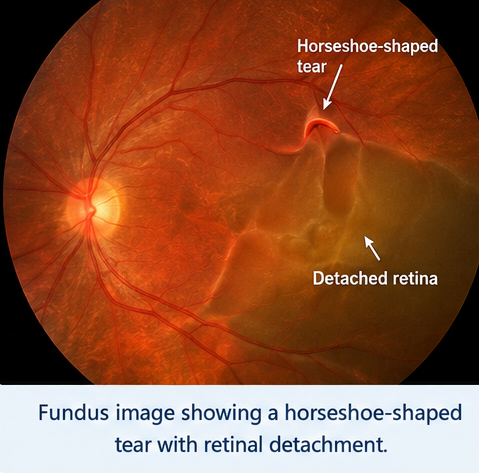
Subretinal fluid extended from the retinal break into the adjacent retina, producing a rhegmatogenous retinal detachment. At presentation, the detachment approached but had not yet involved the foveal center, consistent with a macula-on retinal detachment.

The optic disc appeared structurally preserved, and no major retinal vascular abnormality was identified.
The left eye did not demonstrate retinal detachment, although careful peripheral retinal examination was performed to identify predisposing retinal lesions.
Optical coherence tomography of the macular region confirmed preservation of foveal attachment without significant subfoveal fluid.
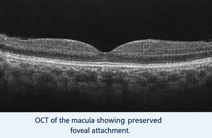
Because the retinal tear and detachment were clearly visualized on dilated fundus examination, ocular ultrasonography was not essential for diagnosis. B-scan ultrasonography would have been appropriate if visualization of the retina had been obscured by vitreous hemorrhage, dense cataract, or other media opacity.
Based on the characteristic symptoms and retinal examination, a diagnosis of acute rhegmatogenous retinal detachment secondary to a superior temporal horseshoe retinal tear was made.
Differential Diagnosis
Posterior Vitreous Detachment
Posterior vitreous detachment frequently causes sudden flashes and floaters and is an important differential diagnosis. It can also create retinal tears that subsequently lead to retinal detachment. However, the presence of an elevated detached retina with subretinal fluid confirmed that the patient's condition had progressed beyond an uncomplicated posterior vitreous detachment.
Vitreous Hemorrhage
Vitreous hemorrhage may produce sudden floaters, hazy vision, or marked visual loss. It may also accompany a retinal tear. In this patient, however, direct fundus examination demonstrated a retinal break and associated retinal detachment as the principal cause of the visual field defect.
Retinal Tear Without Detachment
An isolated retinal tear can produce flashes and floaters and represents an important precursor to retinal detachment. In the present case, subretinal fluid extending beyond the retinal break with visible elevation of the neurosensory retina established the presence of retinal detachment.
Tractional Retinal Detachment
Tractional retinal detachment occurs when fibrovascular membranes exert mechanical traction on the retina and is particularly associated with proliferative diabetic retinopathy. The patient did not have diabetes or significant retinal neovascular disease, and a full-thickness retinal tear was identified, favoring a rhegmatogenous mechanism.
Exudative Retinal Detachment
Exudative or serous retinal detachment occurs because of accumulation of subretinal fluid without a retinal break and may occur in inflammatory, vascular, or neoplastic disorders. Identification of a horseshoe retinal tear in this patient strongly supported rhegmatogenous retinal detachment.
Management and Outcome
The patient was referred urgently to a vitreoretinal surgeon because the detachment remained macula-on and progression could threaten central vision.
Following detailed retinal assessment, surgical repair was recommended. The location and extent of the retinal tear and detachment were considered in selecting the operative approach.
The patient underwent pars plana vitrectomy. During surgery, the vitreous traction associated with the retinal tear was relieved, subretinal fluid was managed, and the causative retinal break was treated with endolaser photocoagulation.
An intraocular gas tamponade was used to support retinal reattachment and maintain contact between the treated retinal break and the underlying retinal pigment epithelium during healing.
Postoperatively, the patient was instructed regarding the prescribed head positioning to optimize the effect of the intraocular gas bubble. He was also advised to avoid air travel and significant altitude changes while intraocular gas remained present.
Topical postoperative therapy and scheduled ophthalmological follow-up were provided.
During subsequent examination, the retina remained attached and the treated retinal break showed appropriate laser reaction. No new retinal tears were detected.
The patient's peripheral visual symptoms improved, and central visual acuity remained relatively well preserved because the foveal center had not detached before surgical repair.
At follow-up, the retina remained anatomically attached without evidence of recurrent detachment. Continued surveillance of both eyes was recommended because retinal breaks or detachments may subsequently occur in predisposed patients.
Discussion
Rhegmatogenous retinal detachment occurs when a full-thickness retinal break allows liquefied vitreous to pass into the subretinal space. Progressive accumulation of fluid separates the neurosensory retina from the retinal pigment epithelium and compromises normal retinal function.
Posterior vitreous detachment is an important mechanism underlying many retinal tears. With aging, the vitreous undergoes liquefaction and gradually separates from the retina. Persistent vitreoretinal adhesion at certain points can exert traction during this process and produce a retinal tear.
Once a break forms, liquefied vitreous may enter through the defect and accumulate beneath the retina, resulting in retinal detachment.
The clinical history can provide an important early clue. Photopsia results from mechanical stimulation of the retina by vitreoretinal traction, while new floaters may reflect vitreous opacities, pigment, or hemorrhage associated with a retinal tear.
As detachment progresses, patients often describe a dark shadow, veil, or curtain spreading across the visual field. The location of the perceived visual field defect generally corresponds to the opposite area of retinal involvement.
The status of the macula is particularly important when evaluating retinal detachment. In macula-on RRD, the fovea remains attached and central visual acuity may therefore remain relatively preserved. Progression into the macula can cause a sudden and substantial reduction in central vision.
Consequently, an acute macula-on retinal detachment generally requires urgent surgical evaluation to reduce the risk of progression and loss of central vision.
Diagnosis is primarily established by a careful dilated retinal examination. Indirect ophthalmoscopy with peripheral retinal assessment helps identify the extent of detachment and locate retinal breaks. Optical coherence tomography can provide detailed assessment of macular involvement.
B-scan ultrasonography is particularly valuable when direct retinal visualization is prevented by vitreous hemorrhage, dense cataract, or other media opacity.
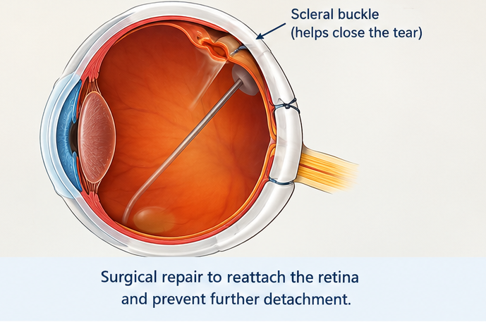
Several surgical approaches can be used to repair rhegmatogenous retinal detachment, including pars plana vitrectomy, scleral buckling, pneumatic retinopexy, or combinations of these techniques. Selection depends on factors such as the patient's age, lens status, number and location of retinal breaks, extent of detachment, vitreoretinal traction, and surgeon assessment.
The fundamental goals of treatment are to identify and close retinal breaks, relieve relevant vitreoretinal traction, and allow the retina to reattach to the retinal pigment epithelium. Laser photocoagulation or cryotherapy can create chorioretinal adhesion around retinal breaks.
Intraocular gas or silicone oil may be used as an internal tamponade in selected surgical repairs. When gas is used, adherence to postoperative positioning and restrictions regarding air travel and altitude is important until the gas has adequately resorbed.
Visual outcome depends on several factors, particularly whether the macula is attached at the time of treatment, the duration of macular detachment when present, and the presence of complications such as proliferative vitreoretinopathy.
In the present case, the classical progression from photopsia and floaters to a curtain-like peripheral visual field defect prompted ophthalmological assessment. Identification of a superior temporal horseshoe retinal tear with macula-on rhegmatogenous retinal detachment allowed urgent surgical intervention before central retinal involvement.
Conclusion
Retinal detachment should be considered an ophthalmic emergency in patients presenting with sudden flashes, new-onset floaters, or a progressive curtain-like defect in the visual field.
A detailed history followed by prompt dilated fundus examination is critical for identifying retinal tears and determining the extent of retinal detachment. Optical coherence tomography can help establish macular status, while B-scan ultrasonography is useful when direct visualization is limited.
Rhegmatogenous retinal detachment requires timely treatment to close the causative retinal break and restore retinal attachment. The urgency is particularly important when the macula remains attached because progression can result in substantial loss of central vision.
In this patient, recognition of the characteristic symptoms, identification of a horseshoe retinal tear with macula-on detachment, and timely vitreoretinal surgery resulted in successful anatomical reattachment with preservation of useful central vision.
References
- Sultan ZN, Agorogiannis EI, Iannetta D, Steel D, Sandinha T. Rhegmatogenous retinal detachment: a review of current practice in diagnosis and management. BMJ Open Ophthalmol. 2020;5(1). https://pmc.ncbi.nlm.nih.gov/articles/PMC7549457/
- Mitry D, Charteris DG, Fleck BW, Campbell H, Singh J. The epidemiology of rhegmatogenous retinal detachment: geographical variation and clinical associations. Br J Ophthalmol. 2010;94(6):678–684. https://pubmed.ncbi.nlm.nih.gov/19515646/
- Lumi X, Hawlina M, Glavač D, et al. Ageing of the vitreous: from acute onset floaters and flashes to retinal detachment. Ageing Res Rev. 2015;21:71–77. https://pubmed.ncbi.nlm.nih.gov/25841656/
- Steel D. Retinal detachment. BMJ Clin Evid. 2014;2014:0710. https://pmc.ncbi.nlm.nih.gov/articles/PMC3940167/
- Schwartz SG, Flynn HW Jr. Primary retinal detachment: scleral buckle or pars plana vitrectomy? Curr Opin Ophthalmol. 2006;17(3):245–250. https://pubmed.ncbi.nlm.nih.gov/16794436/
- Stewart S, Chan W. The relationship between visual acuity and duration of macula-off retinal detachment: a systematic review of the literature. Retina. 2007;27(9):1169–1174. https://pubmed.ncbi.nlm.nih.gov/18046217/
Read more such content on @ Hidoc Dr | Medical Learning App for Doctors
Recommended News For You
Recommended Articles For You
Featured News
Featured Articles
Featured Events
Featured KOL Videos
1.
Regular physical activity before cancer diagnosis may lower progression and death risks
2.
Medication Self-Injection Embraced by Breast Cancer Patients
3.
Financial hardship for cancer survivors due to high-cost immunotherapies, especially for blood cancer patients
4.
FDA Advisors Split on Regimens for Breast, Prostate Cancers
5.
ctDNA-Guided Adjuvant Therapy Cuts Bladder Cancer Costs
1.
RDW-CV vs. MCV: Knowing the Difference Can Help You Better Understand Your Health
2.
Clinical Pathways in Hematology in Clinical Decision-Making
3.
Immune Surveillance Screening for Early Cancer Risk Detection
4.
Unlocking the Mysteries of Lymphoblastic Lymphoma: A Journey Into the Unknown
5.
AI-Driven Digital Pathology in Oncology: From Histology Images to Predictive Treatment Biomarkers
1.
International Conference on Innovations in Critical Care for Oncology and Cardiology
2.
International Conference on Oncology, Cardiology and Critical Care Policy
3.
International Conference on Oncology, Cancer Prevention and Public Health
4.
International Conference on Cancer Nursing and Rehabilitation Strategies
5.
International Conference on Best Practices in Oncology, Cardiology and Critical Care
1.
How Advances in Cancer Research Are Improving Treatment Options
2.
Navigating the Complexities of Ph Negative ALL - Part II
3.
Signs and Symptoms of Leukemia
4.
Updates on Standard V/S High Risk Myeloma Treatment- The Next Part
5.
Virtual Case Study on Elephantiasis of Lower Limb- An Initiative by Hidoc Dr.

Address :
Hidoc Dr. Inc. | Delaware C Corp | 1309 Coffeen Ave. Suite 1200, Sheridan WY, 82801
Phone :
+1-415-463-3094
Email :
anishagadia@hidoc.co
© Copyright 2026 Hidoc Dr. Inc.
Terms & Conditions - LLP | Inc. | Privacy Policy - LLP | Inc. | Account Deactivation

To get started please enter your email ID
Welcome to Hidoc Dr.
Join to enhance your clinical skills and gain specialized in-depth medical knowledge