Actinic Keratosis Presenting With Multiple Hyperkeratotic Lesions: A Case Report
OthersPage Navigation
Abstract
Actinic keratosis is a common premalignant cutaneous lesion resulting from chronic ultraviolet (UV) radiation-induced damage to keratinocytes. It typically presents as rough, scaly, erythematous or hyperkeratotic lesions on chronically sun-exposed areas of the skin. Although individual lesions may remain stable, actinic keratosis can progress to cutaneous squamous cell carcinoma, making early recognition and appropriate treatment essential. We report the case of a 62-year-old man who presented with multiple rough, scaly lesions over the face, scalp, and dorsal aspects of both hands that had progressively increased in number and thickness over several years. Clinical examination revealed multiple erythematous hyperkeratotic plaques with adherent scales over sun-exposed areas. Dermoscopic evaluation demonstrated features suggestive of actinic keratosis, and a lesional biopsy confirmed the diagnosis. The patient was managed with lesion-directed and field-directed therapy, along with strict photoprotection. At follow-up, significant clinical improvement was observed, with no evidence of progression to invasive squamous cell carcinoma. This case highlights the importance of early identification of actinic keratosis, assessment of lesions with suspicious clinical features, and timely treatment to reduce the risk of malignant transformation.
Introduction
Actinic keratosis is a chronic UV radiation-induced disorder of the epidermis characterized by atypical proliferation of keratinocytes. It commonly occurs in individuals with prolonged cumulative sun exposure and is frequently observed on the face, scalp, ears, neck, forearms, and dorsal hands.
The clinical appearance of actinic keratosis varies from subtle roughness or erythematous macules to thick, hyperkeratotic plaques. Lesions may be asymptomatic or associated with itching, tenderness, burning, or discomfort. The condition is considered part of a disease spectrum associated with chronic photodamage and may progress to invasive cutaneous squamous cell carcinoma.
Risk factors include advanced age, fair skin phenotype, cumulative UV exposure, occupational outdoor activity, immunosuppression, and a history of previous skin cancers. Clinical diagnosis is often based on characteristic morphology and distribution, while dermoscopy and histopathological examination may be required in atypical, thick, persistent, or suspicious lesions.
Treatment options include cryotherapy, curettage, excision, topical 5-fluorouracil, imiquimod, diclofenac, tirbanibulin, photodynamic therapy, and other field-directed approaches. Selection of treatment depends on lesion characteristics, number and distribution of lesions, patient factors, and the extent of surrounding photodamage.
We report a case of multiple actinic keratoses presenting as progressive hyperkeratotic lesions over chronically sun-exposed areas, emphasizing the importance of early diagnosis, appropriate treatment, and long-term surveillance.
Case Report
A 62-year-old man presented to the dermatology outpatient department with a 3-year history of multiple rough and scaly skin lesions involving the forehead, scalp, cheeks, and dorsal surfaces of both hands. The lesions had gradually increased in number and thickness over time. The patient reported occasional itching and mild tenderness over some of the thicker lesions but denied spontaneous bleeding, ulceration, or significant pain.
The patient had a history of prolonged occupational sun exposure for more than two decades due to outdoor work. He reported inconsistent use of sunscreen and frequently worked outdoors during peak daylight hours. There was no history of previous skin malignancy, immunosuppressive therapy, or organ transplantation.
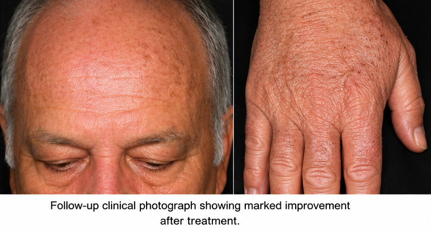
On examination, multiple erythematous to brownish macules, papules, and hyperkeratotic plaques were observed over chronically sun-exposed areas, particularly the forehead, scalp, cheeks, and dorsal hands.
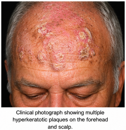
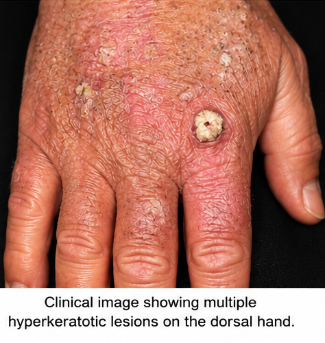
The lesions demonstrated a rough, gritty surface with adherent scales. A few lesions were more prominently hyperkeratotic than the surrounding lesions.
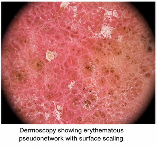
Dermoscopic examination demonstrated features consistent with actinic keratosis, including an erythematous pseudonetwork and surface scale. One particularly thick and persistent lesion on the dorsal aspect of the right hand was considered clinically suspicious and was selected for histopathological evaluation.
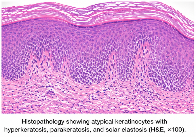
A skin biopsy demonstrated atypical keratinocytes involving the lower epidermal layers, associated with parakeratosis, hyperkeratosis, and solar elastosis in the underlying dermis. There was no evidence of invasion through the basement membrane. The findings were consistent with actinic keratosis.
Based on the clinical presentation, dermoscopic findings, and histopathological examination, a diagnosis of multiple actinic keratoses with prominent hyperkeratotic lesions was established.
Management and Outcome
The patient was counseled extensively regarding the relationship between cumulative UV exposure and actinic keratosis. Strict photoprotection was advised, including regular use of broad-spectrum sunscreen, protective clothing, a wide-brimmed hat, and avoidance of prolonged direct sun exposure.
The thicker, clinically suspicious lesions were managed with lesion-directed therapy. Field-directed treatment was subsequently initiated for areas with multiple lesions and evidence of diffuse photodamage.
The patient tolerated treatment well, with gradual reduction in erythema, scaling, and hyperkeratotic changes. The treated lesions showed significant clinical improvement on follow-up examination.
The patient was advised regarding the chronic nature of actinic keratosis and the possibility of developing new lesions over time. Regular dermatological surveillance was recommended to identify recurrent lesions or any features suggestive of progression to invasive squamous cell carcinoma.
Follow-up
One Month
- The patient reported a reduction in itching and surface roughness.
- Treated lesions demonstrated decreased erythema and scaling.
- No new rapidly growing or ulcerated lesions were observed.
- The patient reported improved adherence to sun-protection measures.
Three Months
- Significant reduction in the number and thickness of visible keratotic lesions was noted.
- Previously treated hyperkeratotic lesions showed marked clinical improvement.
- No evidence of local recurrence at the biopsy site was observed.
- The patient continued regular use of photoprotective measures.
Six Months
- The patient remained clinically stable with no evidence of progression to invasive squamous cell carcinoma.
- Residual photodamaged areas were monitored during routine dermatological examination.
- No new suspicious lesions requiring urgent biopsy were identified.
- Continued periodic dermatological follow-up and long-term photoprotection were advised.
Discussion
Actinic keratosis is a manifestation of chronic cumulative UV-induced epidermal damage and represents an important clinical marker of significant photodamage. It is most frequently observed in older individuals with prolonged exposure to sunlight, particularly those with lighter skin phenotypes.
The clinical presentation can vary considerably. Lesions may appear as rough, scaly macules or papules with varying degrees of erythema and hyperkeratosis. Some patients may experience itching, burning, tenderness, or sensitivity, whereas others may remain asymptomatic.
The distinction between actinic keratosis and early invasive squamous cell carcinoma can sometimes be challenging. Lesions that demonstrate rapid enlargement, marked induration, ulceration, persistent tenderness, bleeding, or excessive hyperkeratosis should be evaluated carefully, and histopathological examination may be required.
Histologically, actinic keratosis is characterized by atypical keratinocytes within the epidermis, abnormal maturation, parakeratosis, and variable degrees of solar elastosis. The absence of invasion through the basement membrane distinguishes actinic keratosis from invasive squamous cell carcinoma.
The risk of progression of an individual actinic keratosis to invasive squamous cell carcinoma is variable and difficult to predict. However, the presence of multiple lesions indicates substantial cumulative photodamage and an increased overall risk of developing keratinocyte malignancies.
Treatment strategies can be broadly categorized into lesion-directed and field-directed therapies. Lesion-directed approaches, such as cryotherapy or curettage, are useful for isolated lesions, whereas field-directed treatments are particularly valuable when multiple lesions and surrounding areas of subclinical photodamage are present.
Topical therapies and photodynamic therapy may be used for field treatment depending on clinical circumstances. The choice of therapy should consider lesion burden, anatomical location, patient preference, treatment tolerability, cosmetic outcomes, and clinician experience.
Photoprotection remains an essential component of long-term management. Regular sunscreen use, protective clothing, and reduction of unnecessary UV exposure may help limit further photodamage and reduce the development of additional actinic keratoses.
In the present case, the patient's prolonged occupational UV exposure, multiple hyperkeratotic lesions over sun-exposed sites, and histopathological findings supported the diagnosis of actinic keratosis. Early recognition and treatment, combined with long-term surveillance and photoprotection, resulted in favorable clinical outcomes.
Prognosis
The prognosis of actinic keratosis is generally favorable when lesions are recognized and treated appropriately. However, the condition is chronic, and new lesions may develop as a consequence of continued cumulative UV-induced skin damage.
Patients with multiple actinic keratoses require ongoing surveillance because of their increased risk of developing cutaneous squamous cell carcinoma. Any lesion demonstrating rapid growth, persistent tenderness, ulceration, bleeding, or significant induration should undergo prompt clinical reassessment and, when indicated, biopsy.
With appropriate treatment, regular dermatological follow-up, and consistent photoprotection, the risk of progression and complications can be reduced.
In this case, timely diagnosis and treatment of multiple actinic keratoses resulted in significant clinical improvement, with no evidence of malignant progression during follow-up.
Conclusion
Actinic keratosis is a common manifestation of chronic UV-induced skin damage and an important precursor lesion for cutaneous squamous cell carcinoma. It may present with subtle roughness or multiple erythematous and hyperkeratotic lesions over chronically sun-exposed areas.
Early clinical recognition, dermoscopic assessment, and histopathological evaluation of suspicious lesions are essential for accurate diagnosis and exclusion of invasive malignancy. Treatment should be individualized according to lesion characteristics and the extent of field cancerization.
This case highlights the importance of recognizing actinic keratosis as a marker of cumulative photodamage rather than an isolated cosmetic condition. Timely treatment, strict photoprotection, patient education, and long-term dermatological surveillance are essential to minimize the risk of progression to cutaneous squamous cell carcinoma.
References
- Eisen DB, Asgari MM, Bennett DD, et al. Guidelines of care for the management of actinic keratosis. J Am Acad Dermatol. 2021;85(4):e209-e233. https://pubmed.ncbi.nlm.nih.gov/33820677/
- Werner RN, Stockfleth E, Connolly SM, et al. Evidence- and consensus-based (S3) Guidelines for the Treatment of Actinic Keratosis. J Eur Acad Dermatol Venereol. 2015;29(11):2069-2079. https://pubmed.ncbi.nlm.nih.gov/26370093/
- Heppt MV, Leiter U, Steeb T, et al. S3 guideline for actinic keratosis and cutaneous squamous cell carcinoma—short version, part 1. J Dtsch Dermatol Ges. 2020;18(3):275-294. https://pubmed.ncbi.nlm.nih.gov/32130773/
- Eisen DB, Asgari MM, Bennett DD, et al. Guidelines of care for the management of actinic keratosis: Executive summary. J Am Acad Dermatol. 2021;85(4):945-955. https://pubmed.ncbi.nlm.nih.gov/34111497/
- Leiter U, Heppt MV, Steeb T, et al. S3 guideline "actinic keratosis and cutaneous squamous cell carcinoma"—update 2023, part 2. J Dtsch Dermatol Ges. 2023;21(11):1422-1433. https://pubmed.ncbi.nlm.nih.gov/37840404/
Read more such content on @ Hidoc Dr | Medical Learning App for Doctors
Recommended News For You
Recommended Articles For You
Featured News
Featured Articles
Featured Events
Featured KOL Videos
1.
Rural colon cancer patients face higher risks, but minimally invasive surgery could narrow gap
2.
Chemotherapy before surgery helps patients with nose and sinus cancer avoid debilitating eye and bone removal
3.
Laryngeal Transplant Helped a Cancer Patient Speak Again in Pioneering Study
4.
The amount of liver tissue removed during gallbladder surgery has no discernible impact on overall survival.
5.
Long-Acting Interferon Therapy Approved for ET
1.
Molecular Network Mapping in Hematologic Disease
2.
Metabolic Reprogramming of the Tumor Microenvironment: Mechanisms, Clinical Relevance, and Emerging Therapeutic Strategies
3.
Hematopoietic Stem Cell Clonal Dynamics Across the Lifespan
4.
Connected Hematology Laboratory Networks for Automated Cross-Institution Data Exchange
5.
Digital Oncology Ecosystems Integrating Longitudinal Patient Trajectories: Transforming Cancer Care through Data-Driven Innovation
1.
International Conference on Innovations in Critical Care for Oncology and Cardiology
2.
International Conference on Oncology, Cardiology and Critical Care Policy
3.
International Conference on Oncology, Cancer Prevention and Public Health
4.
International Conference on Cancer Nursing and Rehabilitation Strategies
5.
International Conference on Best Practices in Oncology, Cardiology and Critical Care
1.
Breaking Ground: ALK-Positive Lung Cancer Front-Line Management - Part V
2.
Common side effects of cancer treatment and how they are managed
3.
Clinical Excellence in Hematology
4.
Best Practices in Hematology
5.
Current Scenario of Blood Cancer- Further Discussion on Genomic Testing & Advancement in Diagnosis and Treatment

Address :
Hidoc Dr. Inc. | Delaware C Corp | 1309 Coffeen Ave. Suite 1200, Sheridan WY, 82801
Phone :
+1-415-463-3094
Email :
anishagadia@hidoc.co
© Copyright 2026 Hidoc Dr. Inc.
Terms & Conditions - LLP | Inc. | Privacy Policy - LLP | Inc. | Account Deactivation

To get started please enter your email ID
Welcome to Hidoc Dr.
Join to enhance your clinical skills and gain specialized in-depth medical knowledge