Meniscal Tear With Knee Joint Effusion: A Case Report
OthersPage Navigation
Abstract
Meniscal tears are common knee injuries that may occur following twisting movements, sports-related trauma, or degenerative changes and can result in pain, swelling, restricted movement, and mechanical symptoms. Knee joint effusion may accompany meniscal injury and can further contribute to discomfort and limitation of function. Clinical assessment, supported by appropriate imaging, is essential for establishing the diagnosis and determining the optimal treatment approach. Magnetic resonance imaging (MRI) is particularly useful for characterizing meniscal pathology and associated intra-articular abnormalities.
We report the case of a 28-year-old male who presented with acute right knee pain and swelling following a twisting injury during recreational sports. He developed difficulty bearing weight, pain during knee flexion, and intermittent clicking. Examination revealed joint-line tenderness, restricted range of motion, and a clinically evident knee effusion. MRI demonstrated a medial meniscal tear associated with moderate knee joint effusion. The patient was initially managed conservatively with activity modification, ice application, analgesic therapy, and a structured physiotherapy program. Progressive improvement in pain, swelling, and knee function was observed during follow-up, with restoration of functional range of motion and return to routine activities.
This case highlights the importance of early clinical assessment, appropriate imaging, and individualized management in patients presenting with meniscal injury and knee joint effusion.
Introduction
Meniscal tears are among the common causes of knee pain and may develop following an acute twisting injury or as a consequence of degenerative changes. Symptoms can include localized joint-line pain, swelling, clicking, catching, locking, and a sensation of instability.
The menisci play an important role in knee biomechanics by contributing to load transmission, shock absorption, joint congruence, stability, and proprioception. Damage to these structures can therefore affect both knee function and mechanical stability.
Knee joint effusion is frequently associated with acute intra-articular injuries. Traumatic causes of knee effusion include meniscal, ligamentous, and osseous injuries. A systematic history and physical examination, together with appropriate imaging, help establish the underlying cause.
Clinical examination may identify joint-line tenderness and mechanical symptoms suggestive of meniscal pathology; however, individual clinical maneuvers may not be sufficiently accurate when used in isolation. MRI provides detailed visualization of the menisci and associated intra-articular structures and is commonly used when additional diagnostic information is required.
Management depends on factors including the type and location of the tear, symptom severity, patient characteristics, and functional requirements. Treatment may include conservative measures, physiotherapy, meniscal repair, or partial meniscectomy in selected patients. Preservation of viable meniscal tissue is an important treatment principle because removal of meniscal tissue can adversely affect knee biomechanics.
We report a case of a traumatic meniscal tear associated with knee joint effusion that showed favorable improvement following conservative management and structured rehabilitation.
Case Report
A 28-year-old male presented to the orthopedic outpatient department with pain and swelling of the right knee following a twisting injury sustained while playing recreational football two days earlier. The patient reported that his foot had remained planted while his body suddenly rotated, followed by immediate pain in the right knee.
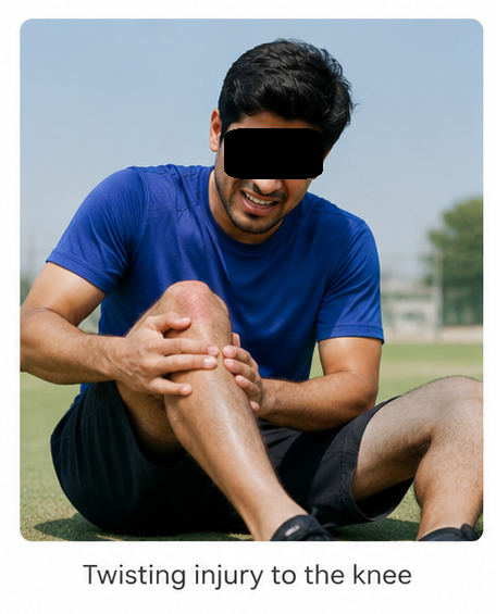
Over the following hours, he developed progressive knee swelling and difficulty walking. He also reported pain during flexion and intermittent clicking within the knee. There was no history of previous knee surgery or significant prior knee injury.
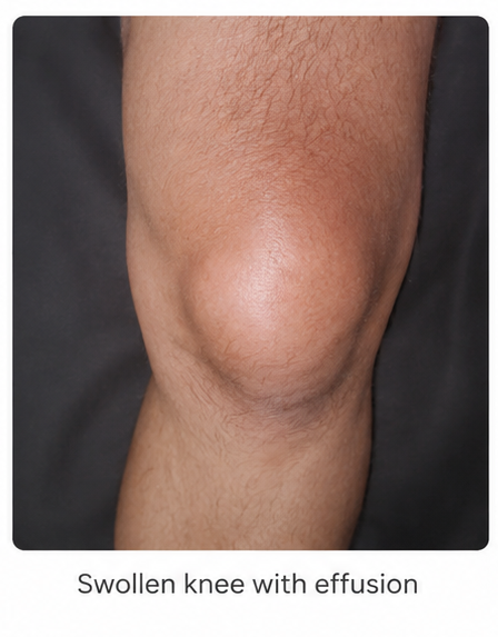
On examination, the patient was ambulatory with an antalgic gait. The right knee demonstrated visible swelling compared with the contralateral knee. A moderate joint effusion was present, with tenderness localized predominantly along the medial joint line.
Active and passive knee movements were restricted because of pain and swelling, particularly during deep flexion. Meniscal provocation produced medial joint-line discomfort. There was no obvious deformity, and distal neurovascular examination was normal.
Plain radiographs of the knee showed no evidence of acute osseous abnormality. Considering the mechanism of injury, joint-line tenderness, mechanical symptoms, and persistent effusion, MRI of the right knee was performed.
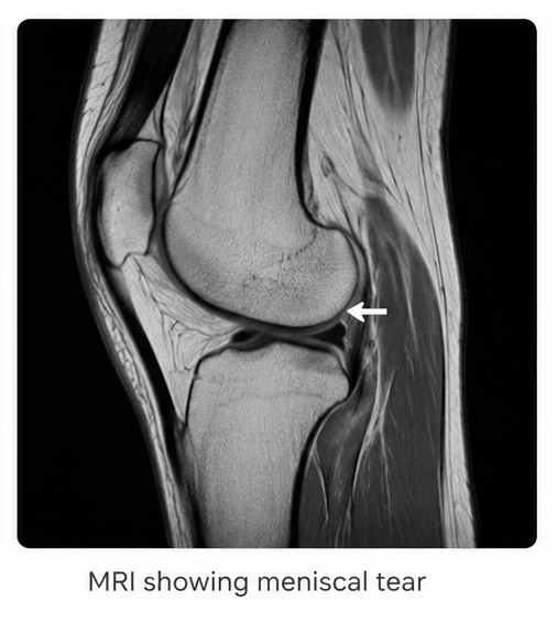
MRI demonstrated a medial meniscal tear with associated moderate knee joint effusion. No evidence of fracture was identified.
Based on the clinical and imaging findings, a diagnosis of traumatic medial meniscal tear with associated knee joint effusion was established.
Management and Outcome
The patient was initially managed conservatively because of the absence of severe mechanical locking or other findings requiring immediate operative intervention.
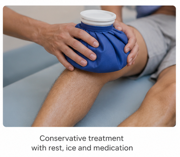
Management included:
- Activity modification and avoidance of twisting or high-impact activities
- Ice application to reduce pain and swelling
- Analgesic medication for symptomatic relief
- Temporary limitation of weight-bearing according to pain tolerance
- Knee range-of-motion exercises
- Progressive quadriceps and lower-limb strengthening
- Structured physiotherapy and functional rehabilitation
The patient was advised to gradually resume activities as pain and swelling improved. Meniscal preservation was emphasized as part of the treatment approach. Conservative management and structured rehabilitation are appropriate for selected patients with meniscal pathology, depending on tear characteristics and clinical presentation.

Serial clinical assessments were performed to monitor pain, knee effusion, range of motion, mechanical symptoms, and functional recovery.
Progressive improvement in symptoms was observed without development of persistent locking, recurrent significant swelling, or other complications.
Follow-up
Two Weeks
- Significant reduction in knee pain and swelling
- Knee effusion had decreased
- Improved ability to walk
- Reduced joint-line tenderness
- Range of motion progressively improved
- Physiotherapy continued
Six Weeks
- Minimal residual knee discomfort
- No significant joint effusion
- Near-complete restoration of knee range of motion
- No recurrent locking or significant clicking
- Improved quadriceps strength
- Gradual return to routine daily activities
Three Months
- Complete resolution of clinically significant knee swelling
- Full functional range of motion
- No recurrent episodes of acute pain or effusion
- Normal walking pattern
- Return to recreational physical activity with appropriate precautions
Discussion
Meniscal tears are common intra-articular knee injuries and may occur following an acute twisting mechanism or develop progressively through degenerative changes. Typical symptoms include joint-line pain, swelling, clicking, catching, locking, and a sensation of the knee giving way.
Knee joint effusion may accompany meniscal injury and represents an important clinical finding during assessment. Traumatic meniscal injuries are among the recognized causes of acute knee effusion, although ligamentous, osseous, and other intra-articular conditions may produce similar findings. Therefore, the mechanism of injury, timing of swelling, physical examination, and appropriate investigations should be considered together.
Clinical examination remains an important component of assessment. Joint-line tenderness and meniscal-specific maneuvers may provide useful information; however, individual clinical tests have limitations and should not be relied upon as the sole basis for diagnosis.
MRI provides detailed assessment of meniscal morphology and associated knee pathology and is particularly useful when the clinical diagnosis requires further clarification. Evidence suggests that experienced clinical examination can be highly informative, while MRI is useful when additional information is required before determining management.
Management should be individualized according to the patient's symptoms, tear characteristics, age, activity level, and functional requirements. Selected patients may improve with conservative treatment incorporating activity modification and structured rehabilitation. Surgical intervention may be considered when symptoms persist or when the tear characteristics and clinical presentation warrant operative treatment.
Current treatment principles increasingly emphasize preservation of the meniscus whenever feasible. Meniscal repair can preserve meniscal tissue and its biomechanical functions, whereas partial meniscectomy may be considered for symptomatic tears that are not amenable to repair.
This case demonstrates that a patient with a traumatic meniscal tear and associated knee joint effusion may achieve favorable functional recovery with appropriate clinical assessment, imaging, conservative management, and structured rehabilitation when there are no indications for immediate surgical intervention.
Prognosis
The prognosis of a meniscal tear depends on the mechanism of injury, tear pattern, location, extent of meniscal involvement, associated knee injuries, symptom severity, and patient-specific factors.
Patients with selected meniscal injuries may experience substantial symptomatic and functional improvement with conservative treatment and rehabilitation. Tears with persistent mechanical symptoms or characteristics that are amenable to repair may require orthopedic intervention. Meniscal preservation remains an important consideration when surgical treatment is required.
Continued clinical follow-up may be appropriate to monitor recurrent pain, swelling, mechanical symptoms, functional limitations, and progression of knee-related pathology.
Conclusion
Meniscal tears can present with knee pain, mechanical symptoms, restricted movement, and associated joint effusion following acute injury. A systematic clinical assessment combined with appropriate imaging is essential for establishing the diagnosis and guiding management.
This case illustrates the favorable recovery of a patient with a traumatic medial meniscal tear and knee joint effusion following conservative treatment and structured rehabilitation. Early recognition, appropriate diagnostic evaluation, symptom-directed management, and preservation of meniscal function are important components of achieving favorable clinical outcomes.
References
- Beaufils P, Becker R, Kopf S, et al. Management of traumatic meniscus tears: the 2019 ESSKA meniscus consensus. Knee Surg Sports Traumatol Arthrosc. 2020;28:1177-1194. https://pubmed.ncbi.nlm.nih.gov/32052121/
- Mordecai SC, Al-Hadithy N, Ware HE, Gupte CM. Treatment of meniscal tears: an evidence based approach. World J Orthop. 2014;5(3):233-241. https://pmc.ncbi.nlm.nih.gov/articles/PMC4095015/
- Johnson MW. Acute knee effusions: a systematic approach to diagnosis. Am Fam Physician. 2000;61(8):2391-2400. https://pubmed.ncbi.nlm.nih.gov/10794580/
- Bhan K. Meniscal tears: current understanding, diagnosis, and management. Cureus. 2020;12(6):e8590. https://pmc.ncbi.nlm.nih.gov/articles/PMC7359983/
- Abram SGF, Beard DJ, Price AJ. Arthroscopic meniscal surgery: a national society treatment guideline and consensus statement. Bone Joint J. 2019;101-B:652-659. https://pubmed.ncbi.nlm.nih.gov/31154847/
Read more such content on @ Hidoc Dr | Medical Learning App for Doctors
Recommended News For You
Recommended Articles For You
Featured News
Featured Articles
Featured Events
Featured KOL Videos
1.
Benefits of perioperative radiotherapy for liver cancer with a high risk of recurrence are investigated in research.
2.
Childhood cancer survivors face new health problems later in life, study shows
3.
Genomic Newborn Screening Could Identify Childhood Cancer Predisposition
4.
Coffee, Tea Tied to Lower Risk of Head and Neck Cancer
5.
Adding Lenvatinib to Pembro Ups PFS in Head and Neck Cancer
1.
Obesity is a major risk factor for cancer
2.
Prevention of Thrombotic Complications in High-Risk Patients
3.
Tailoring the Immune Response: The Dawn of Personalized Vaccines in Cancer Immunotherapy
4.
Megakaryocyte Immune Cell Crosstalk: Mechanisms, Clinical Insights, and Therapeutic Implications
5.
The Truth About TAR Syndrome: Understanding the Rare Genetic Disorder
1.
Asian Symposium on Advancement in Hematology and Oncology (ASAHO)
2.
International Cancer Conference
3.
Asian Symposium on Advancement in Hematology and Oncology (ASAHO)
4.
Asian Symposium on Advancement in Hematology and Oncology
5.
Asian Symposium on Advancement in Hematology and Oncology
1.
Navigating the Complexities of Ph Negative ALL - Part X
2.
Hands-on Workshop: Hematology
3.
Current Scenario of Blood Cancer- A Conclusion on Genomic Testing & Advancement in Diagnosis and Treatment
4.
A Comprehensive Guide to First Line Management of ALK Positive Lung Cancer - Part III
5.
Immunotherapy: A New Era in Cancer Treatment

Address :
Hidoc Dr. Inc. | Delaware C Corp | 1309 Coffeen Ave. Suite 1200, Sheridan WY, 82801
Phone :
+1-415-463-3094
Email :
anishagadia@hidoc.co
© Copyright 2026 Hidoc Dr. Inc.
Terms & Conditions - LLP | Inc. | Privacy Policy - LLP | Inc. | Account Deactivation

To get started please enter your email ID
Welcome to Hidoc Dr.
Join to enhance your clinical skills and gain specialized in-depth medical knowledge