Optic Disc Melanocytoma: A Case Report of an Incidentally Detected Pigmented Optic Nerve Head Lesion
OthersPage Navigation
Abstract
Optic disc melanocytoma is a rare, benign, deeply pigmented tumour arising from melanocytes of the optic nerve head. It is usually discovered incidentally during routine fundus examination and generally follows a stable clinical course. However, its resemblance to malignant melanoma and its potential to cause visual complications or rarely undergo malignant transformation make accurate diagnosis and long-term surveillance important. [1,2]
We describe a representative case of a 42-year-old woman who was referred after a darkly pigmented lesion was detected over the right optic disc during routine ophthalmic examination. Her visual acuity was preserved, and she had no ocular pain, photopsia, or noticeable visual-field loss.
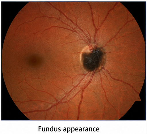
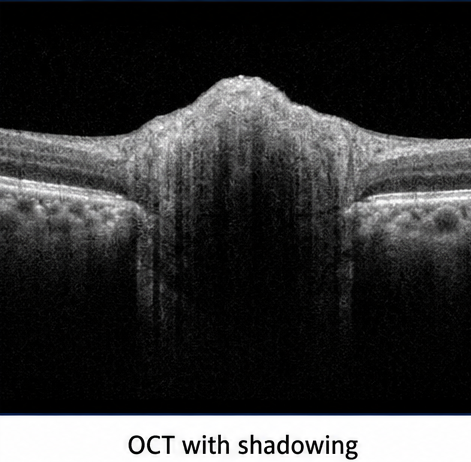
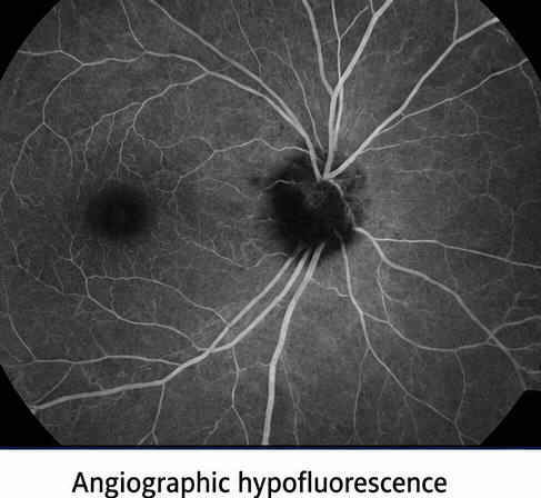
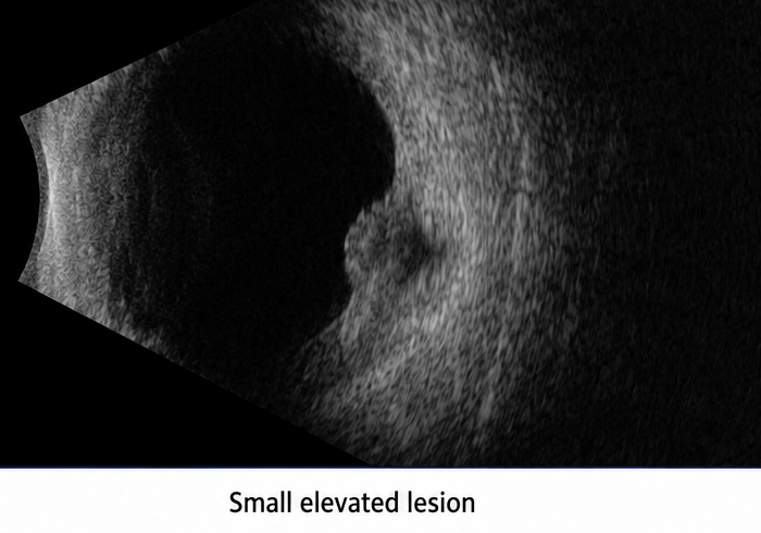
Fundus examination demonstrated a densely pigmented, slightly elevated lesion involving the inferotemporal optic disc with limited extension into the adjacent retina. Optical coherence tomography revealed an elevated hyperreflective lesion with marked posterior shadowing. Fundus fluorescein angiography showed persistent hypofluorescence, while B-scan ultrasonography demonstrated a small acoustically solid lesion without extrascleral extension.
The characteristic clinical and multimodal imaging findings supported a diagnosis of optic disc melanocytoma. Because there were no suspicious features or vision-threatening complications, the patient was managed conservatively with serial fundus photography, optical coherence tomography, ultrasonography, and visual-field assessment. The lesion remained stable during follow-up.
This case highlights the importance of recognizing the characteristic features of optic disc melanocytoma and distinguishing it from malignant melanoma and other pigmented lesions of the optic nerve head.
Introduction
Melanocytoma is a benign melanocytic tumour that may arise anywhere in the uveal tract but most commonly affects the optic disc. It is generally considered a variant of a melanocytic naevus composed of intensely pigmented melanocytes. [1,2]
The lesion typically appears as a dark brown or black mass located on the optic nerve head. It may extend into the adjacent retina or choroid and frequently has feathery margins when retinal nerve-fibre layer involvement is present. Most patients are asymptomatic, and the tumour is detected during routine examination. [1]
Although optic disc melanocytoma usually remains stable, subtle enlargement may occur without indicating malignancy. Visual loss may develop secondary to tumour necrosis, optic nerve compression, retinal vascular obstruction, subretinal fluid, choroidal neovascularisation, or retinal exudation. Malignant transformation is uncommon but remains an important consideration during follow-up. [1–3]
Case Report
A 42-year-old woman was referred to the ophthalmology department after a pigmented lesion was identified over the right optic disc during a routine eye examination. She did not report diminished vision, ocular pain, flashes, floaters, diplopia, or an obvious visual-field defect.
There was no history of ocular trauma, inflammatory eye disease, ocular surgery, cutaneous melanoma, or systemic malignancy. Her medical and family histories were unremarkable.
Best-corrected visual acuity was 6/6 in both eyes. Intraocular pressure was 15 mmHg in the right eye and 14 mmHg in the left eye. Pupillary reactions were normal, with no relative afferent pupillary defect. Colour vision was preserved.
Anterior-segment examination of both eyes was unremarkable. Dilated fundus examination of the right eye revealed a densely pigmented, dark brown-to-black lesion involving the inferotemporal portion of the optic disc. The lesion was slightly elevated and extended minimally into the adjacent peripapillary retina.
The retinal vessels passed over the lesion without significant displacement. There was no orange pigment, retinal haemorrhage, subretinal fluid, hard exudation, vitreous seeding, or prominent feeder vessel. The macula and peripheral retina were normal. Examination of the left fundus was unremarkable.
Investigations
Visual-function Assessment
- Best-corrected visual acuity: 6/6 in both eyes
- Normal colour vision
- No relative afferent pupillary defect
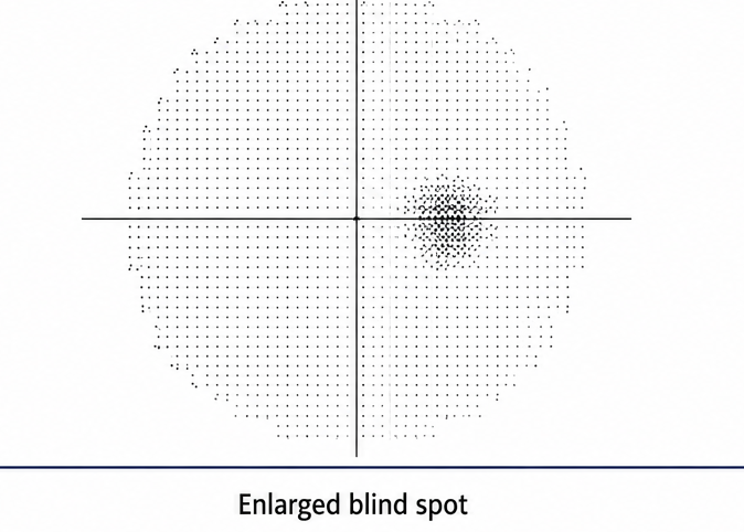
- Automated perimetry demonstrated mild enlargement of the blind spot in the right eye
- Normal visual field in the left eye
Fundus Photography
Colour fundus photography documented:
- A densely pigmented optic disc lesion
- Slight elevation of the lesion
- Limited extension into the adjacent retina
- Feathery margins along the retinal surface
- Absence of haemorrhage, exudation, or subretinal fluid
Optical Coherence Tomography
Spectral-domain optical coherence tomography through the lesion demonstrated:
- An elevated hyperreflective anterior surface
- Dense posterior optical shadowing
- Obscuration of the deeper optic nerve head structures
- Local involvement of the peripapillary retinal nerve-fibre layer
- No intraretinal or subretinal fluid involving the macula
Fundus Autofluorescence
Fundus autofluorescence showed predominantly reduced autofluorescence corresponding to the deeply pigmented lesion.
Fundus Fluorescein Angiography
Fluorescein angiography demonstrated:
- Persistent hypofluorescence over the lesion
- No intrinsic tumour circulation
- No significant late leakage
- No evidence of choroidal neovascularisation
B-scan Ultrasonography
Ocular ultrasonography revealed:
- A small, slightly elevated optic nerve head lesion
- Approximate maximum thickness of 1.2 mm
- Relatively high internal reflectivity
- No extrascleral or retrobulbar extension
Diagnosis
The diagnosis was based on:
- A densely pigmented lesion centred on the optic disc
- Limited extension into the peripapillary retina
- Characteristic feathery margins
- Preserved central visual acuity
- Posterior optical shadowing on optical coherence tomography
- Persistent hypofluorescence on fluorescein angiography
- Absence of orange pigment, subretinal fluid, haemorrhage, or rapid growth
- No evidence of optic nerve or extrascleral extension
The overall clinical and imaging findings were consistent with optic disc melanocytoma.
Differential Diagnosis
The principal differential diagnoses included:
- Juxtapapillary choroidal melanoma
- Choroidal naevus involving the optic disc
- Retinal pigment epithelial hyperplasia
- Combined hamartoma of the retina and retinal pigment epithelium
- Optic disc haemangioma
- Metastatic pigmented tumour
- Pigmented peripapillary choroidal lesion
- Congenital optic disc pigmentation
Juxtapapillary melanoma was the most clinically important differential diagnosis. The absence of progressive growth, orange pigment, subretinal fluid, low internal reflectivity, and invasive features reduced the likelihood of melanoma.
The marked pigmentation, optic disc location, feathery retinal extension, optical coherence tomography shadowing, and angiographic hypofluorescence strongly supported melanocytoma.
Management and Outcome
Because the lesion had characteristic features of optic disc melanocytoma and there were no findings suggestive of malignant transformation or associated vision-threatening complications, active surgical treatment or biopsy was not indicated.
Baseline documentation included:
- Best-corrected visual acuity
- Colour fundus photography
- Optical coherence tomography
- Automated visual-field testing
- Fundus autofluorescence
- Ocular ultrasonography
The patient was counselled regarding the generally benign nature of the lesion and the need for long-term ophthalmic surveillance. She was advised to seek prompt reassessment if she developed reduced vision, a new visual-field defect, flashes, floaters, or ocular discomfort.
At six months, visual acuity remained 6/6, and no change in the lesion’s dimensions or appearance was observed. Optical coherence tomography showed no new retinal or subretinal fluid.
At 12 and 18 months, serial fundus photographs and ultrasonography showed no significant increase in tumour thickness or basal dimensions. The mild blind-spot enlargement remained stable.
Follow-up
Early Follow-up
- Visual acuity remained stable
- No new ocular symptoms developed
- No change in lesion pigmentation or margins
- No retinal haemorrhage or subretinal fluid
- No progression of the visual-field defect
Subsequent Follow-up
- Stable tumour dimensions on fundus photography
- Stable thickness on ultrasonography
- No macular involvement
- No evidence of choroidal neovascularisation
- No optic disc oedema or retinal vascular obstruction
Long-term Follow-up Plan
- Annual dilated fundus examination
- Serial colour fundus photography
- Optical coherence tomography of the lesion and macula
- Periodic automated visual-field testing
- Ultrasonography when clinically indicated
- Earlier review if visual symptoms or lesion enlargement develop
Discussion
Optic disc melanocytoma is a benign, deeply pigmented melanocytic tumour located partly or entirely within the optic nerve head. Histologically, it consists of large, uniform, heavily pigmented cells with small nuclei and minimal mitotic activity. [1,2]
Most melanocytomas are unilateral and are detected incidentally in adults. The characteristic appearance is a dark brown or black, flat or mildly elevated optic disc lesion. Extension into the adjacent retina may produce feathery margins because pigment follows the retinal nerve-fibre bundles. [1]
Visual acuity is usually preserved. Nevertheless, visual-field abnormalities may occur even in otherwise asymptomatic patients. Enlargement of the blind spot is particularly common when the lesion extends beyond the optic disc into the peripapillary retina. Other defects may result from involvement or compression of retinal nerve fibres. [1,3]
Multimodal imaging assists in confirming the diagnosis and establishing baseline measurements. Optical coherence tomography typically demonstrates an elevated hyperreflective lesion with pronounced posterior shadowing caused by dense pigmentation. It can also identify retinal oedema, subretinal fluid, epiretinal membrane formation, or macular complications. [2,4]
Fluorescein angiography usually shows hypofluorescence because the dense melanin blocks background fluorescence. Fundus autofluorescence may similarly demonstrate reduced autofluorescence over the lesion. Ultrasonography is useful for documenting tumour thickness and internal reflectivity, particularly when the lesion is elevated. [2,4]
The principal diagnostic concern is distinguishing melanocytoma from juxtapapillary choroidal melanoma. Features raising concern for malignant transformation include progressive visual loss, rapid enlargement, increasing tumour thickness, extensive optic disc involvement, prominent vascularity, subretinal fluid, haemorrhage, vitreous seeding, and invasive extension. However, slow enlargement alone does not necessarily indicate malignant change because benign melanocytomas may enlarge gradually. [1,3]
Optic disc melanocytoma can occasionally cause visual loss through optic neuropathy, tumour necrosis, retinal vascular obstruction, retinal exudation, subretinal fluid, or choroidal neovascularisation. These complications should be treated according to their underlying mechanism. [1,5]
Observation is the standard management for a typical, asymptomatic lesion. Baseline and serial photography are particularly important because they permit objective comparison of tumour size and margins. Optical coherence tomography, visual-field assessment, and ultrasonography provide complementary structural and functional information.
Biopsy or surgical intervention is generally avoided when the clinical appearance is characteristic. More invasive diagnostic or therapeutic measures may be considered when progressive growth, severe visual deterioration, extrascleral extension, or other findings create substantial concern for malignant transformation.
Prognosis
The prognosis of optic disc melanocytoma is generally favourable. Most lesions remain stable and do not require active treatment.
Mild enlargement may occur over several years without representing malignant transformation. Visual prognosis is usually good when the lesion remains confined and is not associated with optic neuropathy, retinal vascular obstruction, macular involvement, or subretinal fluid.
Malignant transformation is rare, reported in approximately 1–2% of cases. Lifelong surveillance is therefore recommended, even when the lesion has remained stable for many years. [1–3]
Conclusion
Optic disc melanocytoma is a rare, benign pigmented tumour that may resemble malignant melanoma. Recognition of its characteristic funduscopic appearance and multimodal imaging features is essential to prevent unnecessary invasive treatment.
The diagnosis is supported by a deeply pigmented optic disc lesion with feathery retinal extension, posterior shadowing on optical coherence tomography, angiographic hypofluorescence, and absence of suspicious malignant features.
Although most lesions remain stable, periodic surveillance is necessary because melanocytoma may enlarge, produce vision-threatening complications, or rarely undergo malignant transformation. Serial fundus photography, optical coherence tomography, visual-field assessment, and ultrasonography enable reliable long-term monitoring.
References
- Shields JA, Demirci H, Mashayekhi A, Eagle RC Jr, Shields CL. Melanocytoma of the optic disk: a review. Surv Ophthalmol. 2006;51(2):93–104. https://pubmed.ncbi.nlm.nih.gov/16500211/
Giran M, Călugăru D, Călugăru M. Optic disc melanocytoma: a case report. Rom J Ophthalmol. 2021;65(1):91–94. https://pmc.ncbi.nlm.nih.gov/articles/PMC7995506/ - Shields JA, Demirci H, Mashayekhi A, Shields CL. Melanocytoma of the optic disk in 115 cases: the 2004 Samuel Johnson Memorial Lecture, part 1. Ophthalmology. 2004;111(9):1739–1746. https://pubmed.ncbi.nlm.nih.gov/15350332/
Shields CL, Perez B, Benavides R, Materin MA, Shields JA. Optical coherence tomography of optic disk melanocytoma in 15 cases. Retina. 2008;28(3):441–446. https://pubmed.ncbi.nlm.nih.gov/18327136/
Zhang P, Hui YN, Xu WQ, et al. Infrared autofluorescence, short-wave autofluorescence and spectral-domain optical coherence tomography of optic disk melanocytomas. Int J Ophthalmol. 2016;9(5):713–716. https://pmc.ncbi.nlm.nih.gov/articles/PMC4886876/
Read more such content on @ Hidoc Dr | Medical Learning App for Doctors
Recommended News For You
Recommended Articles For You
Featured News
Featured Articles
Featured Events
Featured KOL Videos
1.
The need for more Latinx participants in Alzheimer's trials is urgent.
2.
Study reveals crucial gaps in oral cancer awareness in Middle East and North Africa
3.
Disparities in use of MRI to detect prostate cancer: Race and location may influence testing
4.
In breast-conserving surgery, is micro-CT a useful tool for determining tumor margins?
5.
Study offers clearer picture of childhood brain tumor survival
1.
Revolutionizing Cancer Care: The Promise of Early Detection and Biomarkers
2.
The Oncologist’s Edge: Tools, Trials, and Tech Transforming Treatment
3.
Comprehensive Standards in Oncology for Modern Medicine
4.
Cardio-Oncology: Managing Heart Failure in Survivors of Cancer
5.
Practical Standards in Hematology for Healthcare Excellence
1.
Asian Symposium on Advancement in Hematology and Oncology (ASAHO)
2.
International Cancer Conference
3.
Asian Symposium on Advancement in Hematology and Oncology (ASAHO)
4.
Asian Symposium on Advancement in Hematology and Oncology
5.
Asian Symposium on Advancement in Hematology and Oncology
1.
Targeting Oncologic Drivers with Dacomitinib: A New Approach to Lung Cancer Treatment
2.
Revolutionizing Treatment of ALK Rearranged NSCLC with Lorlatinib - Part II
3.
Efficient Management of First line ALK-rearranged NSCLC - Part VIII
4.
Nimotozumab Plus Chemo-radiotherapy v/s Placebo Plus Chemo-radiotherapy in Locally Advanced Nasopharyngeal Carcinoma
5.
Breaking Ground: ALK-Positive Lung Cancer Front-Line Management - Part V

Address :
Hidoc Dr. Inc. | Delaware C Corp | 1309 Coffeen Ave. Suite 1200, Sheridan WY, 82801
Phone :
+1-415-463-3094
Email :
anishagadia@hidoc.co
© Copyright 2026 Hidoc Dr. Inc.
Terms & Conditions - LLP | Inc. | Privacy Policy - LLP | Inc. | Account Deactivation

To get started please enter your email ID
Welcome to Hidoc Dr.
Join to enhance your clinical skills and gain specialized in-depth medical knowledge