Small Bowel Obstruction Due to Postoperative Adhesions: A Case Report
OthersPage Navigation
Abstract
Small bowel obstruction (SBO) is a common surgical emergency caused by partial or complete interruption of intestinal transit. Postoperative adhesions are among the most frequent causes of mechanical SBO. Patients commonly present with abdominal pain, vomiting, abdominal distension, and inability to pass stools or flatus. Early diagnosis and appropriate management are essential to prevent bowel ischemia, strangulation, necrosis, and perforation. We report the case of a 46-year-old woman with previous abdominal surgery who presented with abdominal pain, repeated vomiting, progressive distension, and obstipation. CT demonstrated dilated small bowel loops with a transition point suggestive of adhesive obstruction, without evidence of ischemia or perforation. Initial conservative management failed, and exploratory laparotomy revealed dense postoperative adhesions causing mechanical obstruction. Adhesiolysis successfully relieved the obstruction, and the patient recovered uneventfully. This case highlights the importance of timely diagnosis, close monitoring, and appropriate surgical intervention in adhesive SBO.
Introduction
Small bowel obstruction results from mechanical or functional impairment of intestinal transit. Common mechanical causes include postoperative adhesions, hernias, malignancy, inflammatory strictures, volvulus, and intussusception. Adhesions following abdominal or pelvic surgery are a frequent cause.
Typical symptoms include colicky abdominal pain, vomiting, abdominal distension, and obstipation. Prolonged obstruction may lead to strangulation, ischemia, necrosis, or perforation. CT imaging helps identify the transition point, underlying cause, and complications. Patients without signs of bowel compromise may initially undergo conservative management, while surgery is indicated for complications or failure of nonoperative treatment.
Case Report
A 46-year-old woman presented with a 2-day history of progressively worsening abdominal pain, repeated vomiting, abdominal distension, and inability to pass stools or flatus for approximately 24 hours. The pain was initially intermittent and colicky but later became persistent.
She had undergone abdominal surgery for a gynecological condition 8 years earlier. There was no history of inflammatory bowel disease, recent trauma, fever, hematemesis, or melena.
On examination, the patient appeared uncomfortable and mildly dehydrated. Her pulse rate was mildly elevated, while blood pressure and respiratory rate were stable. Abdominal examination revealed generalized distension and diffuse tenderness, predominantly in the central and lower abdomen, without guarding or rebound tenderness. Bowel sounds were hyperactive and high-pitched.
Laboratory investigations showed mild hemoconcentration and electrolyte abnormalities consistent with dehydration and vomiting. The white blood cell count was mildly elevated, while serum lactate was normal.
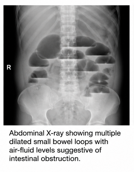
Abdominal radiography demonstrated multiple dilated small bowel loops with air-fluid levels.
Contrast-enhanced CT revealed multiple dilated, fluid-filled small bowel loops with a transition point in the distal ileum, consistent with mechanical obstruction likely caused by postoperative adhesions.
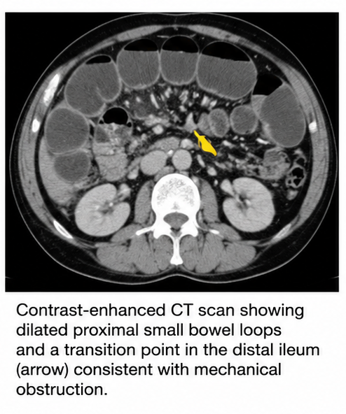
No evidence of perforation, pneumoperitoneum, or advanced bowel ischemia was identified.
A diagnosis of adhesive small bowel obstruction was established.
Management and Outcome
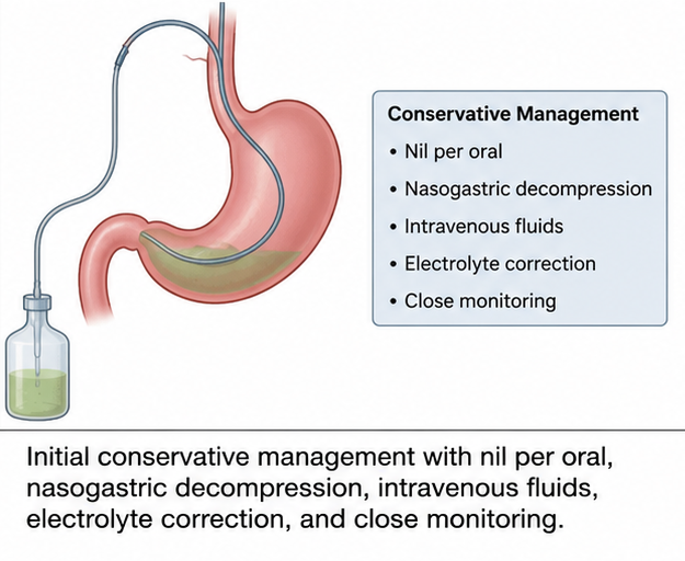
The patient was initially managed conservatively under surgical supervision. Oral intake was withheld, and a nasogastric tube was inserted for decompression. Intravenous fluids were administered, and electrolyte abnormalities were corrected. Serial abdominal examinations and laboratory monitoring were performed.
Although vomiting and abdominal distension partially improved, abdominal pain persisted, and the patient failed to pass flatus or stool. In view of persistent obstructive symptoms, exploratory laparotomy was performed.
Intraoperatively, multiple dense fibrous adhesions involving the small bowel were identified, producing a transition point and mechanical obstruction.
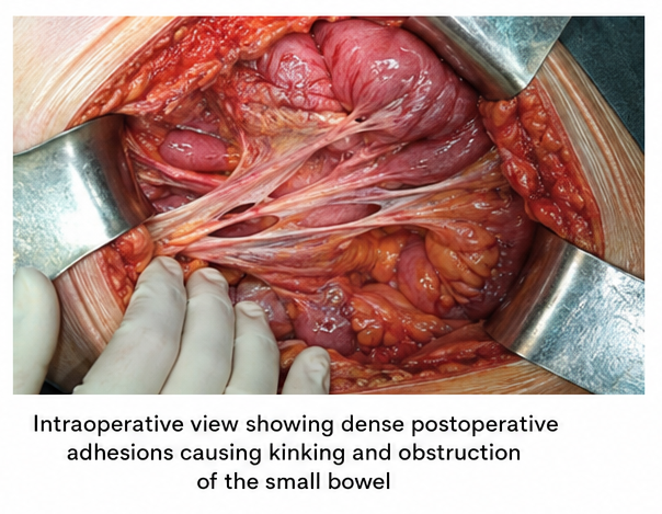
Adhesiolysis was performed, releasing the obstruction.
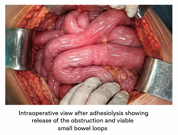
The bowel was viable, with no necrosis or perforation, and bowel resection was not required.
Postoperatively, the patient received intravenous fluids, analgesics, and supportive care. Nasogastric decompression was discontinued after bowel function returned. Oral intake was gradually resumed and well tolerated. The patient passed flatus and subsequently resumed normal bowel movements before being discharged in stable condition.
Follow-up
One Week
-
The surgical wound was healing without infection.
-
Abdominal discomfort had significantly improved.
-
Bowel movements had returned to normal.
-
No recurrent vomiting or distension was reported.
One Month
-
The patient remained clinically well and tolerated a normal diet.
-
No evidence of recurrent obstruction was identified.
-
The surgical incision had healed satisfactorily.
Three Months
-
The patient remained asymptomatic with normal bowel function.
-
No recurrent abdominal pain, vomiting, or obstipation was reported.
-
The patient was counseled regarding the risk of recurrent adhesive obstruction.
Discussion
Small bowel obstruction is a common surgical emergency requiring prompt diagnosis and management. Postoperative adhesions are a major cause of mechanical SBO and can compress or kink bowel loops, impairing intestinal transit.
Patients typically present with abdominal pain, vomiting, distension, and obstipation. Prolonged obstruction may compromise intestinal blood flow, resulting in strangulation, ischemia, necrosis, or perforation. Severe continuous pain, peritoneal signs, fever, tachycardia, leukocytosis, metabolic acidosis, and elevated lactate may indicate bowel compromise.
CT imaging is valuable for identifying dilated bowel loops, the transition point, the underlying cause, and complications such as closed-loop obstruction, ischemia, and perforation.
In patients with adhesive SBO without evidence of strangulation, ischemia, or perforation, nonoperative management with bowel rest, intravenous fluids, electrolyte correction, and nasogastric decompression may be appropriate. Close monitoring is essential.
Surgery is indicated when bowel compromise is suspected or symptoms persist despite conservative treatment. Adhesiolysis is the primary surgical approach, while bowel resection may be necessary when nonviable or perforated bowel is identified.
In the present case, conservative management was initially attempted because there was no evidence of ischemia or perforation. However, persistent symptoms prompted timely surgical exploration. Adhesiolysis successfully relieved the obstruction, and the viable bowel avoided the need for resection.
Prognosis
The prognosis of SBO depends on its cause, duration, severity, and complications. Uncomplicated adhesive obstruction generally has a favorable outcome with timely treatment.
In this case, the absence of ischemia, necrosis, or perforation contributed to a favorable outcome. However, recurrent adhesive SBO remains possible following abdominal surgery. Patients should be advised to seek prompt medical attention for recurrent abdominal pain, vomiting, distension, or inability to pass stools or flatus.
Conclusion
Small bowel obstruction is an important surgical emergency requiring rapid clinical assessment and appropriate imaging. Postoperative adhesions are a common cause of mechanical SBO and may present with abdominal pain, vomiting, distension, and obstipation.
CT helps determine the location and severity of obstruction and identify complications. Conservative management may be appropriate in selected patients without bowel compromise, while persistent symptoms or signs of ischemia, strangulation, or perforation require timely surgical intervention.
In this case, early recognition of failed conservative management and surgical adhesiolysis resulted in successful resolution and an uncomplicated recovery. Close monitoring and timely intervention are essential to prevent potentially life-threatening complications.
References
-
Ten Broek RPG, Krielen P, Di Saverio S, et al. Bologna guidelines for diagnosis and management of adhesive small bowel obstruction. World Journal of Emergency Surgery. 2018;13:24. https://pubmed.ncbi.nlm.nih.gov/29946347/
-
Maung AA, Johnson DC, Piper GL, et al. Evaluation and management of small-bowel obstruction. Journal of Trauma and Acute Care Surgery. 2012;73(5 Suppl 4):S362-S369. https://pubmed.ncbi.nlm.nih.gov/23114494/
-
Bower KL, Lollar DI, Williams SL, et al. Small bowel obstruction. Surgical Clinics of North America. 2018;98(5):945-971. https://pubmed.ncbi.nlm.nih.gov/30275275/
-
Ceresoli M, Coccolini F, Catena F, et al. Water-soluble contrast agent in adhesive small bowel obstruction. American Journal of Surgery. 2016;211(6):1114-1125. https://pubmed.ncbi.nlm.nih.gov/26895669/
-
Amara Y, Leppaniemi A, Catena F, et al. Diagnosis and management of small bowel obstruction in virgin abdomen. World Journal of Emergency Surgery. 2021;16:36. https://pubmed.ncbi.nlm.nih.gov/34217331/
Read more such content on @ Hidoc Dr | Medical Learning App for Doctors
Recommended News For You
Recommended Articles For You
Featured News
Featured Articles
Featured Events
Featured KOL Videos
1.
According to the Lancet, having cancer and VTE at the same time increases mortality risk.
2.
Pooled Cord Blood Transplants Achieve Near-Perfect Results in Early Trial
3.
Experts say oncology, primary care coordination necessary for best cancer patient outcomes
4.
Urinary tract cancer prediction models with high sensitivity and specificity
5.
New guidelines for radiation therapy for HPV-associated head and neck cancer
1.
The Importance of Early Detection in Skin Cancer: Why Regular Skin Exams Matter
2.
Clinical Frameworks in Hematology for Better Care
3.
Bridging Disciplines: Medical Oncology vs Surgical Oncology in the Era of Multidisciplinary Cancer Care
4.
Unveiling the Hidden Mechanisms of Hemolytic Reactions
5.
Practical Applications in Hematology Across Clinical Settings
1.
Asian Symposium on Advancement in Hematology and Oncology (ASAHO)
2.
International Cancer Conference
3.
Asian Symposium on Advancement in Hematology and Oncology (ASAHO)
4.
Asian Symposium on Advancement in Hematology and Oncology
5.
Asian Symposium on Advancement in Hematology and Oncology
1.
Understanding Early Relapse in B-cell ALL: Rates, Risks, and Common Sites
2.
Thromboprophylaxis In Medical Settings- An Update
3.
Recognizing Symptoms That May Require Cancer Evaluation
4.
Updates on Standard V/S High Risk Myeloma Treatment- The Next Part
5.
Recognizing the Signs of Anemia in Adults

Address :
Hidoc Dr. Inc. | Delaware C Corp | 1309 Coffeen Ave. Suite 1200, Sheridan WY, 82801
Phone :
+1-415-463-3094
Email :
anishagadia@hidoc.co
© Copyright 2026 Hidoc Dr. Inc.
Terms & Conditions - LLP | Inc. | Privacy Policy - LLP | Inc. | Account Deactivation

To get started please enter your email ID
Welcome to Hidoc Dr.
Join to enhance your clinical skills and gain specialized in-depth medical knowledge