Strangulate Small Bowel Obstruction Requiring Emergency Bowel Resection
OthersPage Navigation
Introduction
Bowel resection is the surgical removal of a diseased or non-viable segment of the small or large intestine. It may be required for intestinal obstruction, ischemia, perforation, trauma, inflammatory bowel disease, diverticular disease, or malignancy. In emergency practice, one of the most important indications is a strangulated small bowel obstruction, in which impaired venous drainage and arterial blood supply can rapidly progress to transmural necrosis, perforation, peritonitis, sepsis, and death.
Adhesions are a common cause of small bowel obstruction in adults, particularly following previous abdominal surgery. Many uncomplicated adhesive obstructions improve with bowel rest, nasogastric decompression, intravenous fluids, and close observation. However, persistent pain, peritoneal signs, hemodynamic instability, increasing lactate, leukocytosis, metabolic acidosis, or computed tomography findings of a closed-loop obstruction and reduced bowel-wall enhancement suggest ischemia and require urgent surgical exploration.
The surgical objectives are to release the obstruction, preserve viable intestine, resect irreversibly ischemic bowel, and restore gastrointestinal continuity when it is safe to do so.
Case Report
A 56-year-old woman presented to the emergency department with severe, colicky central abdominal pain, progressive abdominal distension, repeated bilious vomiting, and an inability to pass stool or flatus for approximately 24 hours. The pain had initially occurred in waves but had become constant and more intense during the six hours before presentation.
She had undergone an open total abdominal hysterectomy eight years earlier. There was no history of inflammatory bowel disease, abdominal malignancy, hernia, chronic constipation, or previous bowel obstruction. She had hypertension controlled with amlodipine and did not regularly use anticoagulants or nonsteroidal anti-inflammatory drugs.
On examination, the patient appeared dehydrated, anxious, and acutely unwell. Her temperature was 38.1°C, pulse rate was 118 beats/minute, blood pressure was 96/62 mmHg, respiratory rate was 24 breaths/minute, and oxygen saturation was 97% on room air.
The abdomen was markedly distended and diffusely tender, with maximal tenderness in the periumbilical and lower abdominal regions. Guarding and rebound tenderness were present. Bowel sounds were initially high-pitched but became hypoactive during reassessment. No external hernia was identified. Digital rectal examination revealed an empty rectum without blood.
The initial clinical impression was acute small bowel obstruction complicated by strangulation and evolving bowel ischemia. Resuscitation and surgical assessment were initiated immediately.
Investigations
Laboratory testing showed a hemoglobin level of 13.1 g/dL, leukocyte count of 18,400 cells/mm³ with neutrophilia, and platelet count of 312,000 cells/mm³.
Serum sodium was 132 mmol/L, potassium was 3.2 mmol/L, bicarbonate was 18 mmol/L, blood urea nitrogen was 32 mg/dL, and serum creatinine was 1.4 mg/dL. C-reactive protein was elevated at 86 mg/L. Serum lactate was 4.1 mmol/L and remained elevated after initial fluid resuscitation. Liver-function tests, serum amylase, and lipase were not significantly abnormal.
An erect abdominal radiograph demonstrated multiple centrally located dilated small-bowel loops with step-ladder air-fluid levels and a relative absence of colonic gas.
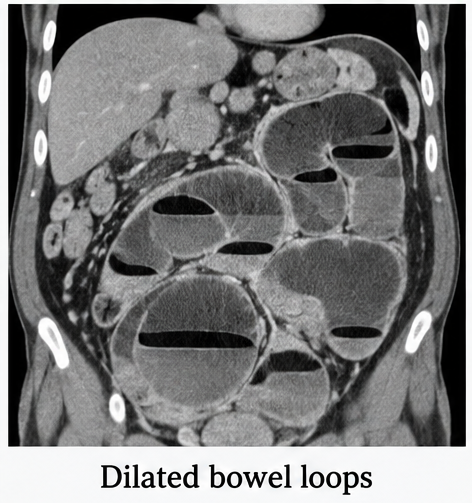
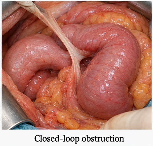
Contrast-enhanced computed tomography of the abdomen and pelvis showed a closed-loop small bowel obstruction in the lower abdomen. Two adjacent transition points created a C-shaped loop of dilated ileum. Mesenteric edema, converging mesenteric vessels, free intraperitoneal fluid, bowel-wall thickening, and reduced mural enhancement raised strong concern for strangulation and ischemia. No free intraperitoneal air was present.
The final preoperative diagnosis was adhesive closed-loop small bowel obstruction with suspected ischemia of the ileum.
Differential Diagnosis
Incarcerated or Strangulated Hernia
A trapped external or internal hernia can cause intestinal obstruction and vascular compromise. Careful examination revealed no groin, umbilical, or incisional hernia. Computed tomography localized the transition points to an adhesive band within the pelvis.
Acute Mesenteric Ischemia
Arterial embolism or thrombosis can cause severe abdominal pain, elevated lactate, and bowel ischemia. In this patient, imaging demonstrated a mechanical closed-loop obstruction without occlusion of the superior mesenteric artery or vein.
Perforated Viscus
Gastrointestinal perforation may produce sudden abdominal pain, guarding, sepsis, and free intraperitoneal air. The absence of pneumoperitoneum and the presence of dilated bowel loops with two transition points favored strangulated obstruction, although impending perforation remained a concern.
Acute Pancreatitis
Acute pancreatitis may cause severe abdominal pain, vomiting, paralytic ileus, and systemic inflammation. Normal pancreatic enzyme levels and the characteristic computed tomography findings excluded this diagnosis.
Large Bowel Obstruction
Colorectal cancer, volvulus, or diverticular stricture may cause abdominal distension, vomiting, and obstipation. Imaging showed a decompressed colon and a transition within the distal small bowel rather than the large intestine.
Management and Outcome
Two large-bore intravenous cannulas were inserted. The patient received balanced crystalloid fluids, supplemental oxygen, electrolyte replacement, urinary catheterization, and continuous hemodynamic monitoring.
A nasogastric tube drained approximately 900 mL of bilious fluid. Broad-spectrum intravenous antibiotics providing gram-negative and anaerobic coverage were administered. Venous thromboembolism prophylaxis was also planned.
Because the patient had peritonitis, persistent hyperlactatemia, and computed tomography evidence of a closed-loop obstruction with suspected ischemia, conservative management was considered unsafe.
After informed consent, the patient underwent emergency exploratory laparotomy. Dense adhesions were found between the distal ileum, pelvic sidewall, and previous operative scar. A fibrous adhesive band had created a closed loop and constricted approximately 35 cm of the distal ileum.
The affected intestinal segment was dark purple, edematous, aperistaltic, and without palpable mesenteric arterial pulsation. Application of warm packs and release of the constricting band did not restore bowel colour, peristalsis, or perfusion, confirming irreversible ischemia.
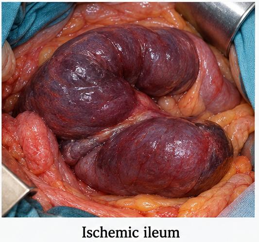
The adhesive band was divided, and careful adhesiolysis was performed. The non-viable ileal segment was resected with margins extending into well-perfused bowel. Approximately 20 cm of healthy terminal ileum was preserved proximal to the ileocecal valve.
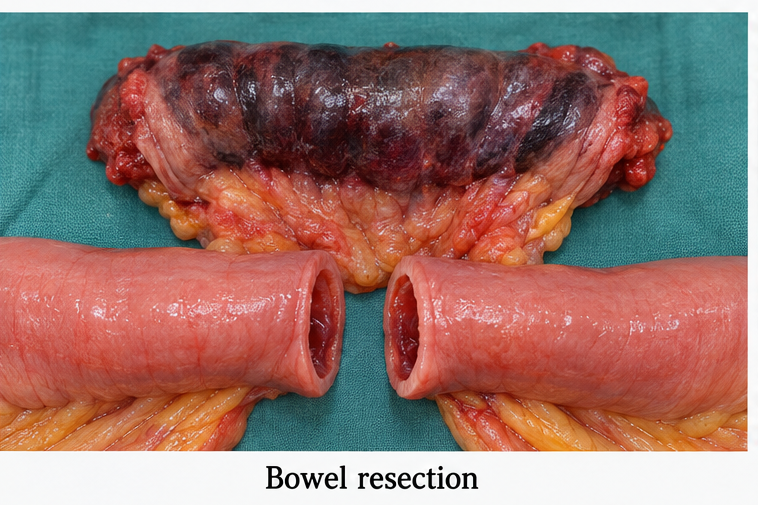
After reassessing bowel perfusion, a stapled side-to-side functional end-to-end anastomosis was created. The mesenteric defect was closed, and hemostasis was secured. The remaining small bowel was examined from the duodenojejunal flexure to the ileocecal junction. No additional ischemic segment or perforation was identified.
A primary anastomosis was selected instead of a stoma because the patient stabilized following resuscitation, the bowel ends were healthy, and there was no gross peritoneal contamination.
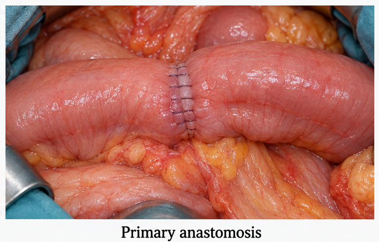
Postoperatively, the patient was monitored in a high-dependency unit. Intravenous fluids, analgesia, antibiotics, nasogastric decompression, thromboprophylaxis, and serial clinical and laboratory assessments were continued.
The serum lactate decreased to 1.8 mmol/L within 12 hours. Urine output improved, and renal function returned to normal. The nasogastric tube was removed on postoperative day two after the drainage decreased and the nausea resolved.
The patient passed flatus on postoperative day three and stool on postoperative day four. Oral intake was gradually advanced from clear liquids to a soft diet. There was no anastomotic leak, intra-abdominal collection, wound infection, prolonged ileus, or recurrent obstruction.
Histopathological examination of the resected bowel showed transmural ischemic necrosis and hemorrhage without evidence of malignancy or inflammatory bowel disease.
The patient was discharged on postoperative day seven in stable condition. At the six-week follow-up, the surgical wound had healed, bowel function was regular, oral intake was adequate, and she had resumed her routine activities.
She was counselled regarding symptoms requiring urgent medical review, including recurrent colicky abdominal pain, persistent vomiting, abdominal distension, fever, wound discharge, or inability to pass stool or flatus.
Discussion
This case demonstrates the progression of adhesive small bowel obstruction to closed-loop strangulation and irreversible intestinal ischemia. A closed-loop obstruction occurs when a bowel segment is obstructed at two points. Continued secretion and gas accumulation increase intraluminal pressure, while mesenteric compression initially impairs venous and lymphatic drainage and subsequently compromises arterial blood supply.
The resulting intestinal edema, hemorrhage, bacterial translocation, and tissue necrosis may progress rapidly. Without timely treatment, bowel perforation, peritonitis, sepsis, and death may occur.
Previous abdominal surgery is the principal risk factor for adhesive small bowel obstruction. The interval between the original operation and the development of obstruction may extend over several years, as demonstrated in this patient.
The diagnosis depends on integrating clinical symptoms, physical examination findings, laboratory trends, and imaging results. No single laboratory value reliably confirms bowel ischemia. Leukocytosis, metabolic acidosis, and elevated lactate levels support concern for tissue hypoperfusion but may be absent during the early stages of ischemia. These abnormalities may also occur because of dehydration or sepsis from other causes.
Computed tomography helps identify the location and cause of obstruction and detect features associated with strangulation. Two transition points, a C-shaped or U-shaped bowel loop, mesenteric vessel convergence, mesenteric edema, free fluid, bowel-wall thickening, pneumatosis intestinalis, portal venous gas, and reduced or absent mural enhancement increase concern for closed-loop obstruction and impaired bowel perfusion.
Imaging should not delay surgery when generalized peritonitis or clinical deterioration is already evident.
Hemodynamically stable patients without peritonitis, strangulation, or ischemia may undergo a closely monitored trial of non-operative management. This approach generally includes fasting, nasogastric decompression when indicated, intravenous fluid and electrolyte correction, and repeated abdominal examinations. Water-soluble contrast may support diagnosis and help predict the likelihood of non-operative resolution in selected patients with adhesive obstruction.
In contrast, this patient had constant abdominal pain, guarding, hypotension, persistent lactate elevation, free intraperitoneal fluid, and reduced bowel-wall enhancement. These findings justified immediate surgical exploration.
Intraoperative assessment of bowel viability is based on intestinal colour, peristalsis, mesenteric pulsation, bleeding from a cut edge, and adjunctive perfusion-assessment methods where available. Clearly necrotic bowel must be resected. However, excessive removal of potentially viable intestine should be avoided because extensive resection may result in short-bowel syndrome and long-term nutritional dependence.
When bowel viability remains uncertain, temporary abdominal closure followed by planned second-look surgery may be appropriate, particularly in patients with extensive ischemia or physiological instability.
The decision between primary anastomosis and stoma formation depends on perfusion of the bowel ends, extent of contamination, anastomotic location, bowel edema, hemodynamic stability, comorbidities, vasopressor requirement, and overall risk of anastomotic leakage.
In this patient, limited bowel resection, healthy resection margins, absence of perforation, and restoration of physiological stability supported primary anastomosis. Postoperative surveillance remained essential because anastomotic leakage, intra-abdominal abscess, ileus, wound complications, and recurrent adhesive obstruction may occur following bowel resection.
Conclusion
Strangulated small bowel obstruction is a surgical emergency in which delayed treatment may result in intestinal necrosis, perforation, sepsis, extensive bowel resection, and death. In this patient, a postoperative adhesive band caused a closed-loop obstruction and irreversible ischemia of the ileum.
Early recognition of peritoneal signs, persistent lactate elevation, and high-risk computed tomography findings prompted urgent exploratory laparotomy. Adhesiolysis, resection of the non-viable ileum, and primary anastomosis resulted in an uncomplicated recovery.
Careful resuscitation, timely operative decision-making, preservation of viable bowel, secure intestinal reconstruction, and structured postoperative monitoring were central to the favourable outcome.
References
- Ten Broek RPG, et al. Bologna guidelines for diagnosis and management of adhesive small bowel obstruction: 2017 update. World Journal of Emergency Surgery. 2018;13:24. https://pubmed.ncbi.nlm.nih.gov/29946347/
- Bala M, et al. Acute mesenteric ischemia: Updated guidelines of the World Society of Emergency Surgery. World Journal of Emergency Surgery. 2022;17:54. https://pubmed.ncbi.nlm.nih.gov/36085246/
- Catena F, et al. Bowel obstruction: A narrative review for all physicians. World Journal of Emergency Surgery. 2019;14:20. https://pubmed.ncbi.nlm.nih.gov/31168315/
- Paulson EK, Thompson WM. Review of small-bowel obstruction: The diagnosis and when to worry. Radiology. 2015;275(2):332–342. https://pubmed.ncbi.nlm.nih.gov/25906300/
- Ten Broek RPG, et al. Burden of adhesions in abdominal and pelvic surgery: Systematic review and meta-analysis. BMJ. 2013;347:f5588. https://pubmed.ncbi.nlm.nih.gov/24092941/
Read more such content on @ Hidoc Dr | Medical Learning App for Doctors
Recommended News For You
Recommended Articles For You
Featured News
Featured Articles
Featured Events
Featured KOL Videos
1.
Regular physical activity before cancer diagnosis may lower progression and death risks
2.
Medication Self-Injection Embraced by Breast Cancer Patients
3.
Financial hardship for cancer survivors due to high-cost immunotherapies, especially for blood cancer patients
4.
FDA Advisors Split on Regimens for Breast, Prostate Cancers
5.
ctDNA-Guided Adjuvant Therapy Cuts Bladder Cancer Costs
1.
RDW-CV vs. MCV: Knowing the Difference Can Help You Better Understand Your Health
2.
Clinical Pathways in Hematology in Clinical Decision-Making
3.
Immune Surveillance Screening for Early Cancer Risk Detection
4.
Unlocking the Mysteries of Lymphoblastic Lymphoma: A Journey Into the Unknown
5.
AI-Driven Digital Pathology in Oncology: From Histology Images to Predictive Treatment Biomarkers
1.
International Conference on Innovations in Critical Care for Oncology and Cardiology
2.
International Conference on Oncology, Cardiology and Critical Care Policy
3.
International Conference on Oncology, Cancer Prevention and Public Health
4.
International Conference on Cancer Nursing and Rehabilitation Strategies
5.
International Conference on Best Practices in Oncology, Cardiology and Critical Care
1.
How Advances in Cancer Research Are Improving Treatment Options
2.
Navigating the Complexities of Ph Negative ALL - Part II
3.
Signs and Symptoms of Leukemia
4.
Updates on Standard V/S High Risk Myeloma Treatment- The Next Part
5.
Virtual Case Study on Elephantiasis of Lower Limb- An Initiative by Hidoc Dr.

Address :
Hidoc Dr. Inc. | Delaware C Corp | 1309 Coffeen Ave. Suite 1200, Sheridan WY, 82801
Phone :
+1-415-463-3094
Email :
anishagadia@hidoc.co
© Copyright 2026 Hidoc Dr. Inc.
Terms & Conditions - LLP | Inc. | Privacy Policy - LLP | Inc. | Account Deactivation

To get started please enter your email ID
Welcome to Hidoc Dr.
Join to enhance your clinical skills and gain specialized in-depth medical knowledge