Pemphigus Vulgaris Presenting With Extensive Mucocutaneous Blistering: A Case Report
OthersPage Navigation
Abstract
Pemphigus vulgaris is a chronic autoimmune blistering disorder characterized by pathogenic autoantibodies directed against desmosomal proteins, resulting in loss of keratinocyte adhesion and intraepidermal blister formation. The disease commonly affects the oral mucosa and skin and may present with painful erosions, flaccid blisters, and mucocutaneous ulceration. Early diagnosis and appropriate treatment are essential to prevent complications such as secondary infection, fluid loss, and nutritional impairment. We report the case of a 42-year-old man who presented with painful oral erosions followed by multiple fragile blisters and erosions over the trunk, face, and extremities. Clinical examination revealed widespread superficial erosions and flaccid bullae involving the skin and oral mucosa, with a positive Nikolsky sign. Histopathological examination demonstrated suprabasal acantholysis, while direct immunofluorescence revealed intercellular immunoglobulin G deposition in a characteristic reticular pattern. The findings confirmed a diagnosis of pemphigus vulgaris. The patient was treated with systemic corticosteroid therapy and steroid-sparing immunosuppressive treatment, along with wound care and supportive management. Significant clinical improvement was observed during follow-up, with healing of mucocutaneous lesions and no development of new significant blisters. This case highlights the importance of early recognition, immunopathological confirmation, and timely systemic treatment of pemphigus vulgaris.
Introduction
Pemphigus vulgaris is a rare, potentially serious autoimmune vesiculobullous disorder characterized by intraepidermal blister formation due to autoantibody-mediated disruption of desmosomal adhesion between keratinocytes. The disease primarily affects the skin and mucous membranes and commonly presents with painful mucosal erosions followed by fragile, flaccid blisters and superficial cutaneous erosions.
The oral mucosa is frequently involved and may represent the initial site of disease manifestation. Patients may develop painful erosions involving the buccal mucosa, palate, gingiva, and tongue, which can interfere with eating, drinking, and oral hygiene. Cutaneous lesions typically appear as flaccid blisters that rupture easily, leaving superficial erosions and crusted areas.
Pemphigus vulgaris results primarily from autoantibodies against desmoglein 3, with additional involvement of desmoglein 1 in patients with mucocutaneous disease. Diagnosis is based on clinical findings supported by histopathology, direct immunofluorescence, and, when appropriate, serological testing for desmoglein-specific autoantibodies.
Treatment aims to achieve rapid disease control, promote healing, prevent new blister formation, and minimize treatment-related complications. Systemic corticosteroids remain an important component of therapy, while steroid-sparing immunosuppressive agents and biologic therapies may be used according to disease severity and patient factors.
We report a case of pemphigus vulgaris presenting with extensive mucocutaneous involvement, emphasizing the importance of early diagnosis, immunopathological confirmation, and prompt treatment.
Case Report
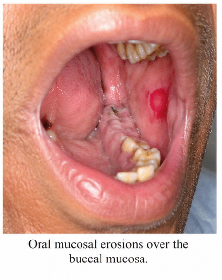
A 42-year-old man presented to the dermatology outpatient department with a 2-month history of painful oral erosions, followed by multiple blisters and erosions over the trunk, face, and extremities. The patient initially developed painful oral ulcers that progressively increased in number and interfered with eating and drinking.
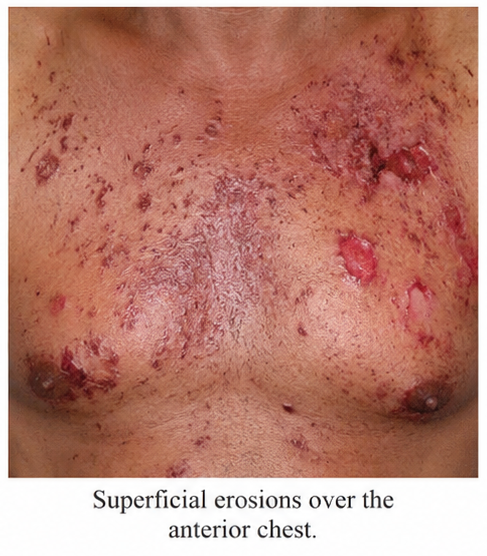
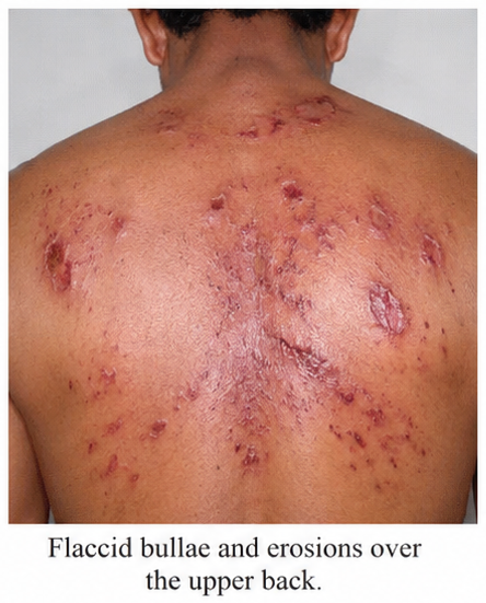
Approximately 3 weeks later, he developed fragile blisters over the chest and upper back that ruptured easily, leaving painful superficial erosions.
The patient had no previous history of autoimmune blistering disease or similar skin lesions. There was no history of recent medication exposure preceding symptom onset, immunosuppressive therapy, or malignancy. Family history was unremarkable for autoimmune or blistering disorders.
On examination, multiple irregular, painful erosions with areas of crusting were observed over the anterior chest, upper back, face, and proximal extremities. A few intact flaccid bullae were present over the trunk. Oral examination revealed multiple irregular erosions involving the buccal mucosa, hard palate, and gingiva. The lesions were tender and caused significant discomfort during eating.
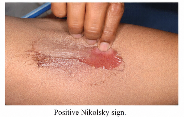
Gentle lateral pressure applied to clinically uninvolved-appearing skin resulted in superficial epidermal separation and extension of erosion, producing a positive Nikolsky sign.
Based on the clinical presentation of painful mucosal erosions, flaccid bullae, superficial cutaneous erosions, and a positive Nikolsky sign, pemphigus vulgaris was suspected.
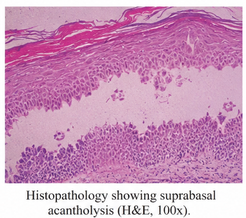
A lesional skin biopsy was performed for histopathological evaluation. Microscopic examination demonstrated suprabasal separation of the epidermis with acantholytic keratinocytes, resulting in an intraepidermal blister. The basal keratinocytes remained attached to the basement membrane, producing the characteristic "row of tombstones" appearance.
Direct immunofluorescence of perilesional skin demonstrated intercellular deposition of immunoglobulin G in a characteristic reticular or fish-net pattern throughout the epidermis. The combined clinical, histopathological, and immunofluorescence findings confirmed the diagnosis of pemphigus vulgaris with mucocutaneous involvement.
Management and Outcome
The patient was counseled regarding the autoimmune nature and chronic course of pemphigus vulgaris, as well as the importance of treatment adherence and regular follow-up. Systemic corticosteroid therapy was initiated to achieve rapid disease control, and a steroid-sparing immunosuppressive agent was subsequently introduced for long-term management.
Supportive wound care was provided for cutaneous erosions, with appropriate dressings applied to affected areas. Oral care measures were advised to reduce discomfort and maintain adequate hygiene. The patient was encouraged to maintain sufficient hydration and nutritional intake because of painful oral lesions.
During treatment, the patient was monitored for new blister formation, secondary infection, and potential adverse effects associated with systemic immunosuppression.
Over the following weeks, the formation of new blisters gradually decreased. Existing cutaneous erosions began to heal, while oral mucosal lesions improved significantly. The patient reported reduced pain and improved ability to eat and drink.
At follow-up, significant clinical improvement was observed, with substantial reduction in mucocutaneous disease activity and no new extensive blister formation.
Follow-up
One Month
-
Significant reduction in oral pain and discomfort was reported.
-
The number of newly developing blisters had markedly decreased.
-
Existing cutaneous erosions demonstrated progressive healing.
-
Oral mucosal erosions showed substantial improvement.
-
No significant secondary bacterial infection was observed.
Three Months
-
Nearly complete healing of previously extensive cutaneous erosions was noted.
-
No significant new flaccid bullae were observed.
-
Oral mucosal lesions had markedly improved, allowing comfortable oral intake.
-
The patient demonstrated good adherence to treatment.
Six Months
-
The patient remained clinically stable with minimal residual mucosal involvement.
-
No significant new blistering episodes were reported.
-
Previously affected areas demonstrated healing with post-inflammatory pigmentary changes.
-
Continued dermatological follow-up and immunosuppressive therapy were advised.
Discussion
Pemphigus vulgaris is a chronic autoimmune blistering disorder caused by pathogenic autoantibodies against desmosomal proteins involved in keratinocyte adhesion. Loss of intercellular adhesion, or acantholysis, results in fragile intraepidermal blisters that rupture easily and leave painful erosions.
The clinical presentation varies according to the distribution of desmosomal antigen involvement. Mucosal disease is common and may precede cutaneous manifestations. Oral erosions can be particularly painful and may interfere with eating, drinking, and oral hygiene.
Cutaneous involvement typically manifests as flaccid blisters that are easily disrupted by minor trauma. Consequently, patients often present with erosions and crusted lesions rather than intact bullae. The Nikolsky sign may be positive because of the fragility of epidermal adhesion.
Diagnosis requires correlation of clinical findings with histopathological and immunopathological investigations. Histopathology typically demonstrates suprabasal acantholysis, with separation of the epidermis above the basal cell layer. Direct immunofluorescence of perilesional skin is an important diagnostic investigation and demonstrates intercellular immunoglobulin G deposition in a characteristic reticular or fish-net pattern.
Serological testing for desmoglein-specific antibodies may provide additional diagnostic and disease-monitoring information. Differential diagnoses include pemphigus foliaceus, mucous membrane pemphigoid, bullous pemphigoid, and paraneoplastic pemphigus. Clinical distribution, histopathological findings, and immunofluorescence patterns help distinguish these disorders.
Treatment aims to suppress autoimmune activity, achieve disease control, promote healing, and prevent complications. Systemic corticosteroids are commonly used for rapid disease control, while steroid-sparing immunosuppressive agents may reduce cumulative corticosteroid exposure. Biologic therapies may be considered in selected patients according to disease severity and treatment response.
Supportive management is also important. Patients with extensive erosions may require wound care, infection prevention, nutritional support, and management of fluid and electrolyte disturbances. Oral lesions may require specialized dental and oral care because pain can interfere with adequate nutrition and hydration.
The prognosis of pemphigus vulgaris has improved with advances in diagnosis and systemic therapy. However, the disease may follow a relapsing-remitting course, requiring long-term monitoring for recurrence and treatment-related adverse effects.
In the present case, painful oral erosions, fragile cutaneous blisters, positive Nikolsky sign, suprabasal acantholysis, and characteristic intercellular immunoglobulin G deposition supported the diagnosis of pemphigus vulgaris. Early diagnosis and systemic treatment resulted in significant improvement in mucocutaneous disease activity and favorable clinical outcomes.
Prognosis
The prognosis of pemphigus vulgaris is generally favorable when diagnosed early and treated appropriately. However, prolonged systemic therapy and regular monitoring may be required because of the potential for relapse.
Patients with extensive mucocutaneous disease may be at risk of secondary infection, fluid and electrolyte imbalance, nutritional deficiencies, and treatment-related adverse effects. Disease recurrence may present with new blisters, erosions, or mucosal lesions after a period of clinical remission.
In this case, timely diagnosis and initiation of systemic immunosuppressive therapy resulted in substantial clinical improvement, with progressive healing of mucocutaneous lesions and no significant disease relapse during follow-up.
Conclusion
Pemphigus vulgaris is a potentially serious autoimmune blistering disorder that commonly presents with painful mucosal erosions and fragile cutaneous blisters. Because its initial manifestations may resemble other mucocutaneous disorders, early recognition and appropriate diagnostic evaluation are essential.
Histopathological examination demonstrating suprabasal acantholysis and direct immunofluorescence showing characteristic intercellular immunoglobulin G deposition are important for confirming the diagnosis.
This case highlights the importance of recognizing the clinical features of pemphigus vulgaris and promptly performing appropriate immunopathological investigations. Early systemic treatment, supportive wound and oral care, patient education, and long-term monitoring are essential for achieving disease control and minimizing complications.
References
-
Schmidt E, Kasperkiewicz M, Joly P. Pemphigus. Lancet. 2019;394(10201):882-894. https://pubmed.ncbi.nlm.nih.gov/31498129/
-
Bystryn JC, Rudolph JL. Pemphigus. Lancet. 2005;366(9479):61-73. https://pubmed.ncbi.nlm.nih.gov/15993232/
-
Murrell DF, Peña S, Joly P, et al. Diagnosis and management of pemphigus: Recommendations of an international panel of experts. J Am Acad Dermatol. 2020;82(3):575-585. https://pubmed.ncbi.nlm.nih.gov/31521135/
-
Hertl M, Jedlickova H, Karpati S, et al. Pemphigus. S2 Guideline for diagnosis and treatment—guided by the European Dermatology Forum. J Eur Acad Dermatol Venereol. 2015;29(3):405-414. https://pubmed.ncbi.nlm.nih.gov/25660157/
-
Harman KE, Brown D, Exton LS, et al. British Association of Dermatologists' guidelines for the management of pemphigus vulgaris 2017. Br J Dermatol. 2017;177(5):1170-1201. https://pubmed.ncbi.nlm.nih.gov/28820197/
Read more such content on @ Hidoc Dr | Medical Learning App for Doctors
Recommended News For You
Recommended Articles For You
Featured News
Featured Articles
Featured Events
Featured KOL Videos
1.
Promising OS at 5 Years With Chemo-Free Combo in Urothelial Cancer
2.
Black Canadians Face Multiple Barriers to Blood Donation
3.
Stop Calling It Cancer; The Alcohol Paradox; Rural Patients Lose Ground in Screening
4.
Experts say oncology, primary care coordination necessary for best cancer patient outcomes
5.
Neural networks and label-free microscopy enable accurate detection of pancreatic tumors
1.
Machine Learning in Hematology: Boosting Diagnostic Precision and Speed in Medical Practice
2.
Advancing Cancer Care: Insights into Oncology Trials, Immunotherapy, and CAR-T Innovations
3.
Clinical Frameworks in Hematology for Better Care
4.
Empowering Lung Cancer Diagnosis Through the Synergy of Advanced Technologies and Artificial Intelligence
5.
The Benefits of Understanding Haptoglobin High: What You Need to Know
1.
Asian Symposium on Advancement in Hematology and Oncology (ASAHO)
2.
International Cancer Conference
3.
Asian Symposium on Advancement in Hematology and Oncology (ASAHO)
4.
Asian Symposium on Advancement in Hematology and Oncology
5.
Asian Symposium on Advancement in Hematology and Oncology
1.
An Eagles View - Evidence-based Discussion on Iron Deficiency Anemia- Panel Discussion
2.
Untangling The Best Treatment Approaches For ALK Positive Lung Cancer - Part II
3.
Newer Immunotherapies for Myeloma- A Comprehensive Overview- Part II
4.
Managing ALK Rearranged Non-Small Cell Lung Cancer with Lorlatinib - Part IV
5.
An Eagles View - Evidence-based discussion on Iron Deficiency Anemia- Important Points to Know

Address :
Hidoc Dr. Inc. | Delaware C Corp | 1309 Coffeen Ave. Suite 1200, Sheridan WY, 82801
Phone :
+1-415-463-3094
Email :
anishagadia@hidoc.co
© Copyright 2026 Hidoc Dr. Inc.
Terms & Conditions - LLP | Inc. | Privacy Policy - LLP | Inc. | Account Deactivation

To get started please enter your email ID
Welcome to Hidoc Dr.
Join to enhance your clinical skills and gain specialized in-depth medical knowledge