Adenomyosis Presenting With Severe Dysmenorrhea, Menorrhagia, and an Enlarged Uterus: A Case Report
OthersPage Navigation
Abstract
Adenomyosis is a benign gynecological disorder characterized by the presence of endometrial glands and stroma within the myometrium, accompanied by surrounding smooth muscle hypertrophy. The condition commonly affects women of reproductive age and may present with heavy menstrual bleeding, progressive dysmenorrhea, chronic pelvic pain, or may remain asymptomatic. Clinical diagnosis can be challenging because symptoms overlap with other gynecological conditions, particularly uterine fibroids and endometriosis. We report the case of a 42-year-old woman who presented with progressively worsening menstrual pain, heavy menstrual bleeding, and chronic pelvic discomfort. Clinical examination revealed an enlarged and tender uterus. Pelvic ultrasonography demonstrated a heterogeneous myometrium with features suggestive of adenomyosis, which was further supported by magnetic resonance imaging. The patient was managed with medical therapy for symptom control, followed by definitive surgical treatment because of persistent symptoms and completed childbearing. Histopathological examination of the hysterectomy specimen confirmed the presence of endometrial glands and stroma within the myometrium, establishing the diagnosis of adenomyosis. The patient showed significant improvement following treatment. This case highlights the importance of considering adenomyosis in women presenting with heavy menstrual bleeding and dysmenorrhea and emphasizes the role of imaging and histopathological evaluation in diagnosis.
Introduction
Adenomyosis is a benign estrogen-dependent gynecological disorder characterized by the presence of endometrial glands and stroma within the uterine myometrium, often associated with reactive smooth muscle hyperplasia. The condition primarily affects women of reproductive age and may cause significant menstrual and pelvic symptoms.
The most common clinical manifestations include heavy menstrual bleeding, severe dysmenorrhea, chronic pelvic pain, and an enlarged or tender uterus. Some women may remain asymptomatic, and the condition may be identified incidentally during imaging or histopathological examination following hysterectomy.
The pathogenesis of adenomyosis remains incompletely understood. Several mechanisms have been proposed, including invagination of the endometrial basalis into the myometrium, tissue injury and repair mechanisms, metaplastic transformation, and alterations in hormonal and inflammatory pathways.
Adenomyosis may coexist with other gynecological disorders, including uterine leiomyomas and endometriosis, making clinical diagnosis challenging. Transvaginal ultrasonography is commonly used as an initial imaging modality, while magnetic resonance imaging may provide additional information when the diagnosis is uncertain or when detailed assessment of the uterus is required.
Treatment depends on symptom severity, age, reproductive plans, disease extent, and patient preference. Medical options include hormonal therapies aimed at reducing menstrual bleeding and pain, while uterine-sparing procedures and hysterectomy may be considered in selected patients. Hysterectomy remains a definitive treatment for women who have completed childbearing and have persistent symptoms despite conservative management.
We report a case of adenomyosis presenting with severe dysmenorrhea, heavy menstrual bleeding, and an enlarged uterus, emphasizing the importance of appropriate clinical evaluation and imaging in establishing the diagnosis.
Case Report
A 42-year-old woman presented to the gynecology outpatient department with a 1-year history of progressively worsening menstrual pain and heavy menstrual bleeding. She reported that her menstrual flow had gradually increased over the preceding months and was associated with severe lower abdominal and pelvic pain, particularly during menstruation.
The patient also complained of chronic pelvic heaviness and discomfort that had progressively affected her daily activities. She reported increased fatigue associated with heavy menstrual blood loss. There was no history of fever, abnormal vaginal discharge, or recent pelvic infection.

The patient had no significant history of previous gynecological malignancy or known endometriosis. Her menstrual cycles were otherwise regular. She had completed her family and did not desire future pregnancies. There was no significant family history of gynecological malignancy.
On clinical examination, the patient was hemodynamically stable. Abdominal examination revealed mild lower abdominal tenderness. Pelvic examination demonstrated an enlarged, globular, and mildly tender uterus. No significant adnexal mass was appreciated.
Based on the clinical presentation of heavy menstrual bleeding, progressive dysmenorrhea, pelvic pain, and an enlarged uterus, adenomyosis was considered among the differential diagnoses.
Laboratory investigations revealed anemia consistent with chronic menstrual blood loss. Other routine biochemical investigations were within acceptable limits.
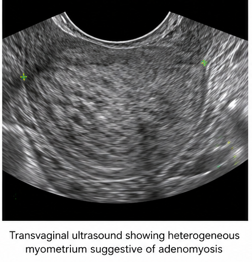
Transvaginal ultrasonography demonstrated an enlarged and heterogeneous uterus with features suggestive of adenomyosis, including heterogeneous myometrial echotexture and an ill-defined endometrial-myometrial junction. No significant adnexal mass was identified.
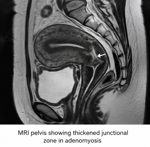
Magnetic resonance imaging of the pelvis was subsequently performed for further characterization. The imaging findings supported the diagnosis of adenomyosis, with thickening of the junctional zone and heterogeneous signal intensity within the myometrium.
Based on the clinical presentation and imaging findings, a diagnosis of symptomatic adenomyosis was made.
Management and Outcome
The patient was counseled regarding the chronic nature of adenomyosis and available treatment options. Because she had completed childbearing and continued to experience significant symptoms, medical treatment was initially considered for symptom control.
Hormonal therapy was initiated to reduce heavy menstrual bleeding and alleviate dysmenorrhea. Analgesic therapy was also provided for pain management.
Despite medical treatment, the patient continued to experience significant menstrual pain and heavy bleeding, which affected her quality of life. Given persistent symptoms and the patient's completed family, definitive surgical management was discussed.
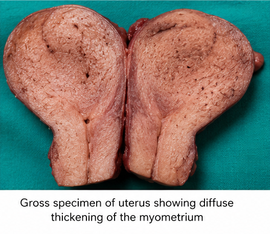
The patient subsequently underwent hysterectomy. The surgical specimen was submitted for histopathological examination.
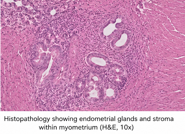
Microscopic examination demonstrated endometrial glands and endometrial stroma located within the myometrium, accompanied by surrounding smooth muscle hypertrophy. These findings confirmed the diagnosis of adenomyosis.
Following surgery, the patient's menstrual bleeding and dysmenorrhea resolved. Her postoperative recovery was uncomplicated, and she reported significant improvement in pelvic discomfort and overall quality of life during follow-up.
Follow-up
One Month
- The patient recovered well following definitive surgical treatment.
- No significant postoperative complications were reported.
- Pelvic pain and heavy menstrual bleeding had resolved.
- The patient reported improvement in daily activities and overall well-being.
Three Months
- The patient remained clinically stable.
- No recurrence of pelvic pain or abnormal uterine bleeding was reported.
- The patient demonstrated significant improvement in quality of life.
Six Months
- The patient remained symptom-free.
- No recurrent pelvic symptoms were noted.
- Continued routine gynecological follow-up was advised.
Discussion
Adenomyosis is a common benign uterine disorder characterized by the presence of endometrial glands and stroma within the myometrium. The condition may produce significant symptoms and can substantially affect physical, emotional, and reproductive health.
The clinical presentation varies considerably. Some women may remain asymptomatic, whereas others develop heavy menstrual bleeding, severe dysmenorrhea, chronic pelvic pain, or pressure-related symptoms. Heavy menstrual bleeding and progressive menstrual pain are among the most frequently reported clinical manifestations.
The uterus in adenomyosis may become enlarged and have a characteristic globular appearance. On pelvic examination, the uterus may be tender, particularly in patients with significant disease activity.
The differential diagnosis includes uterine leiomyomas, endometriosis, endometrial pathology, and other causes of abnormal uterine bleeding. Adenomyosis may also coexist with leiomyomas and endometriosis, further complicating clinical assessment.
Imaging plays an important role in diagnosis. Transvaginal ultrasonography is commonly used as the initial investigation and may demonstrate features such as a heterogeneous myometrium, myometrial cysts, fan-shaped shadowing, and an irregular or ill-defined junctional zone. MRI can provide better visualization of the uterine anatomy and junctional zone and may be particularly useful when ultrasound findings are inconclusive.
Histopathological examination demonstrates endometrial glands and stroma within the myometrium, generally accompanied by surrounding smooth muscle hyperplasia. Although histopathology has traditionally been considered the definitive method of diagnosis, advances in imaging have enabled adenomyosis to be recognized during the patient's lifetime.
Treatment should be individualized according to symptom severity, age, reproductive goals, disease extent, and patient preference. Medical treatment may include hormonal therapies aimed at reducing menstrual bleeding and suppressing symptoms. Nonsteroidal anti-inflammatory drugs may also be used to manage dysmenorrhea.
Uterine-sparing interventions may be considered in selected patients who wish to preserve fertility or avoid hysterectomy. However, hysterectomy remains the definitive treatment for women who have completed childbearing and have persistent symptoms despite conservative management.
In the present case, the combination of heavy menstrual bleeding, progressive dysmenorrhea, pelvic pain, and an enlarged uterus raised clinical suspicion of adenomyosis. Imaging findings supported the diagnosis, while histopathological examination following hysterectomy confirmed the presence of endometrial glands and stroma within the myometrium. Definitive treatment resulted in complete resolution of the patient's symptoms.
Prognosis
The prognosis of adenomyosis is generally favorable, particularly when symptoms are appropriately recognized and managed. However, the condition can significantly affect quality of life because of chronic pelvic pain, dysmenorrhea, and heavy menstrual bleeding.
Patients may experience persistent or recurrent symptoms despite medical therapy, and treatment selection should therefore be individualized. Women who desire future fertility may require conservative or uterus-preserving approaches, whereas hysterectomy provides definitive treatment for those who have completed childbearing.
In this case, timely diagnosis and appropriate management resulted in complete resolution of heavy menstrual bleeding and pelvic pain, with significant improvement in the patient's quality of life.
Conclusion
Adenomyosis should be considered in women presenting with heavy menstrual bleeding, progressive dysmenorrhea, chronic pelvic pain, and an enlarged or tender uterus. Because its clinical manifestations may overlap with other gynecological conditions, appropriate imaging is important for establishing a presumptive diagnosis.
Transvaginal ultrasonography serves as an important initial diagnostic modality, while MRI can provide additional characterization of myometrial involvement. Histopathological examination demonstrates the characteristic presence of endometrial glands and stroma within the myometrium and can confirm the diagnosis.
This case highlights the importance of early recognition of adenomyosis and individualized treatment planning based on symptom severity and reproductive goals. Medical therapy may provide symptom relief, while definitive surgical treatment may be appropriate for women with persistent symptoms who have completed childbearing.
References
- Dason S, Maxim M, Sanders A, et al. Guideline No. 437: Diagnosis and Management of Adenomyosis. J Obstet Gynaecol Can. 2023;45(6):417-429.e1. https://pubmed.ncbi.nlm.nih.gov/37244746/
- Selntigia A, Molinaro P, Tartaglia S, et al. Adenomyosis: An Update Concerning Diagnosis, Treatment, and Fertility. J Clin Med. 2024;13(17):5224. https://pubmed.ncbi.nlm.nih.gov/39274438/
- Moawad G, Fruscalzo A, Youssef Y, et al. Adenomyosis: An Updated Review on Diagnosis and Classification. J Clin Med. 2023;12(14):4828. https://pubmed.ncbi.nlm.nih.gov/37510943/
- Galati G, Ruggiero G, Grobberio A, et al. The Role of Different Medical Therapies in the Management of Adenomyosis: A Systematic Review and Meta-Analysis. J Clin Med. 2024;13(11):3302. https://pubmed.ncbi.nlm.nih.gov/38893013/
- Zeccola AM, Allen SE. Alternative Treatments of Adenomyosis: An Update in Procedural Management and Clinical Outcomes. Curr Opin Obstet Gynecol. 2024;36(4):287-295. https://pubmed.ncbi.nlm.nih.gov/38837727/
- McCaughey T, Mooney SS, Newman M, et al. International Delphi Consensus on the Histopathological Diagnosis of Adenomyosis. J Clin Pathol. 2024;77(7):502. https://pubmed.ncbi.nlm.nih.gov/38429094/
Read more such content on @ Hidoc Dr | Medical Learning App for Doctors
Recommended News For You
Recommended Articles For You
Featured News
Featured Articles
Featured Events
Featured KOL Videos
1.
Rural colon cancer patients face higher risks, but minimally invasive surgery could narrow gap
2.
Chemotherapy before surgery helps patients with nose and sinus cancer avoid debilitating eye and bone removal
3.
Laryngeal Transplant Helped a Cancer Patient Speak Again in Pioneering Study
4.
The amount of liver tissue removed during gallbladder surgery has no discernible impact on overall survival.
5.
Long-Acting Interferon Therapy Approved for ET
1.
Molecular Network Mapping in Hematologic Disease
2.
Metabolic Reprogramming of the Tumor Microenvironment: Mechanisms, Clinical Relevance, and Emerging Therapeutic Strategies
3.
Hematopoietic Stem Cell Clonal Dynamics Across the Lifespan
4.
Connected Hematology Laboratory Networks for Automated Cross-Institution Data Exchange
5.
Digital Oncology Ecosystems Integrating Longitudinal Patient Trajectories: Transforming Cancer Care through Data-Driven Innovation
1.
International Conference on Innovations in Critical Care for Oncology and Cardiology
2.
International Conference on Oncology, Cardiology and Critical Care Policy
3.
International Conference on Oncology, Cancer Prevention and Public Health
4.
International Conference on Cancer Nursing and Rehabilitation Strategies
5.
International Conference on Best Practices in Oncology, Cardiology and Critical Care
1.
Breaking Ground: ALK-Positive Lung Cancer Front-Line Management - Part V
2.
Common side effects of cancer treatment and how they are managed
3.
Clinical Excellence in Hematology
4.
Best Practices in Hematology
5.
Current Scenario of Blood Cancer- Further Discussion on Genomic Testing & Advancement in Diagnosis and Treatment

Address :
Hidoc Dr. Inc. | Delaware C Corp | 1309 Coffeen Ave. Suite 1200, Sheridan WY, 82801
Phone :
+1-415-463-3094
Email :
anishagadia@hidoc.co
© Copyright 2026 Hidoc Dr. Inc.
Terms & Conditions - LLP | Inc. | Privacy Policy - LLP | Inc. | Account Deactivation

To get started please enter your email ID
Welcome to Hidoc Dr.
Join to enhance your clinical skills and gain specialized in-depth medical knowledge