Papular Urticaria Presenting as Recurrent Intensely Pruritic Papules in a Child
OthersPage Navigation
Introduction
Papular urticaria, also known as persistent insect-bite reaction, is a papulovesicular hypersensitivity response to arthropod bites. It occurs more frequently in children, possibly because immunological tolerance to arthropod antigens has not yet developed. Fleas and mosquitoes are common triggers, although bedbugs, mites, gnats, and other insects may also be responsible. The offending bite is often not noticed or recalled by the patient. [1–3]
Papular urticaria typically manifests as recurrent crops of intensely pruritic, erythematous papules on exposed areas of the body. A central punctum or small vesicle may be visible. New lesions can develop while older lesions are resolving. Repeated scratching may result in excoriations, secondary bacterial infection, scarring, and post-inflammatory hyperpigmentation. [1,2]
Case Report
An 8-year-old boy was brought to the dermatology outpatient department with multiple intensely pruritic skin lesions that had appeared intermittently over six weeks. The eruption initially developed over both lower legs and subsequently involved the forearms, ankles, and waist.
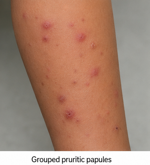
The patient’s mother reported that fresh lesions appeared every few days, particularly after the child played on the floor or with a recently adopted stray kitten. Individual lesions persisted for several days before gradually flattening and leaving dark marks.
The itching was moderate to severe and occasionally disturbed the child’s sleep. However, it was not exclusively nocturnal. There was no associated fever, malaise, joint pain, facial swelling, breathing difficulty, abdominal discomfort, or gastrointestinal complaint.
No other family member had developed similar lesions. There was no history of recent travel, use of a new medication, change in soap or detergent, or exposure to a new topical product. The child had a history of mild allergic rhinitis but no history of chronic eczema, asthma, or previous episodes of urticaria.
General examination revealed a comfortable and afebrile child with stable vital signs. Cutaneous examination demonstrated multiple discrete and grouped erythematous, oedematous papules measuring approximately 3–10 mm.
The lesions were predominantly distributed over the extensor aspects of the forearms, lower legs, ankles, and waistline. Several papules had a visible central punctum or tiny haemorrhagic crust. Excoriations were present over some lesions, while older lesions had evolved into hyperpigmented macules.
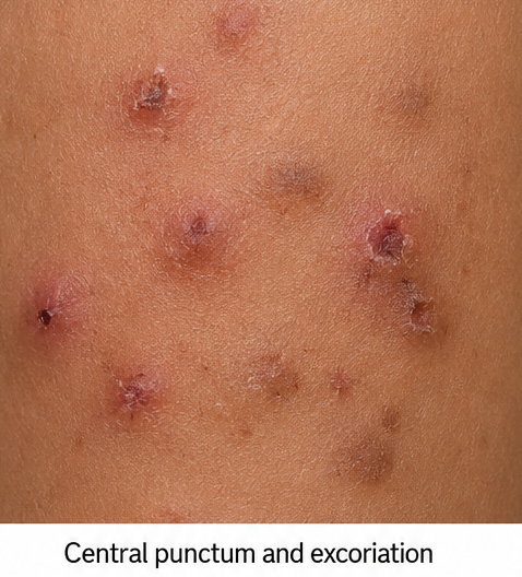
A few papules were arranged in short linear groups. The palms, soles, scalp, genital region, finger webs, and oral mucosa were unaffected. There were no burrows, follicular pustules, palpable purpura, transient wheals, blisters, or enlarged lymph nodes.
Investigations
A complete blood count was normal apart from mild peripheral eosinophilia of 7%. Liver and renal function tests were within normal limits. Extensive laboratory investigations were not considered necessary because the history and clinical findings did not suggest an underlying systemic disorder.
Skin scraping obtained from a representative lesion and the finger webs was negative for mites, eggs, and scybala. This finding reduced the likelihood of scabies.
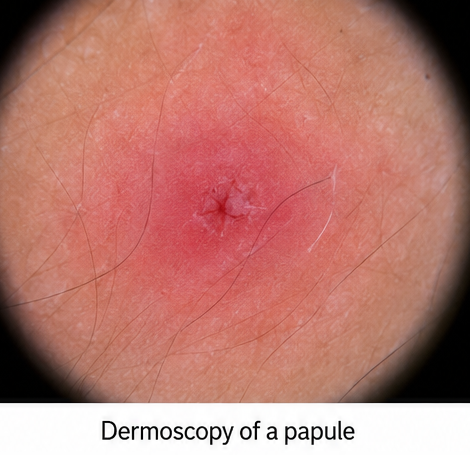
Dermoscopy of a recently developed papule showed a central punctum surrounded by an erythematous, structureless area with superficial crusting.
A punch biopsy was performed because of the recurrent nature of the eruption and parental concern. Histopathological examination revealed mild epidermal spongiosis with superficial and deep perivascular and interstitial inflammatory infiltrates containing lymphocytes and numerous eosinophils.
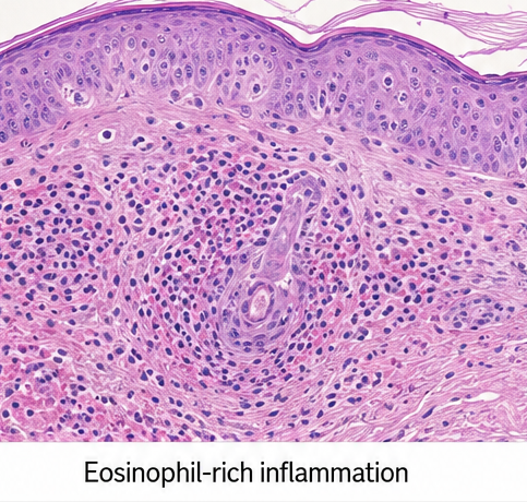
There was no evidence of vasculitis, granulomatous inflammation, or an atypical cellular infiltrate. The histopathological findings were compatible with an arthropod-bite hypersensitivity reaction. [4]
Veterinary examination of the recently adopted kitten subsequently confirmed flea infestation. The recurrent crops of papules, exposed-site distribution, central puncta, lesions at different stages of evolution, eosinophil-rich inflammation, and relevant environmental exposure supported the diagnosis of papular urticaria secondary to flea bites.
Differential Diagnosis
Scabies was considered because of the prominent itching. However, the absence of burrows, sparing of the finger webs and genital region, lack of similarly affected family members, negative skin scraping, and predominance of lesions over exposed areas argued against scabies.
Atopic dermatitis was considered but was unlikely because there was no chronic relapsing eczematous eruption, xerosis, typical flexural distribution, or previous history of atopic dermatitis.
Prurigo simplex may present with persistent pruritic papules. However, the presence of central puncta, lesions developing in crops, grouped distribution, and relevant household exposure favoured an insect-bite hypersensitivity reaction.
Bacterial folliculitis was excluded because the lesions were not centred around hair follicles and there were no pustules, tenderness, or purulent discharge.
Ordinary urticaria was considered less likely because the individual lesions persisted for several days and left residual pigmentation. In contrast, typical urticarial wheals generally resolve within 24 hours without leaving pigmentation. [5]
Management and Outcome
The family was counselled that successful treatment required both symptomatic relief and elimination of the arthropod source.
The kitten received appropriate veterinary treatment for flea infestation. The family was advised to wash bedding at a high temperature and thoroughly clean carpets, rugs, upholstered furniture, sleeping areas, and other locations where the kitten spent time. Professional pest-control measures were also arranged.
A medium-potency topical corticosteroid was prescribed as a thin layer over newly developed inflammatory papules once daily for seven days. The family was advised to avoid applying the corticosteroid to the face, flexures, or unaffected skin.
A non-sedating oral antihistamine was administered at an age-appropriate dose to control pruritus. Regular use of an emollient, application of cool compresses, maintenance of short fingernails, and avoidance of scratching were recommended.
There was no clinical evidence of secondary bacterial infection; therefore, topical or systemic antibiotics were not prescribed.
At the two-week follow-up visit, no new papules had appeared during the preceding five days. Pruritus had decreased substantially, and the excoriated lesions were healing.
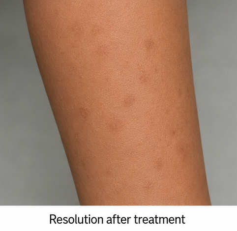
At six weeks, the active eruption had completely resolved. A few scattered post-inflammatory hyperpigmented macules remained over the lower legs and forearms. There was no secondary infection or permanent scarring.
At three months, the child remained free of recurrence following continued flea-control measures. The family was advised that the eruption could return with renewed exposure to fleas or other arthropods and that residual pigmentation might fade gradually over time.
Discussion
Papular urticaria represents an exaggerated hypersensitivity response to antigens present in arthropod saliva rather than conventional transient urticaria. Studies have associated the condition with exposure to fleas, mosquitoes, bedbugs, and mites. It is particularly common among susceptible children living in tropical and subtropical regions. [2,6]
The diagnosis is generally clinical. Important diagnostic features include recurrent crops of papules, intense pruritus, a central punctum or vesicle, predominance over exposed areas, grouped or asymmetric distribution, lesions at different stages of evolution, and residual post-inflammatory pigmentation.
Short linear clusters may suggest flea or bedbug bites. However, this distribution alone cannot reliably identify the responsible arthropod. [7] A focused clinical history should assess pet ownership, recent travel, sleeping arrangements, seasonal occurrence, affected household contacts, and signs of infestation.
Histopathological examination may support the diagnosis when the clinical presentation is atypical. Findings can include epidermal spongiosis, dermal oedema, and superficial and deep perivascular inflammatory infiltrates containing eosinophils. Nevertheless, these findings are not entirely specific and should be interpreted alongside the clinical history and lesion morphology. [4]
Treatment combines avoidance of further bites with symptomatic therapy. Protective clothing and appropriate insect repellents may reduce exposure. Pets and the household environment should be treated when fleas or mites are suspected.
Topical corticosteroids applied early to newly developed lesions can reduce cutaneous inflammation. Oral antihistamines may help control itching and prevent scratching. Secondary bacterial infection should be treated when present. [1,3]
The favourable response in this case followed recognition and elimination of the flea exposure. Treating only the itching without addressing the affected animal and household environment would probably have resulted in continued exposure and recurrent lesions.
Conclusion
Papular urticaria is a common but frequently misidentified hypersensitivity reaction to arthropod bites. Recurrent crops of intensely pruritic papules with central puncta, exposed-site distribution, and post-inflammatory pigmentation are important diagnostic clues.
A targeted exposure history may identify the responsible trigger even when the patient does not recall an insect bite. Management is most effective when topical anti-inflammatory and antipruritic treatment is combined with veterinary care and environmental control.
Early recognition can prevent continued exposure, secondary infection, scarring, pigmentary changes, and unnecessary investigations.
References
-
DermNet. Papular urticaria. https://dermnetnz.org/topics/papular-urticaria
-
Sekerel BE, et al. The many faces of pediatric urticaria. Front Allergy. 2023. https://pmc.ncbi.nlm.nih.gov/articles/PMC10655015/
-
Singh S, Mann BK. Insect bite reactions. Indian J Dermatol Venereol Leprol. 2013;79:151–164. https://pubmed.ncbi.nlm.nih.gov/23442453/
-
DermNet. Urticaria and urticaria-like conditions. https://dermnetnz.org/topics/urticaria-and-urticaria-like-conditions
-
DermNet. Urticaria: An overview. https://dermnetnz.org/topics/urticaria-an-overview
-
Halpert E, et al. Prevalence of papular urticaria caused by flea bites and associated factors in children. World Allergy Organ J. 2017;10:36. https://pmc.ncbi.nlm.nih.gov/articles/PMC5674867760. https://pmc.ncbi.nlm.nih.gov/articles/PMC6106677/
Read more such content on @ Hidoc Dr | Medical Learning App for Doctors
Recommended News For You
Recommended Articles For You
Featured News
Featured Articles
Featured Events
Featured KOL Videos
1.
The need for more Latinx participants in Alzheimer's trials is urgent.
2.
Study reveals crucial gaps in oral cancer awareness in Middle East and North Africa
3.
Disparities in use of MRI to detect prostate cancer: Race and location may influence testing
4.
In breast-conserving surgery, is micro-CT a useful tool for determining tumor margins?
5.
Study offers clearer picture of childhood brain tumor survival
1.
Revolutionizing Cancer Care: The Promise of Early Detection and Biomarkers
2.
The Oncologist’s Edge: Tools, Trials, and Tech Transforming Treatment
3.
Comprehensive Standards in Oncology for Modern Medicine
4.
Cardio-Oncology: Managing Heart Failure in Survivors of Cancer
5.
Practical Standards in Hematology for Healthcare Excellence
1.
Asian Symposium on Advancement in Hematology and Oncology (ASAHO)
2.
International Cancer Conference
3.
Asian Symposium on Advancement in Hematology and Oncology (ASAHO)
4.
Asian Symposium on Advancement in Hematology and Oncology
5.
Asian Symposium on Advancement in Hematology and Oncology
1.
Targeting Oncologic Drivers with Dacomitinib: A New Approach to Lung Cancer Treatment
2.
Revolutionizing Treatment of ALK Rearranged NSCLC with Lorlatinib - Part II
3.
Efficient Management of First line ALK-rearranged NSCLC - Part VIII
4.
Nimotozumab Plus Chemo-radiotherapy v/s Placebo Plus Chemo-radiotherapy in Locally Advanced Nasopharyngeal Carcinoma
5.
Breaking Ground: ALK-Positive Lung Cancer Front-Line Management - Part V

Address :
Hidoc Dr. Inc. | Delaware C Corp | 1309 Coffeen Ave. Suite 1200, Sheridan WY, 82801
Phone :
+1-415-463-3094
Email :
anishagadia@hidoc.co
© Copyright 2026 Hidoc Dr. Inc.
Terms & Conditions - LLP | Inc. | Privacy Policy - LLP | Inc. | Account Deactivation

To get started please enter your email ID
Welcome to Hidoc Dr.
Join to enhance your clinical skills and gain specialized in-depth medical knowledge