Morning Glory Syndrome: A Case Report of Congenital Optic Disc Anomaly, Diagnostic Evaluation, and Visual Management
OthersPage Navigation
Abstract
Morning glory syndrome (MGS), also known as morning glory disc anomaly, is a rare congenital malformation of the optic nerve characterized by a distinctive funnel-shaped excavation of the optic disc, radiating retinal vessels, and central glial tissue. The condition is usually unilateral and may cause variable degrees of visual impairment. Although the characteristic fundus appearance often establishes the diagnosis, multimodal ophthalmic imaging and neuroimaging may be required because MGS can be associated with intracranial and systemic abnormalities.
We report a case of a 12-year-old boy who presented with reduced vision in the right eye and difficulty identifying distant objects. Ophthalmic examination demonstrated reduced visual acuity and a characteristic enlarged, funnel-shaped optic disc excavation with radiating retinal vessels and central glial tissue. Optical coherence tomography demonstrated structural abnormalities involving the optic nerve head and adjacent retina. Neuroimaging did not identify a significant intracranial structural abnormality. The patient was diagnosed with unilateral morning glory syndrome and managed with refractive correction, visual rehabilitation, and regular ophthalmic surveillance.
This case highlights the importance of recognizing the characteristic appearance of MGS, differentiating it from other congenital optic disc anomalies, assessing for associated neurological abnormalities, and monitoring for retinal complications.
Introduction
Morning glory syndrome is a rare congenital optic nerve malformation named for the resemblance of the affected optic disc to the morning glory flower. It is characterized by an enlarged, funnel-shaped optic disc excavation, central glial tissue, and retinal vessels radiating outward from the disc margin. The condition is generally unilateral, although bilateral cases have been reported.
Visual function varies considerably. Patients may present with reduced visual acuity, visual field abnormalities, strabismus, or amblyopia, while some cases may be detected incidentally. The underlying embryological mechanism remains incompletely understood, with abnormalities in embryonic fissure closure and optic nerve development among the proposed mechanisms.
MGS may be associated with intracranial abnormalities, including basal encephalocele and cerebrovascular abnormalities. Neuroimaging is therefore important when the anomaly is identified, particularly in children or patients with neurological or craniofacial findings.
Retinal complications, including serous retinal detachment, retinoschisis-like changes, and macular abnormalities, may further compromise vision. Optical coherence tomography (OCT) provides valuable structural information and assists in identifying these complications.
Case Report
A 12-year-old boy presented with gradually reduced vision in the right eye. His parents had noticed difficulty with distant visual tasks over several months. There was no history of acute visual loss, ocular trauma, previous ocular surgery, recurrent inflammation, or significant headache.
There was no history of seizures, developmental delay, weakness, or other neurological symptoms. The child had no known congenital ocular disease or relevant family history.
On examination, the child was clinically stable, and neurological examination was unremarkable. Best-corrected visual acuity was reduced in the right eye compared with the left. Pupillary examination demonstrated an asymmetric response consistent with reduced optic nerve function in the affected eye.
Anterior segment examination was normal in both eyes.
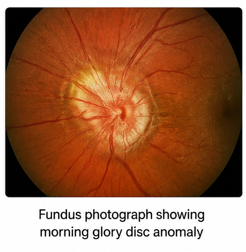
Dilated fundus examination of the right eye revealed an enlarged optic disc with a prominent funnel-shaped excavation. A central tuft of glial tissue was present, with retinal vessels radiating outward from the disc margin. Peripapillary pigmentation and structural abnormalities involving the posterior pole were also noted. The left optic disc was normal.
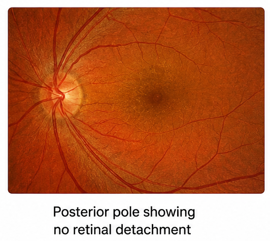
There was no clinical evidence of active retinal detachment.
Investigations
A comprehensive ophthalmic assessment was performed. Refraction was undertaken to identify any correctable component contributing to reduced vision.
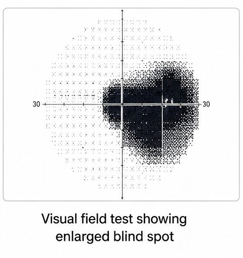
Visual field testing demonstrated an enlarged blind-spot region in the affected eye.
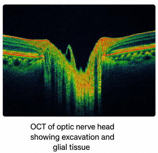
OCT of the optic nerve head and macula demonstrated abnormal excavation of the optic nerve head with associated structural changes involving the adjacent retina. The examination also evaluated for macular schisis, subretinal fluid, and other retinal complications.
Fundus photography documented the characteristic optic disc appearance, including the funnel-shaped excavation, central glial tissue, and radiating retinal vasculature.
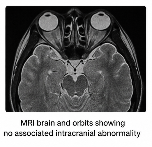
Because MGS may be associated with intracranial abnormalities, magnetic resonance imaging of the brain and orbits was performed. No basal encephalocele or major intracranial structural abnormality was identified.
The clinical and imaging findings supported the diagnosis of unilateral morning glory syndrome.
Diagnosis
The diagnosis was based on:
- Reduced visual acuity in the affected eye
- Enlarged, funnel-shaped optic disc excavation
- Central glial tissue within the optic disc
- Radiating retinal vessels
- Associated posterior pole structural abnormalities
- OCT findings consistent with a congenital optic nerve anomaly
- Visual field abnormality
- Neuroimaging assessment for associated intracranial abnormalities
The findings were consistent with unilateral morning glory syndrome.
Management and Outcome
Management focused on optimizing visual function, identifying associated abnormalities, and monitoring for ocular complications. There is currently no established treatment that reverses the underlying congenital optic disc malformation. [1]
Appropriate refractive correction was prescribed. Because reduced vision during childhood may be compounded by amblyopia, visual rehabilitation was initiated.
The parents were counseled regarding the congenital nature of the condition and the importance of regular ophthalmic follow-up.
Serial dilated fundus examinations were planned to monitor for retinal complications, particularly retinal detachment and macular involvement. OCT was repeated during follow-up to assess the optic nerve head, macula, and any development of intraretinal or subretinal fluid.
No surgical intervention was required because there was no retinal detachment or other sight-threatening retinal complication.
During follow-up, visual function remained stable. The patient continued refractive correction and visual rehabilitation, with no new neurological symptoms.
Follow-up
One Month
- Stable visual acuity
- Appropriate response to refractive correction
- No retinal detachment
- Stable neurological status
- Continued visual rehabilitation
Three Months
- Stable ocular examination
- No significant progression of visual symptoms
- Stable OCT findings
- No significant subretinal fluid
- Continued ophthalmic follow-up
Six Months
- Stable visual function
- No new neurological manifestations
- Stable optic disc appearance
- No significant retinal complications
- Continued surveillance
One Year
- Sustained visual stability
- No evidence of progressive retinal detachment
- No newly identified intracranial abnormality
- Continued visual rehabilitation
- Ongoing fundus and OCT monitoring
Discussion
Morning glory syndrome is an uncommon congenital optic nerve anomaly with a characteristic funduscopic appearance. The enlarged funnel-shaped disc excavation, central glial tissue, and radiating retinal vessels are important diagnostic features.
Visual impairment varies between patients and may result from optic nerve involvement, refractive error, amblyopia, or associated retinal abnormalities. Visual field defects may also occur.
Important differential diagnoses include optic nerve coloboma and peripapillary staphyloma. Optic nerve coloboma generally involves an excavation predominantly affecting the inferior optic disc, whereas peripapillary staphyloma is characterized by excavation surrounding the optic disc. Recognition of the characteristic morphology of MGS helps distinguish it from these conditions.
Multimodal imaging improves diagnostic assessment. OCT provides detailed visualization of the optic nerve head and retinal structures and can identify associated macular abnormalities.
Neuroimaging is important because MGS may coexist with intracranial abnormalities, including basal encephalocele and cerebrovascular abnormalities. Magnetic resonance imaging can help identify these associated developmental abnormalities and determine whether additional neurological assessment is required.
Retinal complications are an important consideration during follow-up. Serous retinal detachment, retinoschisis-like changes, and other forms of retinal detachment have been reported. OCT is particularly useful for detecting these changes at an early stage.
Patients without sight-threatening retinal complications are generally managed conservatively, with correction of refractive errors, visual rehabilitation, amblyopia management where appropriate, and regular monitoring. When retinal detachment or other significant retinal pathology develops, surgical intervention may be required.
This case emphasizes the importance of recognizing the characteristic optic disc appearance of MGS and performing appropriate ocular and systemic evaluation. Early identification allows clinicians to optimize visual function and monitor for potentially vision-threatening complications.
Prognosis
The prognosis depends on baseline visual function, degree of optic nerve involvement, presence of amblyopia, associated neurological abnormalities, and development of retinal complications.
Some patients retain useful vision, whereas others experience persistent visual impairment. Retinal detachment and macular involvement may further compromise visual outcomes.
Patients with associated intracranial abnormalities may require neurological or neurosurgical evaluation. Long-term ophthalmic surveillance remains important even when the initial presentation is uncomplicated because retinal complications may develop during follow-up.
Conclusion
Morning glory syndrome is a rare congenital optic nerve malformation characterized by an enlarged funnel-shaped optic disc excavation, central glial tissue, and radiating retinal vessels.
This case highlights the importance of detailed fundus examination, multimodal ophthalmic imaging, differentiation from other congenital optic disc anomalies, and assessment for associated intracranial abnormalities.
Although the underlying optic disc malformation cannot be reversed, refractive correction, visual rehabilitation, and regular ophthalmic surveillance can help optimize visual function. Continued monitoring for retinal detachment, macular abnormalities, and other ocular complications is essential for long-term management.
References
- American Academy of Ophthalmology EyeWiki. Morning Glory Anomaly. https://eyewiki.org/Morning_Glory_Anomaly
- Jeng-Miller KW, Cestari DM, Gaier ED. Congenital anomalies of the optic disc: insights from optical coherence tomography imaging. Curr Opin Ophthalmol. 2017;28(6):579-586. https://pubmed.ncbi.nlm.nih.gov/28817389/
- Jeng-Miller KW, et al. Morning glory disc anomaly: characteristic MR imaging findings. https://pubmed.ncbi.nlm.nih.gov/23660287/
- Kook MS, et al. Morning glory syndrome: clinical, computerized tomographic, and ultrasonographic findings. J Pediatr Ophthalmol Strabismus. https://pubmed.ncbi.nlm.nih.gov/16250218/
- Li Y, et al. The clinical characteristics and imaging findings of morning glory syndrome. Int J Ophthalmol. https://pubmed.ncbi.nlm.nih.gov/18704313/
- Yamada K, et al. Morning glory syndrome: MR imaging. AJNR Am J Neuroradiol. https://pubmed.ncbi.nlm.nih.gov/10506908/
- Park K, et al. Retinal complications associated with congenital optic disc anomalies determined by swept-source optical coherence tomography. Retina. https://pubmed.ncbi.nlm.nih.gov/29018703/
- Chang MY, et al. Retinal detachment associated with optic disc colobomas and morning glory syndrome. Eye (Lond). https://pubmed.ncbi.nlm.nih.gov/22241012/
Read more such content on @ Hidoc Dr | Medical Learning App for Doctors
Recommended News For You
Recommended Articles For You
Featured News
Featured Articles
Featured Events
Featured KOL Videos
1.
Extended Maintenance with Imatinib Enhances Disease-Free Survival in High-Risk GIST.
2.
Prostate cancer therapies: AI-based image analysis detects early organ damage
3.
Applications are being accepted for TN Health's post-doctoral fellowship program in gynecologic oncology and minimal access surgery; complete details are available here.
4.
OA Tied to Arthritis AE Risk in ICI-Treated Patients
5.
Dr. Prerana S. Nesargi's message for Childhood Cancer Awareness Month 2023 is to increase understanding of pediatric oncology.
1.
The Statistical Paradigm of Leukemia: From Evolving Epidemiology and Molecular Stratification to Precision Therapies and Future Horizons in Leukemia
2.
Hematopoietic Recovery After Childhood Therapy: Mechanisms, Clinical Implications, and Emerging Strategies
3.
Oncology Partnerships: Driving Innovation Through Strategic Collaborations and Pharma Engagement USA
4.
Understanding Verrucous Carcinoma: Symptoms, Diagnosis, and Treatment Options
5.
Genomic Control of Erythropoietic Stem Cell Renewal
1.
Asian Symposium on Advancement in Hematology and Oncology (ASAHO)
2.
International Cancer Conference
3.
Asian Symposium on Advancement in Hematology and Oncology (ASAHO)
4.
Asian Symposium on Advancement in Hematology and Oncology
5.
Asian Symposium on Advancement in Hematology and Oncology
1.
Management of 1st line ALK+ mNSCLC (CROWN TRIAL Update) - Part IV
2.
Summary of The Impact of CDK4/6 Inhibition in HR+/HER2- Metastatic Breast Cancer: Insights from PALOMA-2
3.
Efficient Management of First line ALK-rearranged NSCLC - Part VI
4.
Treatment Paradigm for Patients with R/R Adult B-cell ALL- Expert Discussions
5.
A Continuation to Deep Dive Into EGFR Mutation Positive Non-Small Cell Lung Cancer

Address :
Hidoc Dr. Inc. | Delaware C Corp | 1309 Coffeen Ave. Suite 1200, Sheridan WY, 82801
Phone :
+1-415-463-3094
Email :
anishagadia@hidoc.co
© Copyright 2026 Hidoc Dr. Inc.
Terms & Conditions - LLP | Inc. | Privacy Policy - LLP | Inc. | Account Deactivation

To get started please enter your email ID
Welcome to Hidoc Dr.
Join to enhance your clinical skills and gain specialized in-depth medical knowledge