Necrolytic Acral Erythema Associated with Chronic Hepatitis C Infection
OthersPage Navigation
Introduction
Necrolytic acral erythema is a rare inflammatory dermatosis characterized by recurrent, well-demarcated, hyperkeratotic plaques predominantly affecting the acral surfaces. The dorsal aspects of the feet and toes are the most commonly involved sites, although lesions may occasionally extend to the ankles, legs, knees, hands, and elbows. [1,2]
The condition was initially described as a cutaneous marker of hepatitis C virus infection. However, hepatitis C–negative cases associated with zinc deficiency, nutritional abnormalities, coeliac disease, Crohn’s disease, and other systemic conditions have subsequently been reported. [1–3]
Clinical manifestations vary according to the disease stage. Early lesions may appear as erythematous or violaceous papules, flaccid blisters, erosions, or scaly plaques. Chronic lesions become thick, hyperpigmented, sharply demarcated, and hyperkeratotic. Patients frequently report itching, burning, pain, or tenderness. [1,2]
Because the condition can resemble psoriasis, eczema, acrodermatitis enteropathica, or necrolytic migratory erythema, diagnosis may be delayed. Recognition is important because the eruption may reveal previously undiagnosed hepatitis C infection or an underlying nutritional deficiency.
Case Report
A 46-year-old man presented to the dermatology outpatient department with recurrent, painful, dark-coloured lesions over the dorsal surfaces of both feet for approximately eight months.
The eruption initially appeared as small reddish papules over the toes. These lesions gradually enlarged and merged to form scaly plaques extending across the dorsum of both feet. The patient reported intermittent burning, itching, and tenderness, particularly while wearing closed footwear or walking for prolonged periods.
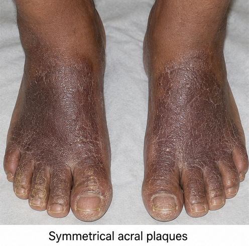
During the preceding three months, the lesions had become thicker and darker. Several superficial fissures had developed within the plaques, occasionally causing discomfort while walking. The patient denied lesions over the palms, soles, scalp, flexural areas, genital region, or oral mucosa.
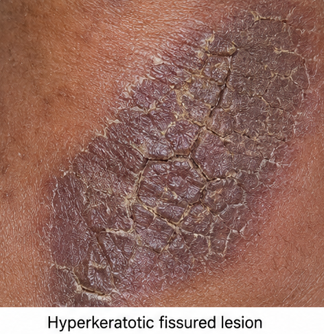
He had previously used topical corticosteroid and antifungal preparations prescribed elsewhere, with only temporary improvement. The lesions repeatedly returned after treatment was discontinued.
The patient reported fatigue and reduced appetite but denied fever, jaundice, abdominal pain, weight loss, diarrhoea, steatorrhoea, recurrent oral ulcers, or gastrointestinal bleeding. He had no known history of psoriasis, diabetes mellitus, inflammatory bowel disease, coeliac disease, pancreatic disease, or chronic nutritional disorder.
He reported receiving a blood transfusion approximately 15 years earlier following a road traffic accident. There was no history of intravenous drug use or previously diagnosed viral hepatitis.
General examination revealed a moderately built and nourished man with stable vital signs. There was no pallor, icterus, cyanosis, generalized lymphadenopathy, oedema, or clinically evident ascites.
Cutaneous examination demonstrated bilateral, symmetrical, well-demarcated hyperpigmented plaques over the dorsal aspects of the feet and toes. The plaques had an erythematous-to-violaceous peripheral margin, adherent scale, central hyperpigmentation, and areas of marked hyperkeratosis.
Several shallow fissures and superficial erosions were present within the thicker plaques. Mild tenderness was noted on palpation, but there was no purulent discharge, active bleeding, increased local temperature, or evidence of secondary bacterial infection.
The soles, interdigital spaces, nails, palms, scalp, elbows, and genital region were unaffected. No oral lesions, angular cheilitis, glossitis, or alopecia were observed.
The symmetrical acral distribution, recurrent course, characteristic hyperpigmented and hyperkeratotic plaques, and associated burning and tenderness raised suspicion of necrolytic acral erythema.
Investigations
A complete blood count showed mild normocytic anaemia. Fasting blood glucose, glycated haemoglobin, renal function, thyroid profile, and serum electrolytes were within normal limits.
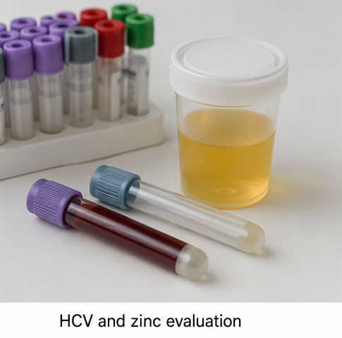
Liver function testing revealed mildly elevated serum aminotransferase levels. Serum albumin was slightly reduced. Serum zinc was below the laboratory reference range.
Screening for hepatitis C antibodies was positive. Hepatitis C virus RNA testing confirmed active infection. Screening tests for hepatitis B virus and human immunodeficiency virus were negative.
Serum glucagon was within the normal range. No clinical or laboratory findings suggested glucagonoma syndrome. Screening for coeliac disease was negative, and the patient had no gastrointestinal manifestations suggesting inflammatory bowel disease or significant malabsorption.
A potassium hydroxide examination of skin scrapings was negative for fungal elements.
A punch biopsy was obtained from the active margin of a plaque. Histopathological examination demonstrated hyperkeratosis, focal parakeratosis, epidermal pallor, spongiosis, and focal necrosis involving the superficial epidermis. A mild superficial perivascular inflammatory infiltrate was present within the dermis.

The clinical distribution, histopathological findings, low serum zinc level, and confirmed hepatitis C infection supported the diagnosis of necrolytic acral erythema associated with chronic hepatitis C infection.
Differential Diagnosis
Psoriasis was considered because of the well-demarcated scaly plaques. However, the absence of typical lesions over the scalp, elbows, knees, and sacral region, together with the acral distribution and necrolytic histopathological changes, favoured necrolytic acral erythema.
Chronic eczema can cause itching, scaling, lichenification, and fissuring. However, the symmetrical involvement of the dorsal feet, sharply defined hyperpigmented plaques, and associated hepatitis C infection were more consistent with necrolytic acral erythema.
Acrodermatitis enteropathica and acquired zinc-deficiency dermatosis were considered because the patient had a reduced serum zinc level. These disorders commonly affect acral and periorificial areas and may be accompanied by alopecia, diarrhoea, or impaired wound healing. The absence of periorificial disease and the characteristic dorsal foot distribution favoured necrolytic acral erythema.
Necrolytic migratory erythema was excluded because there were no migratory annular lesions involving the intertriginous, perineal, perioral, or lower abdominal regions. Serum glucagon was normal, and there were no systemic features suggestive of glucagonoma syndrome.
Tinea corporis was considered but was unlikely because fungal examination was negative and previous antifungal therapy had produced no sustained response.
Management and Outcome
The patient was counselled regarding the association between necrolytic acral erythema, zinc deficiency, and chronic hepatitis C infection. He was informed that successful management required treatment of both the cutaneous lesions and the underlying systemic condition.
Oral zinc supplementation was initiated under medical supervision. Baseline nutritional status was assessed, and periodic monitoring was planned to avoid complications associated with prolonged or excessive zinc supplementation.
The patient was advised to apply a bland emollient regularly to reduce dryness, scaling, and fissuring. Gentle cleansing was recommended, and he was instructed to avoid harsh soaps, repeated friction, tight footwear, and unnecessary use of topical preparations.
An appropriate dressing was used over the superficial erosions. The patient was advised to monitor for increasing pain, redness, warmth, swelling, or discharge that could indicate secondary infection.
He was referred to a hepatologist for evaluation and treatment of chronic hepatitis C infection. Further assessment of hepatic status and initiation of suitable antiviral therapy were planned according to the viral genotype, disease stage, potential drug interactions, and prevailing clinical recommendations.
At the four-week follow-up, burning and tenderness had reduced substantially. The erythematous margins had flattened, scaling had decreased, and no new erosions had appeared.
After eight weeks, the plaques showed marked reduction in thickness and hyperkeratosis. The fissures had healed, walking was more comfortable, and serum zinc had improved. Residual post-inflammatory hyperpigmentation remained over the dorsal feet.
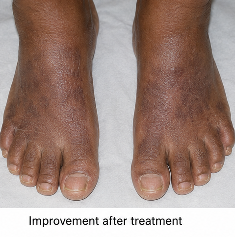
The patient was advised to continue follow-up for monitoring of the skin lesions, nutritional status, hepatitis C infection, and potential recurrence.
Discussion
Necrolytic acral erythema is recognized as a distinct member of the group of necrolytic erythemas. It is most strongly associated with chronic hepatitis C infection, although seronegative cases have also been documented. [1–3]
The precise pathogenesis remains uncertain and is probably multifactorial. Proposed mechanisms include zinc deficiency, abnormal zinc metabolism, reduced serum albumin, amino-acid deficiency, hepatic dysfunction, altered glucagon metabolism, and inflammatory or immune-mediated effects associated with hepatitis C infection. [1,2]
Zinc is essential for epidermal integrity, protein synthesis, wound healing, immune regulation, and several enzymatic processes. Zinc deficiency or impaired tissue availability may contribute to epidermal necrosis and the characteristic cutaneous changes. Importantly, some patients respond to zinc supplementation even when their serum zinc concentration is within the normal range. [1]
The eruption usually develops symmetrically over the dorsal surfaces of the feet and toes. The palms, soles, and nails are generally spared. Less commonly, lesions may appear over the ankles, legs, knees, hands, elbows, buttocks, or genital region. [1,4]
Early lesions may present as erythematous or violaceous papules, vesicles, flaccid blisters, erosions, or scaly plaques. During the established phase, individual lesions coalesce into sharply demarcated, thick, hyperpigmented plaques with adherent scale. Late lesions may become thinner and psoriasiform, with a dark red peripheral margin and residual pigmentation. [1,4]
Histopathological findings depend on the stage and biopsy site. Early lesions may demonstrate spongiosis, superficial epidermal pallor, and necrosis. Established lesions can show psoriasiform hyperplasia, hyperkeratosis, parakeratosis, epidermal necrosis, and a superficial inflammatory infiltrate. These findings are supportive but not entirely specific; clinical correlation is therefore essential. [1,2]
Evaluation should include testing for hepatitis C infection and assessment of liver function, serum zinc, albumin, glucose, and relevant nutritional parameters. Additional investigations for malabsorption, inflammatory bowel disease, or glucagonoma may be considered when indicated by the clinical presentation.
Oral zinc is the most consistently reported treatment and may improve lesions regardless of the initial serum zinc concentration. Treatment of an associated systemic disorder is equally important. In patients with hepatitis C infection, antiviral therapy may contribute to improvement or resolution of the eruption. [1,5]
Necrolytic acral erythema may follow a relapsing course, with recurrence after zinc supplementation is discontinued. Continued clinical monitoring and correction of the underlying metabolic or infectious abnormality are therefore important.
The improvement observed in this patient following zinc supplementation, together with management of the underlying hepatitis C infection, supported the diagnosis. Residual pigmentation persisted after the inflammatory and hyperkeratotic components improved, which is consistent with the chronic nature of the condition.
Conclusion
Necrolytic acral erythema should be considered in patients presenting with recurrent, symmetrical, painful, or pruritic hyperkeratotic plaques over the dorsal feet and toes.
Recognition of its characteristic distribution is important because the eruption may be an early cutaneous indicator of previously undiagnosed hepatitis C infection or an underlying nutritional abnormality.
Diagnosis requires clinicopathological correlation, testing for hepatitis C, assessment of zinc and nutritional status, and exclusion of clinically similar dermatoses.
Oral zinc supplementation and treatment of the associated systemic condition can produce substantial clinical improvement. Continued follow-up is necessary because recurrence may occur after treatment is withdrawn.
References
- Inamadar AC, Shivanna R, Ankad BS. Necrolytic acral erythema: current insights. Clinical, Cosmetic and Investigational Dermatology. 2020;13:275–281. https://pmc.ncbi.nlm.nih.gov/articles/PMC7147628/
- Abdallah MA, Ghozzi MY, Monib HA, Hafez AM, Hiatt KM, Smoller BR, Horn TD. Necrolytic acral erythema: a cutaneous sign of hepatitis C virus infection. Journal of the American Academy of Dermatology. 2005;53(2):247–251. https://pubmed.ncbi.nlm.nih.gov/16021118/
- Das A, Kumar P, Gharami RC. Necrolytic acral erythema in the absence of hepatitis C virus infection. Indian Journal of Dermatology. 2016;61(1):96–99. https://pmc.ncbi.nlm.nih.gov/articles/PMC4763711/
- Bansal S, Pathania S, Sawatkar GU, Jindal R. Dermoscopy in necrolytic acral erythema: a case report. Indian Dermatology Online Journal. 2024. https://pmc.ncbi.nlm.nih.gov/articles/PMC10868904/
- Kumar R, Arora S, Ranjan E, Das N. Necrolytic acral erythema in a seronegative hepatitis C patient with vitamin B12 deficiency. Indian Dermatology Online Journal. 2020;11(2):278–279. https://pmc.ncbi.nlm.nih.gov/articles/PMC7001408/
Read more such content on @ Hidoc Dr | Medical Learning App for Doctors
Recommended News For You
Recommended Articles For You
Featured News
Featured Articles
Featured Events
Featured KOL Videos
1.
Prostate cancer death risk is higher in late teens and early 20s weight gain.
2.
Bayer is transforming pharmaceutical research into women's health
3.
Biomarker panel offers hope for early pancreatic cancer detection
4.
A small rise in cancer was observed following the 1986 Chernobyl accident.
5.
Added Plinabulin Boosts OS as Later-Line Treatment for EGFR Wild-Type NSCLC
1.
Beyond the Brain Fog: The Complex Neurological Challenges and Therapeutic Advances
2.
Cancer Trials 2025: Vaccines, Metabolic Targets, and the Microbiome in Oncology
3.
Immune Microenvironment Biomarkers for Early Tumor Evolution
4.
Transformative Methods in Oncology and Quality Improvement
5.
Unveiling the Mystery of Epstein-Barr Virus: What We Know So Far

Address :
Hidoc Dr. Inc. | Delaware C Corp | 1309 Coffeen Ave. Suite 1200, Sheridan WY, 82801
Phone :
+1-415-463-3094
Email :
anishagadia@hidoc.co
© Copyright 2026 Hidoc Dr. Inc.
Terms & Conditions - LLP | Inc. | Privacy Policy - LLP | Inc. | Account Deactivation

To get started please enter your email ID
Welcome to Hidoc Dr.
Join to enhance your clinical skills and gain specialized in-depth medical knowledge