Elephantiasis of the Lower Limb Secondary to Lymphatic Filariasis
OthersPage Navigation
Introduction
Elephantiasis represents an advanced stage of chronic lymphoedema characterized by massive swelling, skin thickening, fibrosis, and functional impairment. In endemic regions, lymphatic filariasis is an important cause. It is transmitted through mosquito bites and is most commonly caused by Wuchereria bancrofti, while Brugia malayi and Brugia timori account for a smaller proportion of infections. [1,2]
Chronic lymphatic obstruction, recurrent inflammation, and secondary bacterial infections contribute to progressive tissue enlargement. The lower limbs are commonly affected, although the upper limbs, breasts, and genital organs may also be involved. Advanced disease can cause disability, recurrent infections, reduced mobility, psychological distress, and social stigma. [1–3]
Case Report
A 48-year-old man from a region endemic for lymphatic filariasis presented to the dermatology outpatient department with gradually progressive swelling of the left lower limb for approximately 12 years.
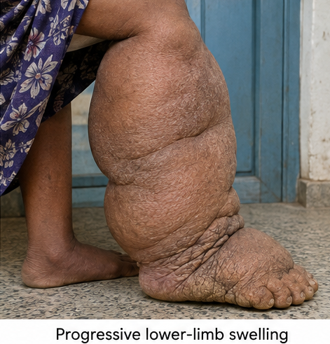
The swelling initially developed around the ankle and dorsum of the foot. It was intermittent during the early years and partially subsided after overnight rest. Over time, the swelling became persistent and progressively extended upward to involve the leg and lower thigh.
During the preceding four years, the patient had noticed increasing thickening and roughness of the overlying skin. He also reported repeated episodes of fever, chills, pain, redness, and warmth involving the affected limb. These episodes occurred three to four times annually and usually lasted several days.
The patient experienced difficulty walking, wearing regular footwear, and performing his occupational activities. He denied any history of trauma, surgery, radiotherapy, malignancy, tuberculosis, prolonged immobilization, or previous deep-vein thrombosis.
There was no similar swelling involving the opposite limb, upper limbs, or genital region. He had no history of breathlessness, orthopnoea, reduced urine output, facial puffiness, abdominal distension, or symptoms suggestive of cardiac, renal, or hepatic disease.
General examination revealed an afebrile patient with stable vital signs. There was no pallor, icterus, cyanosis, generalized lymphadenopathy, or clinically evident systemic oedema.
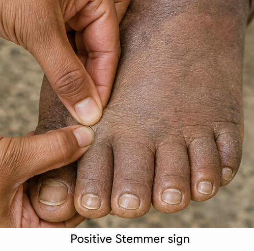
Local examination demonstrated massive non-pitting swelling of the left lower limb extending from the toes to the lower thigh. The dorsum of the foot was prominently involved, with squaring of the toes and inability to pinch the skin at the base of the second toe, indicating a positive Stemmer sign.
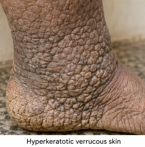
The overlying skin was markedly thickened, dry, hyperpigmented, and hyperkeratotic. Multiple verrucous and papillomatous projections produced a cobblestone-like appearance over the lower leg and ankle. The normal contours of the ankle were obscured.
Several deep skin folds were present, particularly around the ankle and posterior aspect of the leg. Mild maceration and superficial fissuring were observed within some folds. There was no active ulceration, purulent discharge, or evidence of acute cellulitis at presentation.
Peripheral pulses were palpable, and capillary refill was normal. Sensation was preserved. Movement of the ankle was restricted because of the size and weight of the limb, while the hip and knee joints were clinically normal.
Investigations
A complete blood count revealed mild eosinophilia. Liver function, renal function, serum albumin, thyroid profile, fasting blood glucose, and urinalysis were within normal limits.
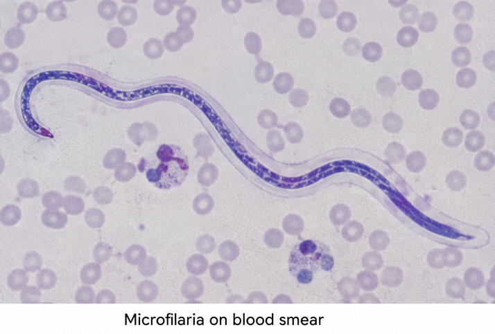
A nocturnal peripheral blood smear demonstrated microfilariae morphologically consistent with Wuchereria bancrofti. A circulating filarial antigen test was also positive, supporting active or recent bancroftian filarial infection. [2,4]
Venous Doppler ultrasonography showed no evidence of deep-vein thrombosis or significant chronic venous obstruction. Soft-tissue ultrasonography demonstrated diffuse thickening of the skin and subcutaneous tissues with interstitial fluid accumulation.
Abdominal and pelvic ultrasonography did not reveal a mass, enlarged lymph nodes, or another structural lesion causing proximal lymphatic obstruction. Chest radiography and electrocardiography showed no abnormality suggestive of cardiac disease.
The longstanding unilateral swelling, positive Stemmer sign, characteristic cutaneous changes, history of recurrent inflammatory episodes, residence in a filariasis-endemic region, and positive filarial investigations supported the diagnosis of advanced elephantiasis secondary to lymphatic filariasis.
Differential Diagnosis
Chronic venous insufficiency was considered because it can produce persistent lower-limb swelling and skin pigmentation. However, the prominent involvement of the foot and toes, positive Stemmer sign, marked verrucous thickening, and absence of significant venous abnormalities on Doppler examination favoured lymphatic obstruction.
Deep-vein thrombosis was excluded by the chronic progressive course and normal venous Doppler findings.
Primary lymphoedema was considered, but the patient’s adult-onset disease, recurrent inflammatory attacks, endemic exposure, eosinophilia, positive circulating filarial antigen, and identification of microfilariae supported secondary filarial lymphoedema.
Elephantiasis nostras verrucosa may develop from chronic non-filarial lymphoedema and can have similar hyperkeratotic and verrucous changes. However, no alternative cause of chronic lymphatic obstruction was identified in this patient, while the parasitological investigations supported lymphatic filariasis. [5]
Systemic causes such as cardiac failure, nephrotic syndrome, chronic liver disease, and severe hypoalbuminaemia were considered unlikely because the swelling was unilateral and the relevant clinical and laboratory findings were normal.
Management and Outcome
The patient was counselled that antifilarial treatment could reduce parasitic infection and transmission but might not reverse the established structural changes associated with advanced elephantiasis.
Following clinical assessment and exclusion of relevant contraindications, antifilarial therapy with diethylcarbamazine was prescribed under physician supervision. Treatment decisions were individualized according to regional recommendations and the possibility of co-endemic filarial infections. [2,4]
Long-term morbidity management was emphasized. The patient was taught to wash the affected limb and deep skin folds daily with soap and clean water. The limb was dried carefully, particularly between the toes and within the folds.
A topical antifungal preparation was prescribed for interdigital maceration. Emollients were advised for dry and fissured skin, while direct application within persistently moist interdigital spaces was avoided. The patient was instructed to inspect the limb daily for cuts, abrasions, entry lesions, redness, warmth, or discharge.

Limb elevation, regular movement, graded walking, and gentle exercises were recommended to support lymphatic drainage and preserve joint mobility. Compression therapy was initiated cautiously after confirming adequate arterial circulation and excluding acute infection.
The patient was advised to wear clean, appropriately fitted footwear and avoid walking barefoot. Weight management and mosquito-bite prevention measures, including protective clothing, insect repellents, and bed nets, were discussed.
At the four-week follow-up, there had been no acute inflammatory episode. The interdigital maceration had resolved, the skin folds were cleaner, and the patient reported reduced heaviness of the limb.
At three months, the limb remained enlarged, but there was a modest reduction in circumference and improvement in skin texture. Walking had become easier, and no new episodes of fever, cellulitis, or lymphangitis were reported.
The patient was advised that continued skin care, exercise, limb elevation, compression where appropriate, and prompt treatment of entry lesions would be necessary to reduce recurrent attacks and prevent further progression.
Discussion
Lymphatic filariasis is a mosquito-borne neglected tropical disease that damages the lymphatic system. Adult filarial worms reside in lymphatic vessels, where they may cause lymphatic dilatation and dysfunction. Clinical disease may appear years after the initial infection. [1,2]
Elephantiasis develops through a complex interaction between lymphatic damage, persistent lymph stasis, chronic inflammation, tissue fibrosis, and recurrent secondary infections. The skin becomes thickened, hyperkeratotic, papillomatous, and increasingly vulnerable to fissures and microbial entry.
Acute dermatolymphangioadenitis episodes are important contributors to disease progression. Many of these attacks are related to secondary bacterial infection entering through damaged skin or interdigital lesions rather than direct parasite activity. Repeated attacks further impair lymphatic function and increase swelling. [1,3]
Diagnosis is based on the clinical appearance, epidemiological exposure, and appropriate laboratory investigations. Circulating filarial antigen testing is particularly useful for W. bancrofti. Microfilariae may be demonstrated on peripheral blood examination, with the timing of blood collection guided by their periodicity. [2,4]
Negative parasitological testing does not always exclude previous filarial infection in a patient with chronic lymphoedema because microfilaraemia may be absent during advanced disease. Alternative causes of lymphatic obstruction must therefore be evaluated according to the clinical context.
Antifilarial medicines target infection but have limited ability to reverse advanced fibrosis and established elephantiasis. Consequently, morbidity management is central to long-term care. Daily washing, meticulous drying, skin and nail care, treatment of entry lesions, exercise, limb elevation, appropriate footwear, and compression therapy can decrease acute attacks and help prevent progression. [1,3,6]
The improvement observed in this patient was primarily reflected by fewer inflammatory episodes, healthier skin, reduced limb heaviness, and better mobility rather than complete reversal of the enlargement. This highlights the importance of combining antiparasitic treatment with sustained lymphoedema care.
Conclusion
Elephantiasis is a severe and disabling manifestation of chronic lymphatic dysfunction. In endemic regions, lymphatic filariasis should be considered in patients presenting with longstanding limb swelling, a positive Stemmer sign, recurrent inflammatory attacks, and hyperkeratotic or verrucous skin changes.
Diagnosis requires clinical evaluation, assessment of endemic exposure, targeted filarial investigations, and exclusion of venous, systemic, malignant, and other causes of lymphatic obstruction.
Although advanced structural changes may be irreversible, early diagnosis and consistent morbidity management can reduce recurrent infections, limit disease progression, improve mobility, and enhance quality of life.
References
- World Health Organization. Lymphatic filariasis. https://www.who.int/news-room/fact-sheets/detail/lymphatic-filariasis
- Centers for Disease Control and Prevention. Clinical overview of lymphatic filariasis. https://www.cdc.gov/filarial-worms/hcp/clinical-overview/index.html
- World Health Organization. Lymphatic filariasis: managing morbidity and preventing disability. https://www.who.int/publications/i/item/9789241505291
- Centers for Disease Control and Prevention. Clinical treatment of lymphatic filariasis. https://www.cdc.gov/filarial-worms/hcp/clinical-care/index.html
- Kharroubi A, et al. Elephantiasis nostras verrucosa of the lower limb. Cureus. 2024. https://pmc.ncbi.nlm.nih.gov/articles/PMC11018103/
- Medeiros ZM, et al. Lymphatic filariasis: a systematic review on morbidity and its repercussions in countries in the Americas. International Journal of Environmental Research and Public Health. 2022;19(1):316. https://www.mdpi.com/1660-4601/19/1/316
Read more such content on @ Hidoc Dr | Medical Learning App for Doctors
Recommended News For You
Recommended Articles For You
Featured News
Featured Articles
Featured Events
Featured KOL Videos
1.
The need for more Latinx participants in Alzheimer's trials is urgent.
2.
Study reveals crucial gaps in oral cancer awareness in Middle East and North Africa
3.
Disparities in use of MRI to detect prostate cancer: Race and location may influence testing
4.
In breast-conserving surgery, is micro-CT a useful tool for determining tumor margins?
5.
Study offers clearer picture of childhood brain tumor survival
1.
Revolutionizing Cancer Care: The Promise of Early Detection and Biomarkers
2.
The Oncologist’s Edge: Tools, Trials, and Tech Transforming Treatment
3.
Comprehensive Standards in Oncology for Modern Medicine
4.
Cardio-Oncology: Managing Heart Failure in Survivors of Cancer
5.
Practical Standards in Hematology for Healthcare Excellence
1.
Asian Symposium on Advancement in Hematology and Oncology (ASAHO)
2.
International Cancer Conference
3.
Asian Symposium on Advancement in Hematology and Oncology (ASAHO)
4.
Asian Symposium on Advancement in Hematology and Oncology
5.
Asian Symposium on Advancement in Hematology and Oncology
1.
Targeting Oncologic Drivers with Dacomitinib: A New Approach to Lung Cancer Treatment
2.
Revolutionizing Treatment of ALK Rearranged NSCLC with Lorlatinib - Part II
3.
Efficient Management of First line ALK-rearranged NSCLC - Part VIII
4.
Nimotozumab Plus Chemo-radiotherapy v/s Placebo Plus Chemo-radiotherapy in Locally Advanced Nasopharyngeal Carcinoma
5.
Breaking Ground: ALK-Positive Lung Cancer Front-Line Management - Part V

Address :
Hidoc Dr. Inc. | Delaware C Corp | 1309 Coffeen Ave. Suite 1200, Sheridan WY, 82801
Phone :
+1-415-463-3094
Email :
anishagadia@hidoc.co
© Copyright 2026 Hidoc Dr. Inc.
Terms & Conditions - LLP | Inc. | Privacy Policy - LLP | Inc. | Account Deactivation

To get started please enter your email ID
Welcome to Hidoc Dr.
Join to enhance your clinical skills and gain specialized in-depth medical knowledge